3636
Axon-mimicking hydrophilic fibre phantoms for diffusion MRI1Quantitative Biomedical Imaging Laboratory, The University of Manchester, Manchester, United Kingdom, 2School of Materials, The University of Manchester, Manchester, United Kingdom, 3Developmental Imaging and Biophysics, UCL Great Ormond Street Institute of Child Health, London, United Kingdom, 4National Physical Laboratory, Teddington, London, United Kingdom, 5College of Textile and Clothing Engineering, Soochow University, Suzhou, China, 6Bioxydyn Limited, Manchester, United Kingdom
Synopsis
We report the development of an axon-mimicking phantom composed of hydrophilic hollow microfibres, and evaluate its potential for validating clinical diffusion MRI. Microfibers were fabricated by the co-electrospinning (co-ES) of polycaprolactone (PCL)-polysiloxane-based surfactant (PSi) mixture as shell and polyethylene oxide (PEO) as core, and characterized by scanning electron microscopy (SEM). Three material samples were constructed and included in the phantom within a water bath. SEM images reveal that PCL-PSi fibres in the samples were uniaxially aligned and hollow, with a similar distribution of pore sizes to axons in vivo. MR measurement shows similar anisotropic diffusion behaviour in each sample.
Introduction
We have previously created hollow polymeric microfibres from polycaprolactone (PCL)1 and constructed them into white matter (WM),2 grey matter (GM),3 and cardiac phantoms 4 for validating diffusion MRI (dMRI). These phantoms were filled with cyclohexane due to PCL hydrophobicity. They demonstrated excellent performance, in terms of MR sensitivity to fibre orientation and sizes and reproducibility of MR measurement.2,4,5 However, the use of cyclohexane causes practical problems, including its evaporation and its potential hazard, leading to complications when transporting phantoms in multi-centre studies. We have recently shown that adding a hydrophilic surfactant (PSi) into PCL fibres can improve their water wettability.6 This work develops a clinical MR phantom from hollow PCL-polysiloxane-based surfactant (PSi) microfibres and assesses its potential as a tool for validating dMRI.Methods
Microstructural characterisation and phantom construction
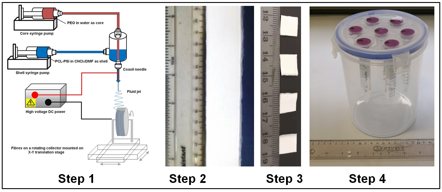
Hollow PCL-PSi microfibres were produced in strip form (thickness: ~0.5 mm) by co-electrospinning (co-ES) (Fig. 1). Pore diameter was calculated from 5 SEM images for each sample using ImageJ.7 A phantom sample was constructed by packing ~15 fibre layers (length: ~15 mm; width: ~10 mm) into a 15 ml general centrifuge plastic tube filled with water. Three samples were created from three fibre strips produced under identical process parameters and assembled into a round plastic container (Fig. 1, inner diameter: ~140 mm; height: ~180 mm) that can house up to 7 samples.
Wettability of PCL-PSi microfibres
Water wettability was evaluated using Krϋss DSA 100 Drop Size Analyzer (Krüss GmbH, Germany).
MR acquisition and analysis
The phantom container was filled with water and scanned on a 3T Siemens Prisma with a 64-channel head coil. A 60-direction DTI scan was performed, with b = 1000 and 2000 s/mm2, ∂ =19 ms, ∆ = 28 ms, TR = 3050 ms, TE = 60 ms, and voxel size = 2 x 2 x 2.2 mm3. Images were corrected with Topup and Eddy using TractoR,8 and MD and FA maps were obtained with TractoR using FSL and a weighted least squares fit. Median and inter-quartile range (IQR) were calculated over 4 slices per sample.
Results
SEM characterisation
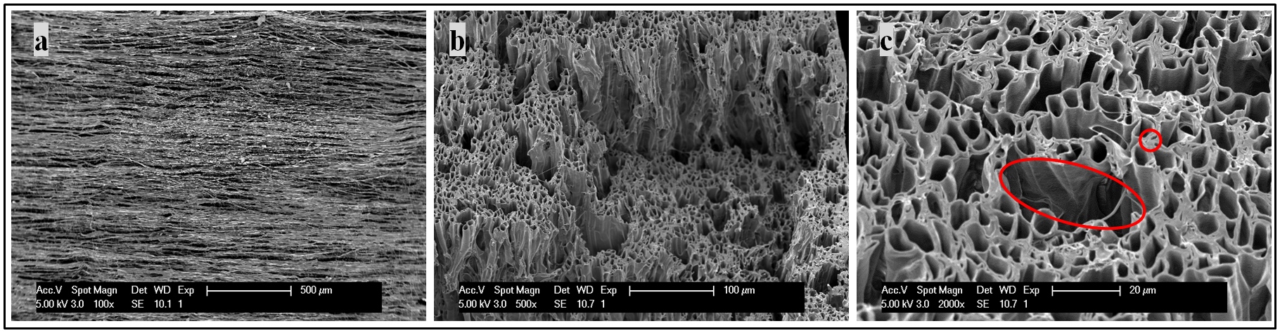
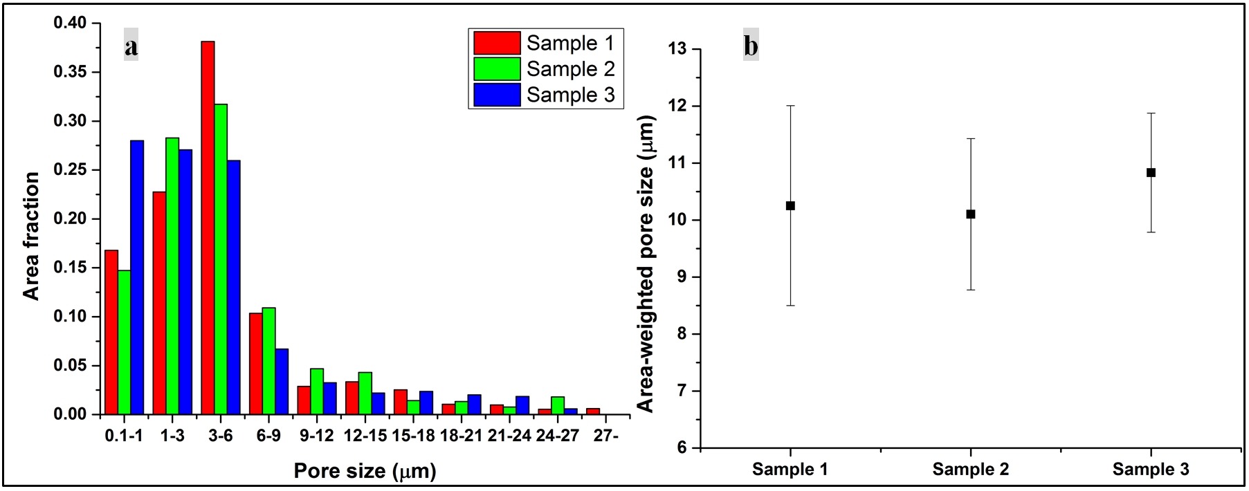
Fig. 2 reveals that PCL-PSi fibres are uniaxially aligned and porous, and look similar to previous PCL fibres.2,7 Fig. 3a shows the range of inner diameters for each of these phantoms. There is a broad range of fiber inner diameters within the phantom, reminiscent of axonal distributions.9 The distributions of pore sizes were found to be not significantly different across samples (Kruskal-Wallis, p = 0.99). Fig. 3b shows area-weighted pore size of the three samples, (10.3 ± 1.7, 10.1 ± 1.3 and 10.8 ± 1.0 μm, for samples 1, 2 and 3, respectively), which were not significantly different (One Way ANOVA, p = 0.69).
Water wettability
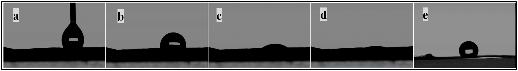
The addition of PSi rendered PCL microfibres hydrophilic, as evidenced by the fact that water droplet spread completely on PCL-PSI microfibres in ~10 seconds (Fig. 4a-d), in comparison with relatively intact water droplet on PCL fibres over a longer period of time (Fig. 4e).
MR measurement
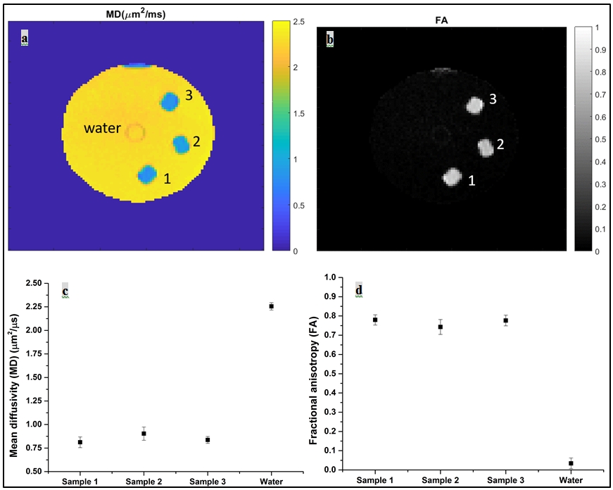
Fig. 5a-b shows MD and FA maps, from which MD and FA values (Fig. 5c-d) of the three samples are 0.81 ± 0.06, 0.90 ± 0.07, 0.84 ± 0.04 µm2/ms and 0.78 ± 0.03, 0.74 ± 0.04, 0.78 ± 0.03, respectively.
Discussion
The development of clinical phantoms requires not only appropriate microstructures, but also sufficient bulk material to be used with clinical imaging protocols. The macro- and microstructures of PCL-PSi microfibres were effectively controlled using co-ES,7 and have similar microstructure to PCL microfibres but, crucially, are hydrophilic. We found that the pore sizes and diffusion results are consistent across the three samples, indicating good manufacturing repeatability. The small difference in pore size distributions is possibly due to the sampling variability in the semi-automated SEM sampling and measurement process or due to the freeze-fracture process used in SEM preparation; however, there is no significant difference in area-weighted pore sizes among the three samples. A Kruskal-Wallis test identified differences between the samples for MD and FA, but these differences were small (a maximum of 0.09 µm2/ms and 0.04 for MD and FA, respectively). This phantom also has the potential to include multiple different samples, such as developed aligned,2 random,3 and crossing fibres,10 and tumour cell-mimicking microspheres.11,12Conclusion
A novel hydrophilic axon-mimicking clinical dMRI phantom has been developed, reflecting in vivo diffusion. The phantom exhibits diffusivity and anisotropy that are in the range expected for white matter, indicating that they can provide a helpful standard for diffusion measurements on clinical scanners.Acknowledgements
This research was supported by "CONNECT”, the FET Programme (FET-Open grant number: 238292) and the CRUK and EPSRC Cancer Imaging Centre in Cambridge and Manchester (C8742/A18097). FL Zhou received a travel grant from the Wellcome Trust [105610/Z/14/Z] for the collaboration with ZX Li on hydrophilic polymers. GJM Parker has a shareholding and part time appointment and directorship at Bioxydyn Ltd. which provides MRI services.References
1. Zhou FL, Hubbard PL, Eichhorn SJ, et al. Coaxially electrospun axon-mimicking fibers for diffusion magnetic resonance imaging. ACS Appl. Mater. Interfaces. 2012; 4: 6311-6316.
2. Hubbard PL, Zhou FL, Eichhorn SJ, et al. Biomimetic phantom for the validation of diffusion magnetic resonance imaging. Magn. Reson. Med. 2015; 73: 299-305.
3. Ye AQ, Cristinacce Hubbard PL, Zhou FL, et al. In Diffusion tensor MRI phantom exhibits anomalous diffusion. 36th Annual International Conference of the IEEE EMBS, pp. 746-749. 26-30 Aug. 2014.
4. Teh I, Zhou FL, Hubbard Cristinacce PL, et al. Biomimetic Phantom for Cardiac Diffusion MRI. J. Magn. Reson. Imaging. 2015; 43: 594-600.
5. Grech-Sollars M, Zhou FL, Waldman AD, et al. Stability and reproducibility of co-electrospun brain-mimicking phantoms for quality assurance of diffusion MRI sequences. NeuroImage. 2018; 181: 395-402.
6. Zhou FL, Li ZX, Gough JE, et al. Axon mimicking hydrophilic hollow polycaprolactone microfibres for diffusion magnetic resonance imaging. Mater. Design. 2018; 137: 394-403.
7. Zhou FL, Eichhorn SJ, Parker GJM, et al. Production and cross-sectional characterization of aligned co-electrospun hollow microfibrous bulk assemblies. Mater. Charact. 2015; 109: 25-35.
8. Clayden JD, Muñoz Maniega S, Storkey AJ, et al. TractoR: Magnetic resonance imaging and tractography with R. J. Statist. Softw. 2011; 44: 1-18.
9. Assaf Y, Blumenfeld-Katzir T, Yovel Y, et al. AxCaliber: a method for measuring axon diameter distribution from diffusion MRI. Magn. Reson. Med. 2008;1354: 1347-1354.
10. Hubbard PL, Zhou FL, Eichhorn SJ, et al. A crossing fibre phantom for diffusion MRI composed of co-electrospun fibres. ISMRM-ESMRMB 2014, No. 2653. Milan, Italy, 10-16 Jun. 2014.
11. McHugh DJ, Zhou FL, Cristinacce Hubbard PL, et al. Ground Truth for Diffusion MRI in Cancer: A Model-Based Investigation of a Novel Tissue-Mimetic Material. IPMI 2015, pp. 179-190. Isle of Skye, UK, 28 June 28 - 3 July, 2015.
12. McHugh DJ, Zhou FL, Wimpenny I, et al. A biomimetic tumour tissue phantom for validating diffusion-weighted MRI measurements. Magn. Reson. Med. 2018; 33:13262-13271.
Figures