3632
Tunable diffusion kurtosis in lamellar vesicle suspensions toward development of quantitative phantom surrogate of tumor microenvironment1Radiology, University of Michigan, Ann Arbor, MI, United States
Synopsis
A set of materials based on nanostructured lamellar vesicles with restricted diffusion compartments is constructed to achieve tunable diffusion kurtosis behavior. The observed apparent diffusion (Dapp) and kurtosis (Kapp) model parameters span the range of values found in vivo. Effect of vesicle population, size, and porosity is studied for estimated diffusion parameters. These nanostructured systems provide an ideal platform for a diffusion kurtosis phantom used to validate quantitative imaging protocols and results.
Introduction
Recent studies show that Diffusion Kurtosis Imaging (DKI) (1) may be useful in characterizing and monitoring changes in cellularity associated with cancer progression and therapy response (2-4). These studies highlight the need for quantitative DKI phantoms that validate results in multisite clinical settings. The synthetic DKI phantoms developed to date lack range of diffusion parameters observed in vivo (9). This abstract describes a system of tunable nanostructured materials (5-8) that generate diffusion and kurtosis values observed in vivo. These nanostructures provide an ideal platform for a quantitative diffusion kurtosis phantom.Methods
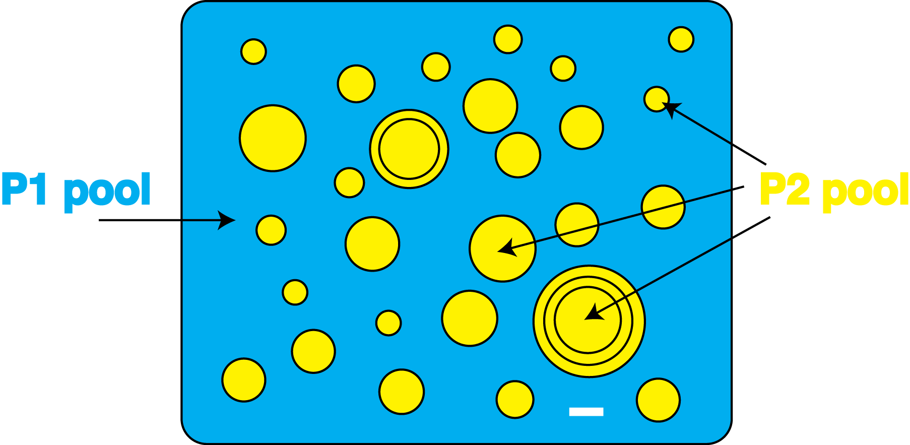
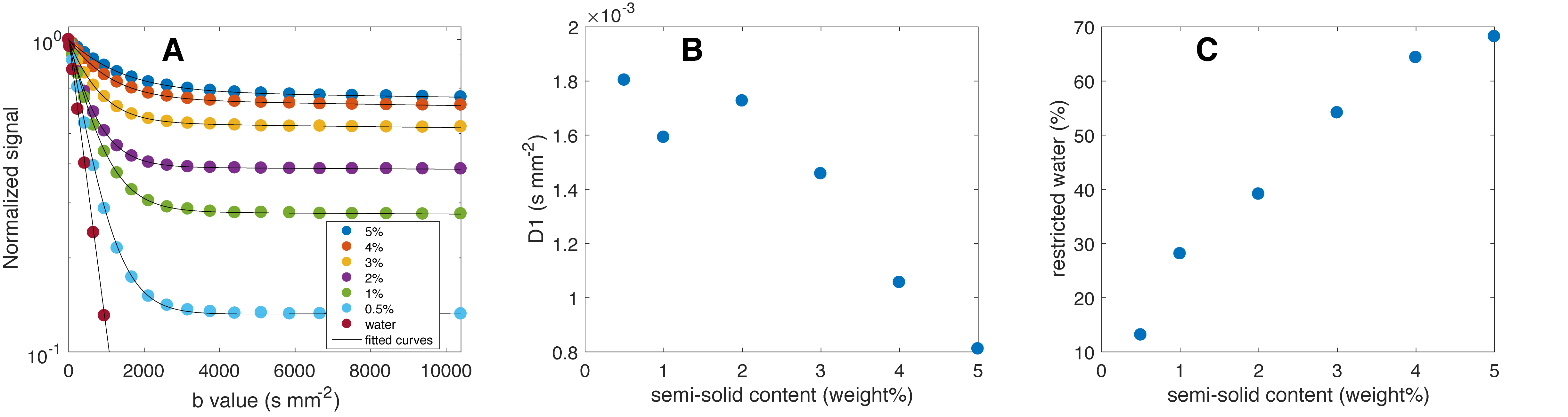
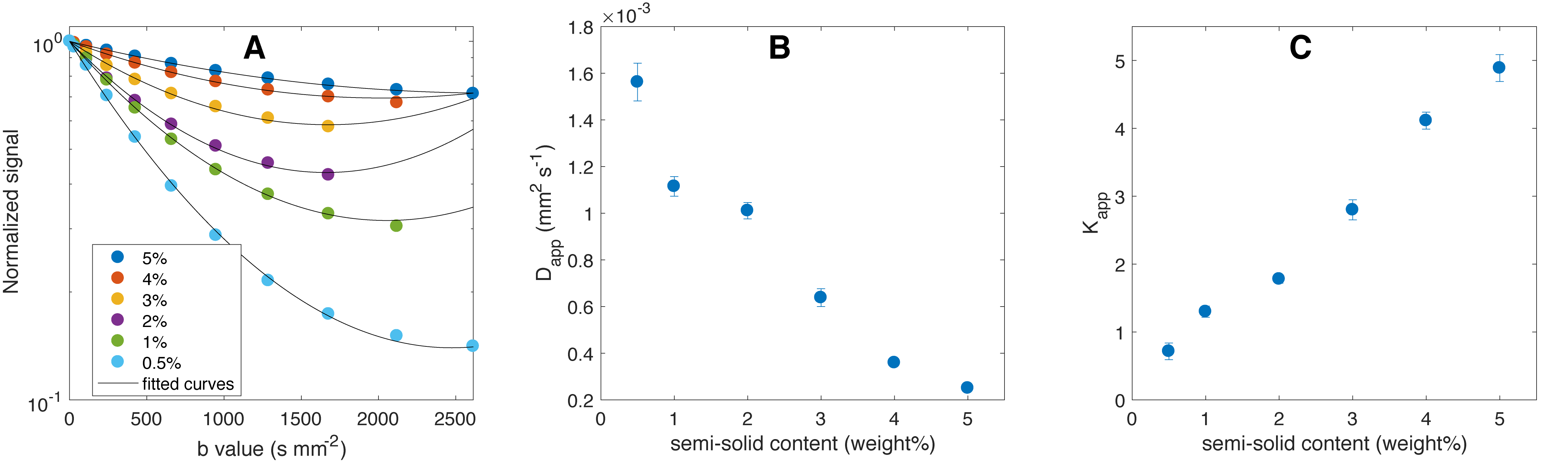
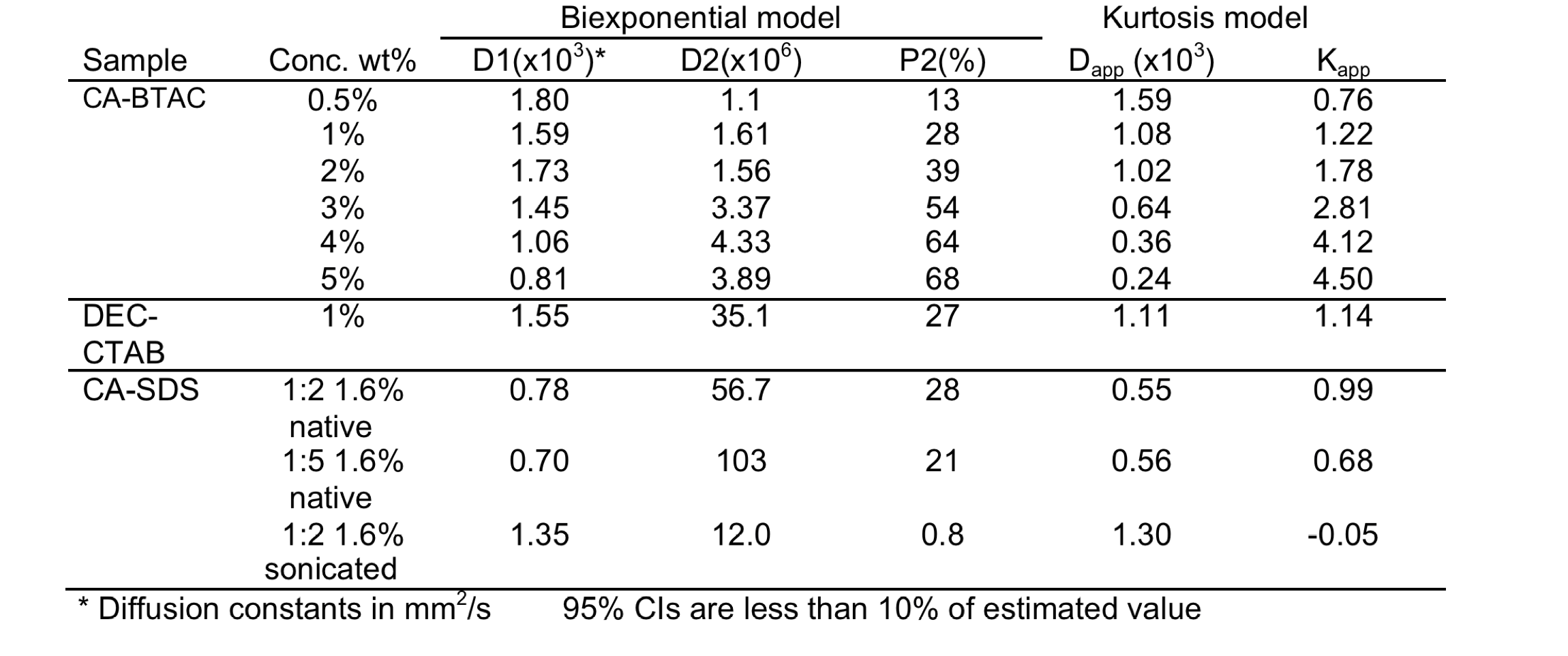
Lamellar vesicle samples (Figure 1) were made by combining a surfactant – either behentriamonium chloride (BTAC), sodium dodecyl sulfate (SDS), or cetrimonium bromide (CTAB) – with a high molecular weight alcohol – either cetearyl alcohol (CA) or 1-decanol (DEC) – at 80 °C and mixing the components to create a homogeneous suspension. After cooling and aging, samples were studied on a 7T Varian/Agilent system at 22 °C using a 1D spin-echo sequence in projection mode. Parameters were δ = 5ms, Δ=100ms, TR/TE = 8000/120 ms, and b-values from 0 to 10,000 s/mm2. The normalized log-signal intensity as a function of b-value was fit with both a biexponential curve (derived D1, D2 diffusion and P2 restricted fraction parameters) and a DKI model (derived apparent diffusion, Dapp, and kurtosis, Kapp, parameters constrained to a valid description range of b-value < 3/(Kapp × Dapp) (1).Results
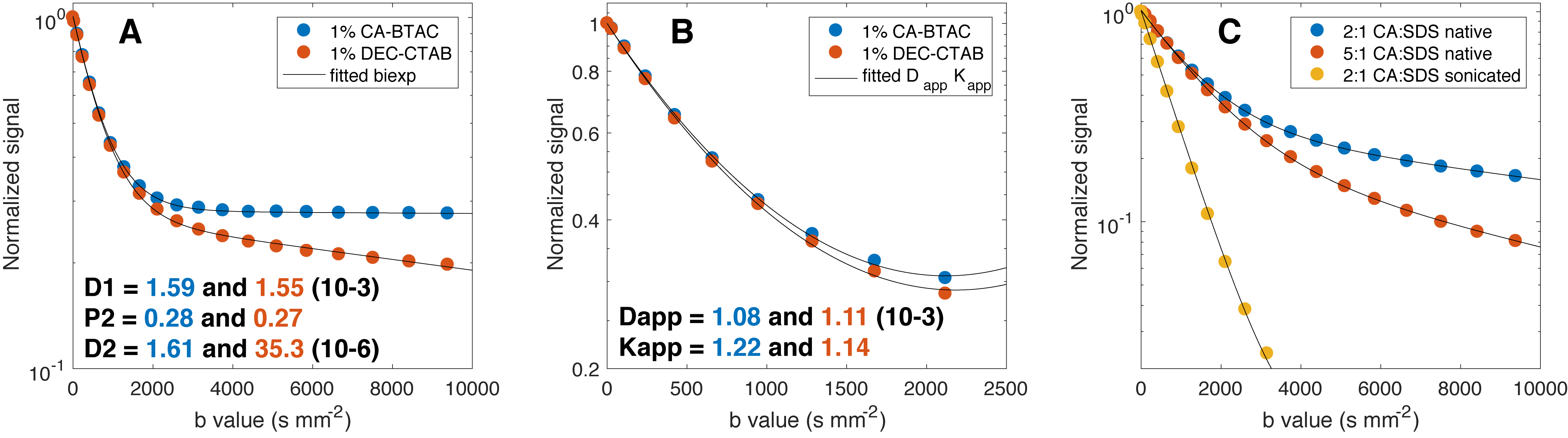
Lamellar vesicles of CA-BTAC from 0.5 to 5% (w/w) provide regions of relatively free and restricted diffusion (Figure 2). Kapp values could be tuned to a desired range by adjusting the weight percent of CA-BTAC suspensions contained in the sample (Figure 3). Vesicles made from DEC-CTAB are more porous that those made from CA-BTAC (Figure 4A) which results in similar Dapp but different Kapp values (Fig 4B). Sonication of a CA-SDS sample reduces the micron sized vesicles (determined by Coulter counter (not shown)) to 100s of nanometers and hence reduces the volume fraction of restricted water molecules from 28% to 0.8%. Constructing the CA-SDS vesicles with a different alcohol:surfactant ratio does not change Dapp(= 0.55 10-3mm2/s) but does reduce Kapp from 0.99 to 0.68 (Fig 4C). Figure 3 show that varying the weight percent of CA-BTAC generates a nearly linear increase in Kapp (Fig 3C, Table 1). In addition, Dapp also decreases with increasing CA-BTAC concentration. Ideally, one would be able to adjust Kapp and keep Dapp constant. Figure 4 shows that we are beginning to gain independent control of Kapp and Dapp by keeping the weight percent of material constant and adjusting either the vesicle size (Fig 4C native vs sonicated) or adjusting the vesicle compositions (Fig 4A and Fig 4C 2:1 vs 5:1 CA:SDS, Table 1).Discussion
Lamellar vesicles provide an ideal platform from which to construct a quantitative DKI phantom. They have many desirable properties. First, they can be assembled from relatively inexpensive and stable materials. Second, the T2 of all samples is greater than 700 ms (r2 in CA-BTAC was determined to be 0.276 s-1wt%-1) providing high SNR over broad b-value range. Third, the ionic surfactants impart charge to the vesicles (a large zeta potential) so that the particles repel each other and stay suspended. And most important, the value of Kapp can be easily tuned over the range found in in cancer tissues. In addition to creating tunable kurtosis parameters, sample stability is of paramount importance for quantitative MRI phantoms. We have shown over a 6 month study of CA-BTAC that, provided the samples remain near room temperature, the combination of low solid concentrations and charged ionic vesicles result in a stable preparation.Conclusion
Nanostructured lamellar vesicles provide a promising framework for quantitative DKI phantoms.Acknowledgements
This work is supported by National Institutes of Health Grants: U01CA166104 and P01CA085878References
1. Jensen JH, Helpern JA. MRI quantification of non-Gaussian water diffusion by kurtosis analysis. NMR in Biomedicine. 2010;23(7):698-710
2. Jansen JF, Stambuk HE, Koutcher JA, Shukla-Dave A. Non-gaussian analysis of diffusion-weighted MR imaging in head and neck squamous cell carcinoma: A feasibility study. AJNR American journal of neuroradiology. 2010;31(4):741-8
3. Rosenkrantz AB, Sigmund EE, Johnson G, Babb JS, Mussi TC, Melamed J, et al. Prostate Cancer: Feasibility and Preliminary Experience of a Diffusional Kurtosis Model for Detection and Assessment of Aggressiveness of Peripheral Zone Cancer. Radiology. 2012;264(1):126-35.
4. Rosenkrantz AB, Padhani AR, Chenevert TL, Koh DM, De Keyzer F, Taouli B, et al. Body diffusion kurtosis imaging: Basic principles, applications, and considerations for clinical practice. Journal of Magnetic Resonance Imaging. 2015;42(5):1190-202.
5. Callaghan PT, Soderman O. Examination of the Lamellar Phase of Aerosol Ot-Water Using Pulsed Field Gradient Nuclear Magnetic-Resonance. Journal of Physical Chemistry. 1983;87(10):1737-44.
6. Karger J. NMR Self-Diffusion Studies in Heterogeneous Systems. Advances in Colloid and Interface Science. 1985;23(1-4):129-48.
7. Le Bihan D. Molecular diffusion, tissue microdynamics and microstructure. NMR Biomed. 1995;8(7-8):375-86.
8. Price WS. NMR studies of translational motion / William S. Price: Cambridge University Press; 2009.
9. Portakal ZG, Shermer S, Jenkins C, Spezi E, Perrett T, Tuncel N, et al. Design and characterization of tissue-mimicking gel phantoms for diffusion kurtosis imaging. Medical physics. 2018;45(6):2476-85
Figures