3630
Delineation of thalamic substructures in ultra-high b-value DWI-measurement with reasonable acquisition time1Diagnostic and interventional Neuroradiology, University Hospital Tuebingen, Tuebingen, Germany
Synopsis
The purpose of this study was to evaluate the capability of optimized and faster ultra-high b-value DWI in separating and identifying intrathalamic substructures compared to a previously described protocol. 7 subjects underwent MR-imaging, including T1 MPRAGE and two DWI sequences at 0 and 5000 s/mm2. DWI was performed with 64 directions and 5 averages (17:56 min) and with 6 directions and 25 averages (8:23 min). Intrathalamic substructures were semi-automatically delineate 4mm above AC/PC line. Accordance between the original sequence and the new speeded-up measurement was high. Acquisition time was reduced by more than 50 % with comparable results.
Purpose
As shown in recent publications, deep brain stimulation (DBS) is becoming more and more important in therapies of various neurological and psychological diseases(1-3). A lack of reliable pre-interventional and individual target identification presents a problem for implantation. Pre-interventional MR-imaging could serve as a new and promising technique to localize new target regions(4). It has been shown, that ultra-high b-value diffusion weighted imaging reveals new intrathalamic substructures which correlate very well with the histological anatomy found in stereotactic atlases(5). Nevertheless, the proposed protocols are time consuming and therefore vulnerable to movement artefacts especially when applied in patients with neurological movement disorders.The purpose of this study was to evaluate the capability of optimized and faster ultra-high b-value DWI in separating and identifying intrathalamic substructures and to compare the new technique with the previously described protocol and histological data.Finally the intention was to move another step into the direction of introducing a measurement protocol that could allow the pre-interventional and personalized stereotactic planning of DBS implantations.Material and Methods
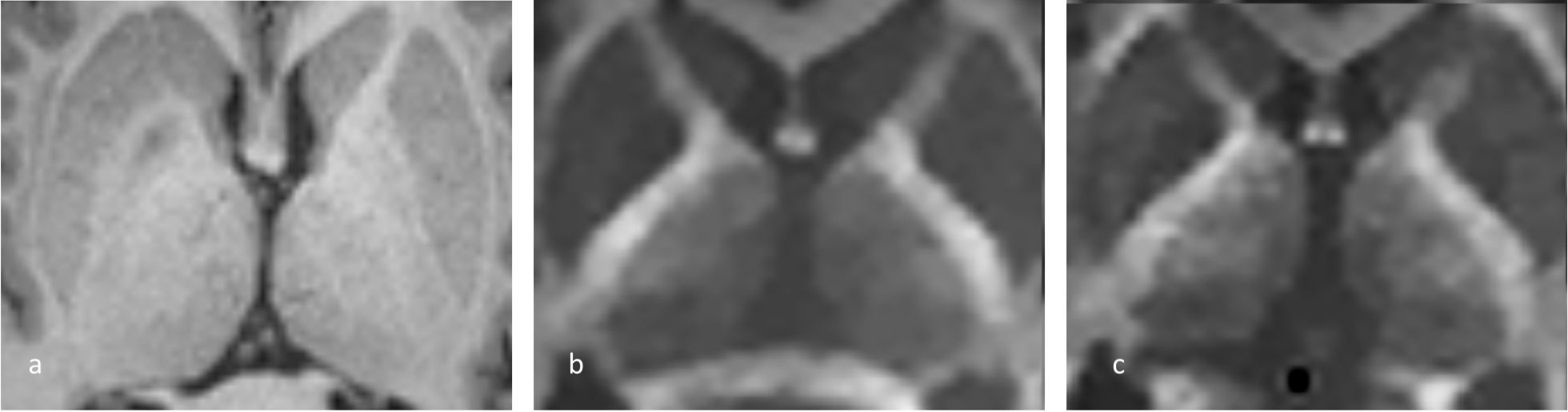
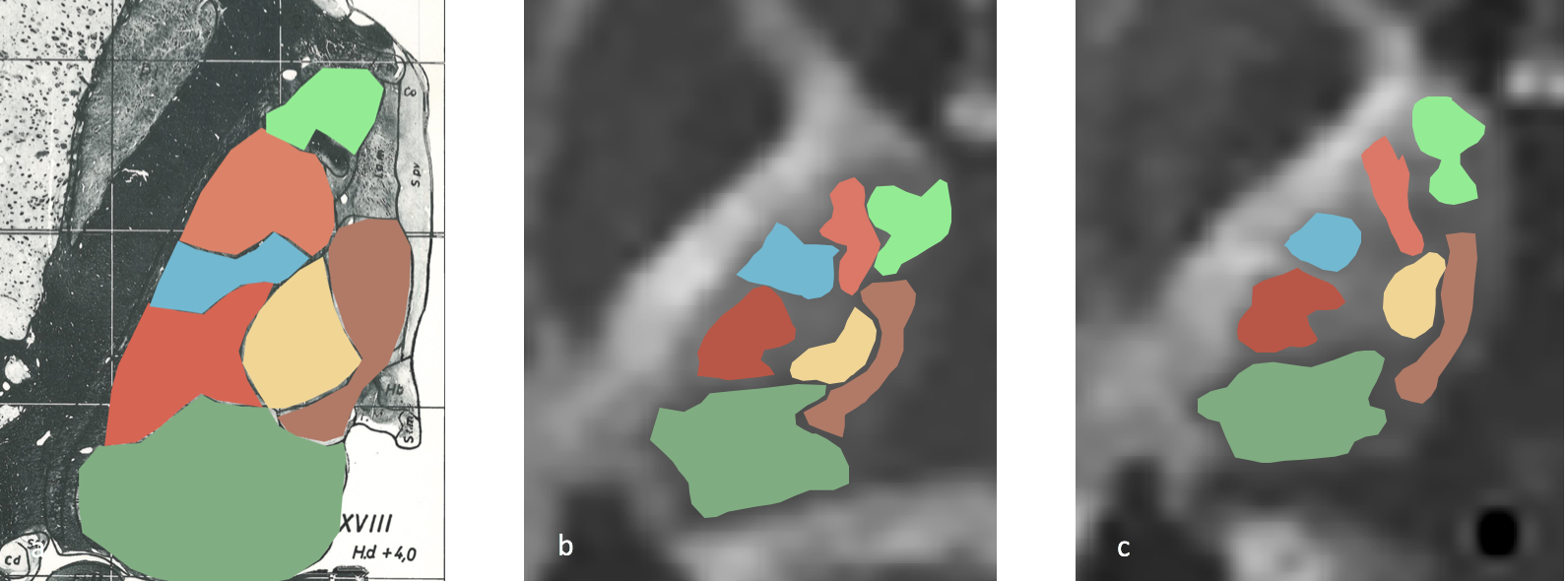
For this prospective MRI-study, approved by the local institutional review board of our university hospital, 7 healthy subjects (4 male, 3 female) were recruited. All subjects provided written informed consent, had to be healthy without any cerebral illnesses or injuries, never underwent surgical intervention in the brain region and didn't receive steroid therapy at that time. No contrast medium was used. Measurements were performed on a 3T-MRI-scanner, able to obtain ultra-high b-values with good noise and vibration levels (PRISMA Fit, Siemens, Erlangen). MPRAGE 3D sequence was applied to reconstruct anatomical images of the subjects’ brains and to plan acquisition of diffusion-weighted images (DWI) in AC/PC orientation. Optimized spin-echo echo-planar imaging DWI sequence with b-values of 0 (b0) and 5000 s/mm2(b5000) was used and diffusion was encoded in 5 directions. For b0, two and for b5000, 25 averages were measured (8:23 minutes acquisition time). A second previously described sequence with the same b-values, encoded in 64 directions with 5 averages at b5000 was added to the protocol (17:56 minutes acquisition time) (5). DWI acquisition was not performed as full brain imaging but focused on thalamic and subthalamic regions. Following to the MRI-Scan, images were denoised using total generalized variation. Intrathalamic substructures, defined prior to the evaluation, were semi-automatically identified in the slice 4 mm above the AC/PC line. Later, the defined substructures were compared between the two sequences and with a histological stereotactic atlas of the brain.Results
In all subjects and in both protocols, all seven intrathalamic substructures could be identified due to different signal variations. The accordance between the original sequence and the new speeded-up measurement was high. The acquisition time was reduced by more than 50% (8:23 minutes compared to 17:56 minutes). Nuclei, which were delineated based on DWI in both sequences, corresponded very well with the histological data from the atlases. In addition, high concordance between the drawn in nuclei in the different subjects and the two acquired sequences could be shown. No movement artefacts were identified on the images and no noise and vibration problems were declared by the patients as it is sometimes the case in ultra-high b-value MR-scanners.Discussion/Conclusion
Ultra-high b-value DWI showed great potential in identifying different thalamic substructures. In this study, a significantly shorter and therefore more easily applicable protocol showed comparable results to the previously described method. Especially in patients with motoric disorders, the acquisition times need to be as short as possible. Reducing them by 9:30 minutes, more than 50% of the original acquisition time, with comparable results therefore is a great step towards fast and reliable identification of thalamic target structures in ultra-high b-value DWI. In conclusion, ultra-high b-value DWI, encoded in only five directions with 25 averages showed comparable potential in determining thalamic nuclei with high signal-to-noise ratio and high concordance with histological data. Further investigations need to be done to evaluate the described protocols in DBS-patients and to correlate anatomical identification with clinical outcome.Acknowledgements
No acknowledgement found.References
1. Koeppen JA, Nahravani F, Kramer M, Voges B, House PM, Gulberti A, et al. Electrical Stimulation of the Anterior Thalamus for Epilepsy: Clinical Outcome and Analysis of Efficient Target. Neuromodulation. 2018.
2. Vyas DB, Ho AL, Dadey DY, Pendharkar AV, Sussman ES, Cowan R, et al. Deep Brain Stimulation for Chronic Cluster Headache: A Review. Neuromodulation. 2018.
3. Habets JGV, Heijmans M, Kuijf ML, Janssen MLF, Temel Y, Kubben PL. An update on adaptive deep brain stimulation in Parkinson's disease. Mov Disord. 2018.
4. Bender B, Wagner S, Klose U. Optimized depiction of thalamic substructures with a combination of T1-MPRAGE and phase: MPRAGE. Clin Neuroradiol. 2016.
5. Nuessle N, Bender B, Klose U. Identification of thalamic substructures in ultra-high b-value DWI. Presented at ISMRM 2018, Paris, France, http://archive.ismrm.org/2018/2057.html
Figures