3616
Metabolite diffusion weighted imaging with golden angle radial echo planar spectroscopic imaging1Danish Research Centre for Magnetic Resonance, Copenhagen University Hospital Hvidovre, Hvidovre, Denmark, 2C.J. Gorter Center for High Field MRI Research, Department of Radiology, Leiden University Medical Center, Leiden, Netherlands, 3Philips Healthcare, Copenhagen, Denmark, 4Center for Magnetic Resonance, Department of Electrical Engineering, Technical University of Denmark, Lyngby, Denmark
Synopsis
Diffusion weighted spectroscopy (DWS) is a promising tool for investigating compartment specific microstructure in heterogeneous tissues. Unlike water abundant in all cellular spaces, the mobility of metabolites provides a window into the microstructure of specific cell types. Multi-shot sequences for diffusion spectroscopic imaging suffer from translation induced phase fluctuations. This has previously been addressed with additional phase navigators. In this work we propose self navigated metabolite diffusion weighted spectroscopic imaging using golden angle radial echo planar gradient readouts with semi-LASER voxel localization. Initial data shows good spatial localization and spectral quality.
Introduction
Diffusion weighted spectroscopy (DWS) is a promising tool for investigating compartment specific microstructure in heterogeneous tissues [1]. Unlike water, which is abundant in all cellular spaces, the mobility of metabolites provides a window into the microstructure of specific cell types. DWS is typically performed using single voxel techniques, but recent work has demonstrated the potential for spectroscopic imaging approaches using conventional chemical shift imaging (CSI) or faster echo planar spectroscopic imaging (EPSI) approaches [2,3]. One challenge for multi-shot diffusion weighted techniques is the need for correcting for phase fluctuations induced by interaction from motion and strong diffusion weighting gradients. This has in previous work been resolved by acquiring a separate navigator representing the centre of k-space between the diffusion weighting and the image readout. An alternative to this approach with the benefit of a simpler sequence layout and a faster acquisition is the use of radial readouts where a phase reference is obtained within the signal readout for each excitation. In this work we propose diffusion weighting combined with radial echo planar spectroscopic imaging (DREPSI) and sLASER volume selection. We present initial data acquired in a human subject on a 7T MRI system.Methods
All experiments were performed on in one healthy male subject on a 7T MRI system (Achieva, Philips Healthcare, Best, Netherlands), using a 32 channel receive coil. Experiments were performed following procedures approved by the local ethics committee and with the participant’s written consent.
The new DREPSI sequence diagram is shown in figure 1 and comprised the following elements: Volume selection: sLASER using FOCI refocusing was used to minimize in-plane chemical shift artifacts [4] (excitation volume 100x100x20mm3, TE/TR=120/3700ms). Diffusion weighting: 8 bipolar diffusion weighted gradients where applied around the four FOCI refocusing pulses. Each gradient lobe was 6.5 ms and applied in 3 orthogonal gradient directions at 0, 15 and 60 mT/m resulting in b = 0, 350 and 5600 s/mm2. Spectroscopic imaging readout: 2D golden angle radial EPSI readout [5,6] was performed (FOV=320x320 mm, voxel size=10x10 mm2, 34 blades, gradient strength = 9.4 mT/m, slew rate = 190 mT/m/ms).
Data were collected with and without partially de-optimized VAPOR water suppression to allow sufficient water signal for phase correction as previously described [2]. Non-water suppressed data was only acquired for the two lowest diffusion conditions. Total imaging time 24 minutes.
Data reconstruction: The overall data processing pipeline is outlined in figure 2.
Results and discussion
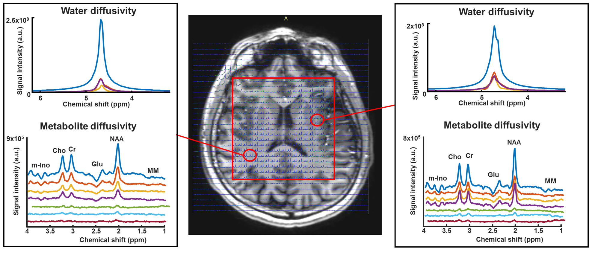
Data quality was considerably improved after correction of k=0 phase fluctuations. Data from one subject is shown in figure 3. The ventricles are well outlined with good spectral quality and bandwidth achievable without interleaving temporal profiles. Spectral peaks strongly depended on b-value as expected. Signal attenuation levels were slightly larger than expected both from the water signal and from metabolite diffusivities reported in earlier studies [1]. Possible explanation could be contamination in the b=0 measurement or insufficient correction of phase errors in the diffusion weighted data. Improvements with regards to this could be sparse sampling reconstruction to correct for data rejection, for improving the point spread function and for reducing the acquisition time. Online re-acquisition of rejected data-points could further improve robustness [2]. Drift in the read out trajectory could also provide an imperfect estimation of the k=0 phase that could be addressed with improved calibration techniques or gradient field monitoring.Conclusion
We demonstrate the feasibility for diffusion weighted spectroscopic imaging with large coverage, small chemical shift and fast self navigated readout with radial echo planar spectroscopic imaging combined with sLASER volume selection. Future work will address robust reconstruction techniques and further experiments on humans and well defined phantoms.Acknowledgements
This research is supported by the Danish Council for Independent Research [grant no. 6111-00349A], the Danish Agency for Science, Technology and Innovation grant no. 0601-01370B, and The John and Birthe Meyer Foundation.References
1. Palombo, M., Shemesh, N., Ronen, I. & Valette, J. Insights into brain microstructure from in vivo DW-MRS. Neuroimage 182, 97–116 (2017).
2. Ercan, A. E., Techawiboonwong, A., Versluis, M. J., Webb, A. G. & Ronen, I. Diffusion-weighted chemical shift imaging of human brain metabolites at 7T. Magn. Reson. Med. 73, 2053–2061 (2015).
3. Fotso, K. et al. Diffusion tensor spectroscopic imaging of the human brain in children and adults. Magn. Reson. Med. (2017). doi:10.1002/mrm.26518
4. Arteaga de Castro, C. S. et al. Improved efficiency on editing MRS of lactate and γ-aminobutyric acid by inclusion of frequency offset corrected inversion pulses at high fields. NMR Biomed. (2013). doi:10.1002/nbm.2937
5. Feng, L. et al. Golden-angle radial sparse parallel MRI: combination of compressed sensing, parallel imaging, and golden-angle radial sampling for fast and flexible dynamic volumetric MRI. Magn. Reson. Med. (2014). doi:10.1002/mrm.24980
6. Posse, S., Tedeschi, G., Risinger, R., Ogg, R. & Bihan, D. Le. High Speed1H Spectroscopic Imaging in Human Brain by Echo Planar Spatial‐Spectral Encoding. Magn. Reson. Med. (1995). doi:10.1002/mrm.1910330106
Figures