3614
Reproducibility of tractography based on fibre orientation distribution and anatomical constraints applied on paediatric diffusion MRI.1ExpORL, KU Leuven, Leuven, Belgium, 2Research and Development, icometrix, Leuven, Belgium
Synopsis
Tractography is known to be sensitive to technical variations but might also be sensitive to issues related to paediatric data, such as head motion. We assessed the reproducibility in paediatric data of the probabilistic tractography algorithm based on fibre orientation distribution and anatomical constraints that enables to deal with crossing fibres and to reconstruct tracts with more anatomical accuracy. Our results showed that the reproducibility of this approach when it is applied on paediatric data is negatively affected by younger age and by head motion but can still achieve good reproducibility for selected tracts.
Introduction
Diffusion MRI (dMRI) has been widely used to study the developing brain by providing information on the structural organisation of the white matter (WM) tracts1. Probabilistic tractography algorithms based on Fibre Orientation Distribution (iFOD2)2 and anatomical constraints (ACT)3 enable to resolve complex structures, such as crossing fibres, and to reconstruct WM tracts with more anatomical accuracy. One of the limitations of tractography is its sensitivity to variations in the technical implementation of the fibre-tracking algorithm, which affects the reproducibility of outcome results4. In addition, the reproducibility might also be affected by issues specific to paediatric data (e.g. age effect, head motion)5. Therefore, our aim is to assess the reproducibility of this tractography approach when it is applied on paediatric data.Methods
Whole brain tractography: The dMRI was processed with MRtrix36. After preprocessing (denoising, Gibbs correction, bias correction, motion correction using NiftyReg), the dMRI was non-rigidly co-registered based on inverse contrast normalisation and up-sampled to the corresponding T1-weighted image for anatomical correspondence and distortion correction7. The fibre orientation distribution (FOD) are then computed with constrained spherical deconvolution. Whole brain tractography was performed with iFOD2 and ACT based on fraction maps of cortical grey matter (GM), WM, deep GM, cerebrospinal fluid that were provided by an age-specific brain segmentation of the T1-weighted images8. The parameters for the tractography were set as follows: minimum tract length=40mm, number of selected streamlines=5 millions, cut-off threshold of FOD amplitude=0.01, seeding and cropping streamline endpoint at GM and WM interface and filtering streamlines to 1 million with SIFT algorithm9.
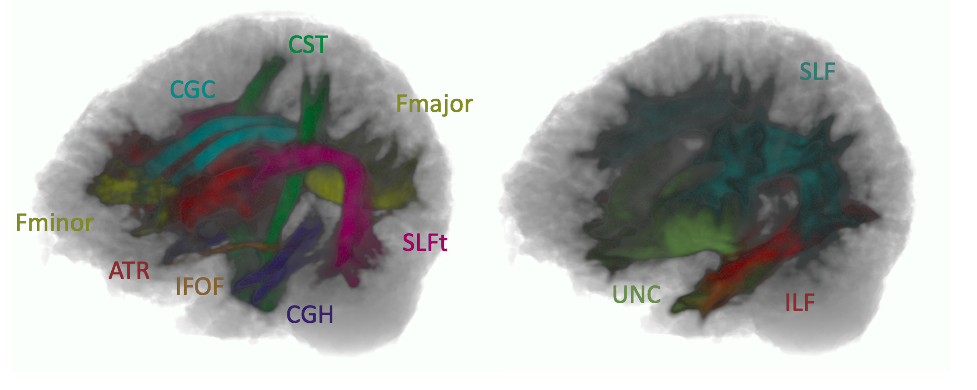
Individual white tract extraction: Region of interests (ROIs) defined on a fractional anisotropy (FA) template were propagated to the subject image with affine and non-rigid registration using NiftyReg. The streamlines of the whole brain tractogram were filtered as streamlines included in the ROIs, such as described in the protocol of Wakana et al10. The validation was made on the following tracts (see Fig. 1): the anterior thalamic radiation (ATR), the cortico-spinal tract (CST), the cingulate gyrus (CGC) with its hippocampal part (CGH), the inferior longitudinal fasciculus (ILF), the inferior fronto-occipital fasciculus (IFOF), the superior longitudinal fasciculus (SLF) and its temporal component (SLFt), the uncinate tract (UNC), the forceps minor and the forceps major.
Results
Data: The reproducibility was assessed based on test-retest scans from the publicly available dataset of Nathan-Kline Institute11. Fifty-four dMRI and the corresponding T1-weighted images representing the brain of children between 6-18 years old were used for the validation. DMRI were acquired with 137 directions at b-value=1500 s/mm2 (resolution: 2 mm3) and T1-weighted images with MPRAGE sequence (resolution: 1mm3).
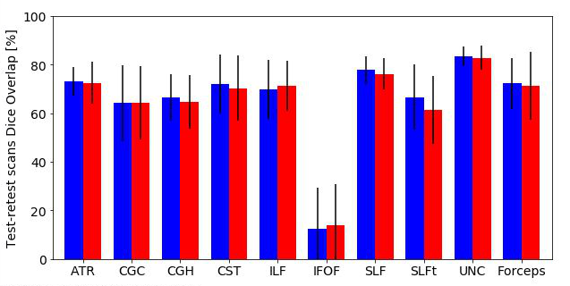
Reproducibility of WM tract segmentation: The reproducibility of the segmentation was measured with the Dice Overlap of WM tracts segmentation between test and retest scans, with a high reproducibility corresponding to a Dice Overlap close to 100% (see Fig.2). The results showed that the Dice Overlap was on average ranged from 12.41% (IFO left) up to 83.34% (UNC left).
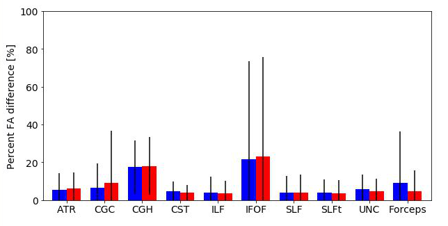
Reproducibility of fractional anisotropy (FA): The reproducibility of FA was assessed with the percent FA difference between test and retest scans, with a high reproducibility corresponding to a percent difference close to 0% (see Fig. 3). The results showed that the percent FA difference was on average ranged from 3.54% (SLFt right) up to 23.01% (IFO right).
Correlation with age and head motion: The Pearson correlation (r) of the reproducibility of WM tracts segmentation with age was significant (|r| > 0.40, with Bonferroni correction) for the bilateral CGC, the left IFOF, the left SLFt, the bilateral UNC and the forceps major. The correlation with the head motion measured with root mean square deviation (RMS) was significant for all tracts except for the right ATR, the left CGH, the bilateral IFOF and the left UNC. Age and head motion were significantly correlated (r=-0.41).
Discussion
The reproducibility of iFOD2 with ACT on paediatric data was comparable to manual virtual dissection based on diffusion tensor on adult data10. A general lower reproducibility is related to younger age and more severe head motion, which indicates that tractography would be less reproducible in younger children because they tend to move more. A particularly low reproducibility was observed for the IFOF tract. A possible explanation is that the anatomical constraints cut off streamlines of IFOF passing through regions of GM-WM partial volume leading to bad tract reconstruction.Conclusion
When applied on paediatric dMRI, the reproducibility of the tractography based FOD and ACT is negatively affected by younger age and by head motion. Nevertheless, good reproducibility is achievable for selected tracts, such as UNC and SLF.Acknowledgements
This research has been supported by the European Union H2020 MSCA-ITN-2014-ETN Programme, Advancing brain research in children’s developmental neurocognitive disorders-project (ChildBrain, #641652).References
1. Morgan S., White S., Bullmore E. et al. A network neuroscience approach to typical and atypical brain development. Biol Psychiatry Cogn Neurosci Neuroimaging. 2018; 3(9):754-766
2. Tournier J.-D., Calamante F., Connelly A. Improved probabilistic streamlines tractography by 2nd order integration over fibre orientation distributions. Proc Intl Soc Mag Reson Med (ISMRM). 2010; 18:1670
3. Smith R., Tournier J.-D., Calamante F. et al. Anatomically-constrained tractography: Improved diffusion MRI streamlines tractography through effective use of anatomical information. Neuroimage. 2012; 62(3):1924-1938.
4. Maier-Hein K., Neher P. Houde J. et al. The challenge of mapping the human connectome based on diffusion tractography. Nat Commun, 2017; 8: 1349
5. Moeded A., Huisman T., Casamassima M. et al. The structural connectome in children: basic concepts, how to build it, and synopsis of challenges for the developing pediatric brain. Neuroradiology. 2017; 59: 445-460.
6. Tournier J.-D., Calamante F. & Connelly A. Mrtrix: Diffusion tractography in crossing fiber regions. Int J Imag Syst Techn. 2012; 22(1): 53-66
7. Bhushan C., Haldar J., Choi S. et al. Co-registration and distortion correction of diffusion and anatomical images based on inverse contrast normalization. Neuroimage. 2015; 15(115): 269-280.
8. Phan T.V., Sima D. Beelen C. et al. Evaluation of methods for volumetric analysis of pediatric brain data: the childmetrix pipeline versus adult-based approaches. Neuroimage Clin. 2018; 19: 734-744
9. Smith R., Tournier J. Calamante F. et al. SIFT: Spherical-deconvolution informed filtering of tractograms. Neuroimage. 2013; 67: 298-312
10. Wakana S., Caprihan A., Panzenboeck M. et al. Reproducibility of quantitative tractography methods applied to cerebral white matter. Neuroimage. 2007; 36(3):630-644.
11. Zuo X., Anderson J., Bellec P. et al. An open science resource for establishing reliability and reproducibility in functional connectomics. Scientific data. 2014.
Figures