3609
Does cerebrospinal fluid pulsation affect DWI thermometry?: healthy volunteer study1KPUM, Kyoto, Japan, 2KPUM Hospital, Kyoto, Japan
Synopsis
Diffusion-weighted imaging (DWI) based thermometry has a potential to be a non-invasive method of temperature measurement for the deep inside of human brain. Nevertheless, the DWI at lateral ventricle in the brain might be influenced by the pulsation flow of cerebrospinal fluid (CSF), which is motivated by heartbeat. The purpose of this study was to investigate the influence of pulsation flow on brain DWI thermometry for healthy subjects. Comparisons were performed among ΔT at three CSF speed selections (slow vs. fast vs. random). There was no significant difference in ΔT among the CSF speed and volume on healthy subjects.
PURPOSE
Diffusion-weighted imaging (DWI) based thermometry has a potential to be a non-invasive method of temperature measurement for the deep inside of human brain [1-12]. Nevertheless, the DWI at lateral ventricle (LV) in brain might be influenced by the pulsation flow of cerebrospinal fluid (CSF), which is motivated by heartbeat. The purpose of this study was to investigate the influence of pulsation flow on brain DWI thermometry for healthy subjects.MATERIALS AND METHODS
Subject: This study was approved by the ethics committee at our institution. Written informed consent for MR examinations was obtained from all subjects prior to participation in this study. A total of 57 healthy subjects (30 men, 27 women; mean (± standard deviation) age, 41.3 ± 15.2 years; range 21 - 69 years) voluntarily participated by leaflets displayed in our hospital. DWI data were used for assessing deep brain temperature.
DWI acquisition: All DWIs were obtained using a 3.0 T whole-body scanner (MAGNETOM Skyra; Siemens Healthcare, Erlangen, Germany). Single-shot echo-planar imaging was used for acquisition (repetition time, 3500 ms; echo time, 92 ms) with a motion-probing gradient in 10 orientations, b values of 1000 s/mm2, and averaging of two images. The field of view was 230 mm. A SENSE technique was used (128 × 53 data points) and reconstructed to 128 × 106 matrices (zero-filled, resolution of 128 × 128). A total of 45 slices with a thickness of 2 mm each were obtained without inter-slice gaps (trans-axial slices, parallel to AC-PC line).
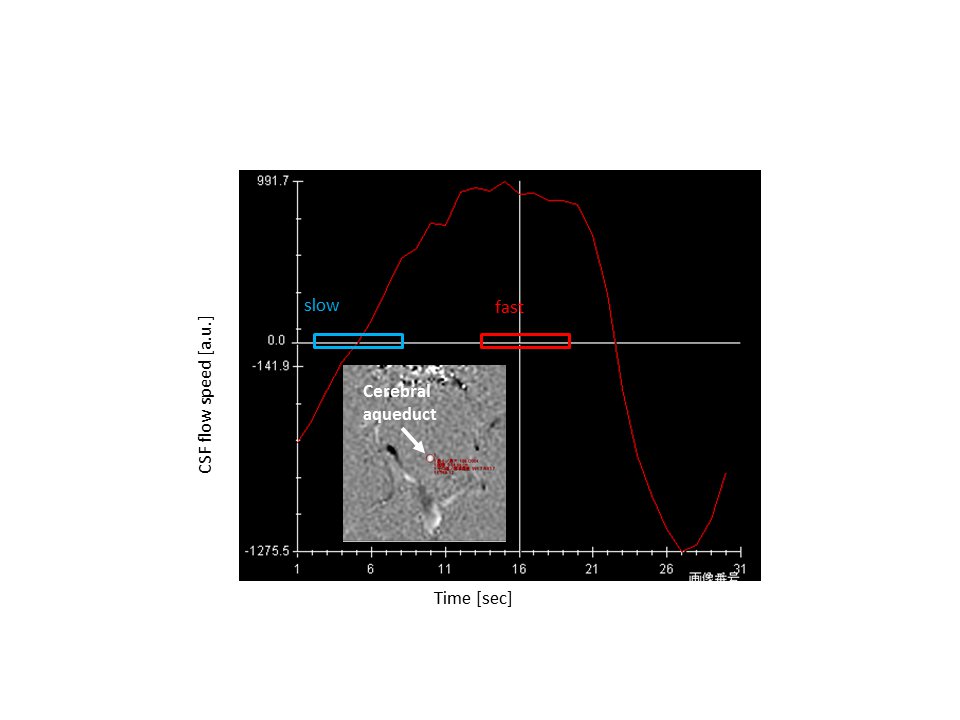
Heat Gating: The cardiac contraction was detected by peripheral pulse transducer. In addition, CSF pulsation flow at cerebral aqueduct was measured by phase contrast method and three DWIs were acquired at the timing of the maximum, the minimum, and at random ascending flow of CSF (Figure 1).
Temperature calculation: The diffusion coefficient along the direction of motion probing gradient Di [mm2/s] was converted to temperature [13]; Ti [°C] = 2256.74 / ln (4.39221 / Di) - 273.15. The temperature within the LV and the mean temperature were calculated by the histogram curve-fitting method [3]. The difference between brain temperature and eardrum temperature (ΔT), measured by infrared thermometer (M30; Terumo, Tokyo, Japan), were used for the comparison.
Statistics: Comparisons were performed among ΔT at three CSF speed selections (slow vs. fast vs. random) by Wilcoxon rank sum test. Spearman correlation coefficient was calculated for a linear correlation between both ΔT and speed ΔT and volume of CSF (Matlab; The Mathworks, Natick, MA, USA). The correlation was evaluated as significant for P values <0.05.
RESULTS
DWI thermometry along with pulsation: Figure 2 shows ΔT along the CSF flow speed at cerebral aqueduct. There was no significant difference among the CSF speed selections (P > 0.05).
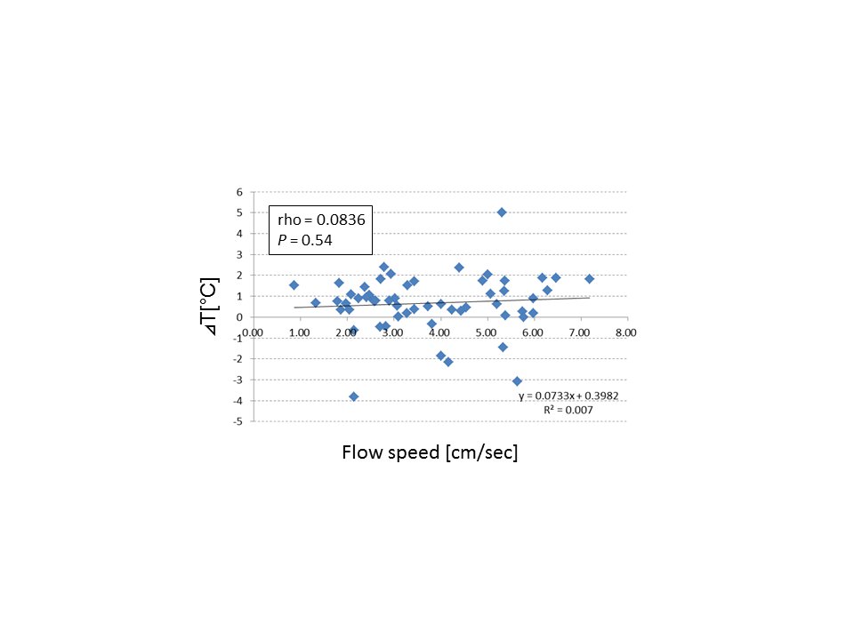
ΔT along with flow speed of CSF: Figure 3 shows ΔT along the CSF flow speed at cerebral aqueduct. There was no significant linear correlation between ΔT and CSF flow speed (P > 0.05).
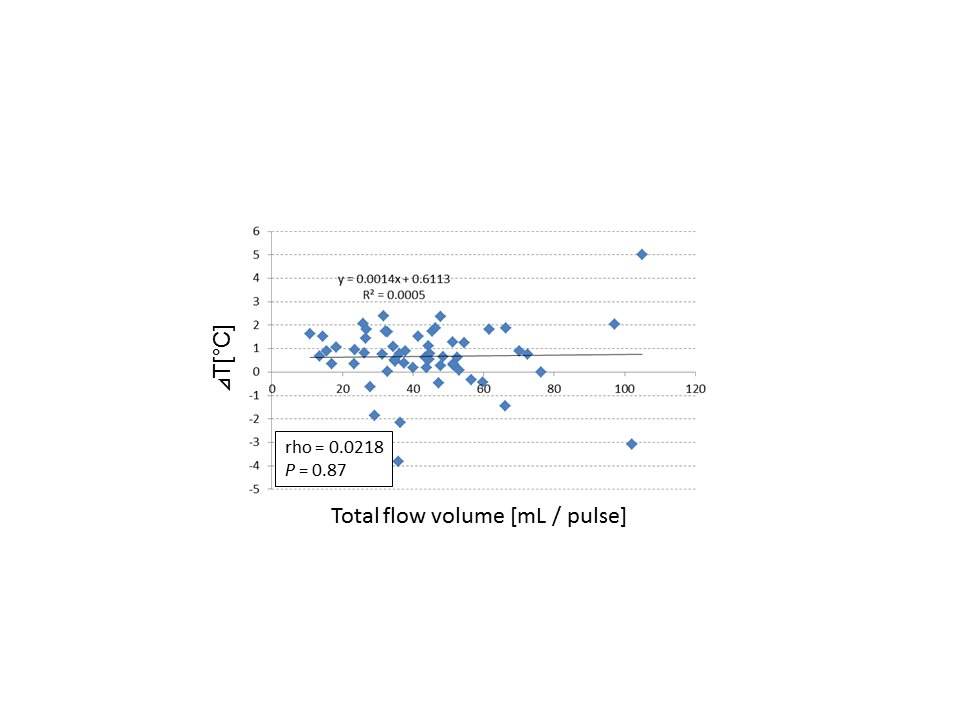
ΔT along with total flow volume of CSF: Figure 4 shows ΔT along the total CSF flow volume at one heartbeat. There was no significant linear correlation between ΔT and CSF flow volume (P > 0.05).
From Fig. 2 to Fig. 4, there was no significant difference in ΔT among the CSF speed and volume on healthy subjects.
LIMITATIONS
For better statistical reliability, further investigation with a larger number of participants is needed. Further investigations are needed before clinical applications can be countenanced.CONCLUSION
The CSF pulsation into the lateral ventricle during measurement of DWI does not significantly affect the measurement of DWI thermometry.Acknowledgements
This work was supported by JSPS KAKENHI Grant Number JP17K10413 and JP17K10415.References
1. Yamada K, et al., Moyamoya patients exhibit higher brain temperatures than normal controls, NeuroReport, 2010; 21: 851-855.
2. Sakai K, et al., Age-dependent brain temperature decline as assessed by DWI thermometry, NMR in Biomedicine, 2011; 24(9): 1063-1067.
3. Sakai K, et al., Calculation methods for ventricular DWI thermometry: phantom and control studies, NMR in Biomedicine, 2012; 25(2): 340-346.
4. Sai A, et al., Diffusion-weighted imaging thermometry in multiple sclerosis, JMRI, 2014; 40(3): 649-654.
5. Ota M et al., Altered coupling of regional cerebral blood flow and brain temperature in schizophrenia compared with bipolar disorder and healthy subjects, Journal of Cerebral Blood Flow & Metabolism, 2014; 34(12): 1868-1873.
6. Tazoe J, et al., Brain core temperature of patients with mild traumatic brain injury as assessed by DWI-thermometry, Neuroradiology, 2014; 56(10): 809-815.
7. Sumida K, et al., Intraventricular cerebrospinal fluid temperature analysis using MR diffusion-weighted imaging thermometry in Parkinson’s disease patients, multiple system atrophy patients and healthy subjects, Brain and Behavior, 2015; 35: 5(6): e00340.
8. Kuriyama N, et al., Ventricular temperatures in idiopathic normal pressure hydrocephalus (iNPH) using DWI-based MR thermometry, Magnetic Resonance in Medical Sciences, 2015; 14(4): 305-312.
9. Tsukamoto T, et al., Assessment of brain temperatures during different phases of the menstrual cycle using diffusion-weighted imaging thermometry, Japanese Journal of Radiology, 2016: 34(4): 277-283.
10. Sumida K, et al., Intraventricular temperature measured by diffusion-weighted imaging compared with brain parenchymal temperature measured by magnetic resonance spectroscopy in vivo, NMR in Biomedicine, 2016; 29(7): 890-895.
11. Sparacia G, et al., Assessment of brain core temperature using MR DWI-thermometry in Alzheimer disease patients compared to healthy subjects, Japanese Journal of Radiology, 2017; 35: 168-171.
12. Sparacia G, et al., Brain core temperature of patients before and after orthotopic liver transplantation assessed by DWI-thermometry, Japanese Journal of Radiology, 2018; 36: 324-330.
13. Kozak LR et al., Using diffusion MRI for measuring the temperature of cerebrospinal fluid within the lateral ventricles, Acta Paediatr. 2010; 99(2): 237–243.
Figures