3606
Diffusional Kurtosis Imaging of Temporal Lobe Gray Matter as a Biomarker of Neurological Disease: Schizophrenia and Autism Spectrum Disorder1Radiology, Center for Biomedical Imaging, New York, NY, United States, 2Sackler Institute of Graduate Biomedical Sciences, New York, NY, United States, 3Psychiatry, New York University School of Medicine, New York, NY, United States
Synopsis
In this study, we employed diffusion kurtosis imaging (DKI) to test for differences in gray matter (GM) microstructure in schizophrenia (SZ) and autism spectrum disorder (ASD). Significantly increased metrics of DKI were found in SZ compared to HC participants, while significantly decreased metrics of DKI were found in ASD compared to HC in the temporal lobe and sub-lobar temporal regions of interest (ROIs). In vivo DKI metrics appear to be sensitive to GM microstructural pathology in SZ and ASD and may provide new information on the neural underpinnings of these disorders.
Background
Prior histological post-mortem studies have shown gray matter (GM) microstructural abnormalities in the temporal lobe (TL) to be a pathological feature of both schizophrenia (SZ) and autism spectrum disorder (ASD).1–3 Despite offering a unique view of microstructural integrity, histological studies are often restricted to small sample sizes and isolated brain regions, and may be confounded by damage to the tissue caused by the fixation process. Evaluation of microstructure in vivo may thus provide essential means to circumvent these limitations. However, examination of gray matter microstructure has remained to date scarce due to the relative lack of non-invasive methods to assess it. The aim of this work is to evaluate the feasibility of employing diffusional kurtosis imaging (DKI) to describe gray matter abnormalities in schizophrenia and autism. DKI is an extension of DTI that accounts for non-Gaussian water diffusion contributions to the diffusion MRI (dMRI) signal and provides several kurtosis indices that reflect tissue microstructural complexity.4 Examining microscopic GM changes across SZ and ASD may help build a more comprehensive model of these diseases and lead to a better understanding of how DKI indexes tissue cytoarchitecture.Methods
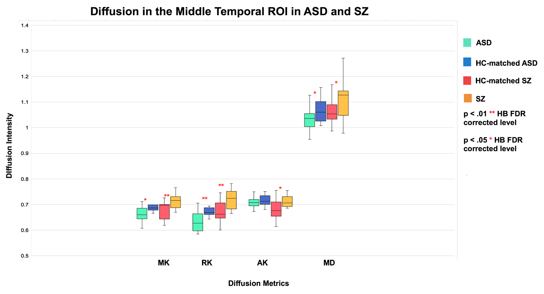
DKI data was acquired in two data sets: 17 SZ patients and 18 matched healthy comparison controls (HC) (right-handed males, 30-55 years old), and 16 ASD patients and 17 matched HC (males, 18-25). Mean (MK), axial (AK), radial (RK) kurtosis and mean diffusivity (MD) metrics were calculated for the temporal lobe and 18 sub-lobar regions of interest (ROIs) delineated by the Desikan-Killiany atlas.5 Analyses used independent-samples t-test to compare diffusion metrics (MK, RK, AK and MD) between patients and HCs in the two data sets in each ROI. The Benjamini-Hochberg (HB) procedure was employed in each analysis to correct for multiple comparisons and decrease the false discovery (FDR) rate.6 Differences were considered significant for p<0.05 HB FDR corrected and at trend level for p<0.10 HB FDR corrected.Results
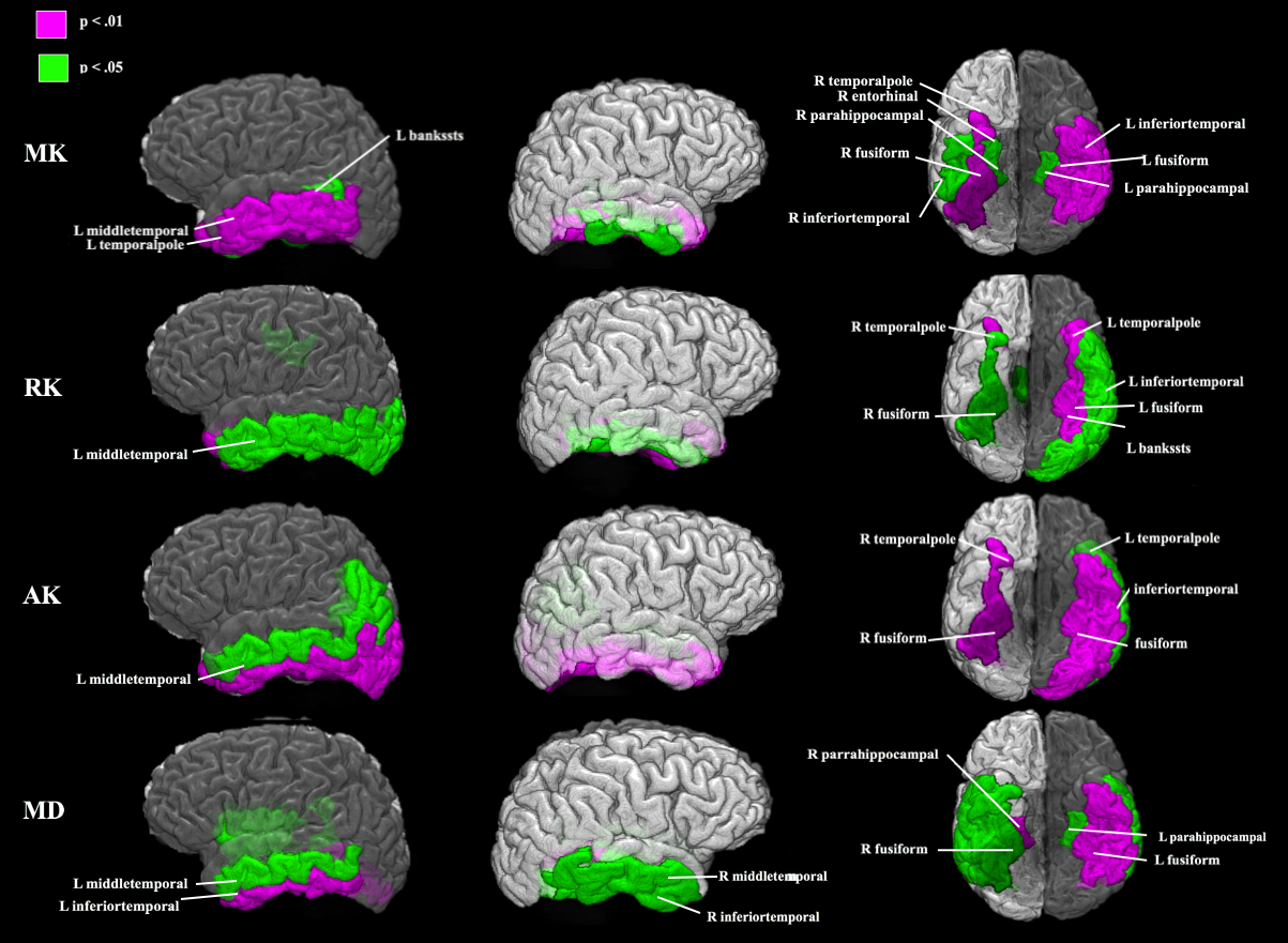
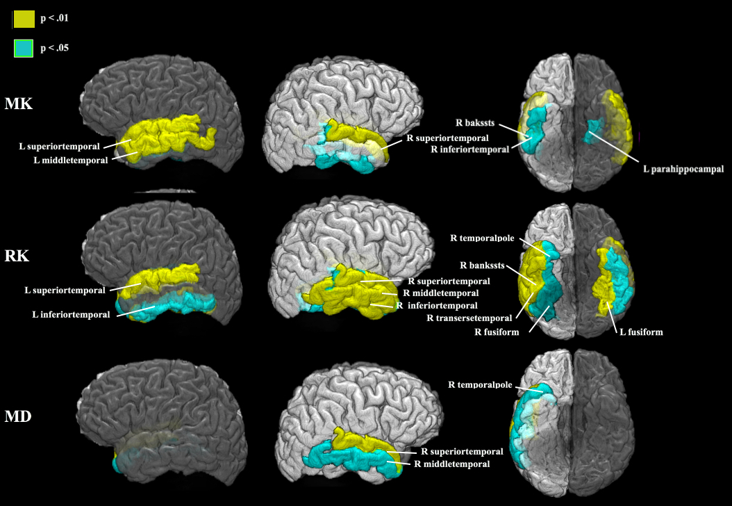
Increases were found in MK, RK, AK and MD in SZ compared to HC in the whole temporal lobe and sub-temporal ROIs: banks of the superior temporal sulcus (bankssts), entorhinal cortex, fusiform and parahippocampal gyri, inferior and middle temporal gyri, and temporal pole ROIs (Figure 1-2). Decreases were found in MK, RK and MD in ASD compared to HC in the whole temporal lobe and sub-temporal ROIs: bankssts, fusiform, parahippocampal, inferior temporal, middle temporal and superior temporal gyri, transverse temporal cortex, and temporal pole ROIs (Figure 1; Figure 3).Discussion
Anatomical T1-weighted MRI studies have documented macroscale GM volume loss and cortical thinning in the TL in SZ with both increase and decrease reported in ASD.7,8 Results presented here suggest that DKI may be sensitive to disease-specific changes in GM tissue microstructure and can be used to create distinct pathology profiles of each disease. The increased kurtosis in SZ may be due to a more restrictive microstructural arrangement (increased microglia and cell packing, iron and protein deposits noted in GM ex vivo), while decreased kurtosis in ASD may reflect abnormalities that create a less restrictive microstructural environment (decreased neuronal density and size, astrogliosis demonstrated in GM ex vivo). 1–3,9–11Conclusion
In vivo DKI metrics appear to be sensitive to GM microstructural pathology in SZ and ASD and may provide useful biomarkers of abnormal cortical structure and function in psychiatric and neurological disorders.Acknowledgements
This work was supported by the National Institute of Mental Health awards R21 MH085228, R01 MH108962 and R03-MH076180 . We greatly thank all of our participants for their help with this study and Researchmatch for supporting our recruitment efforts.References
1. Van Kesteren, C. F. M. G. et al. Immune involvement in the pathogenesis of schizophrenia: A meta-analysis on postmortem brain studies. Transl. Psychiatry 7, (2017).
2. Van Kooten, I. A. J. et al. Neurons in the fusiform gyrus are fewer and smaller in autism. Brain 131, 987–999 (2008).
3. Casanova, M. F. The neuropathology of autism. in The Molecular Basis of Autism 153–171 (2015). doi:10.1007/978-1-4939-2190-4_8
4. Jensen, J. H. & Helpern, J. A. MRI quantification of non-Gaussian water diffusion by kurtosis analysis. NMR in Biomedicine 23, 698–710 (2010).
5. Desikan, R. S. et al. An automated labeling system for subdividing the human cerebral cortex on MRI scans into gyral based regions of interest. Neuroimage 31, 968–980 (2006).
6. Hochberg, B. Controlling the False Discovery Rate: a Practical and Powerful Approach to Multiple Testing. J. R. Stat. Soc. 57, 289–300 (1995).
7. Zipursky, R. B., Lim, K. O., Sullivan, E. V., Brown, B. W. & Pfefferbaum, A. Widespread Cerebral Gray Matter Volume Deficits in Schizophrenia. Arch. Gen. Psychiatry 49, 195–205 (1992).
8. Greimel, E. et al. Changes in grey matter development in autism spectrum disorder. Brain Struct. Funct. 218, 929–942 (2013).
9. Gong, N. J., Wong, C. S., Hui, E. S., Chan, C. C. & Leung, L. M. Hemisphere, gender and age-related effects on iron deposition in deep gray matter revealed by quantitative susceptibility mapping. NMR Biomed. 28, 1267–1274 (2015).
10. Sokolov, B. P., Tcherepanov, A. A., Haroutunian, V. & Davis, K. L. Levels of mRNAs encoding synaptic vesicle and synaptic plasma membrane proteins in the temporal cortex of elderly schizophrenic patients. Biol. Psychiatry 48, 184–196 (2000).
11. Cho, K. I. K. et al. Microstructural Changes in Higher-Order Nuclei of the Thalamus in Patients With First-Episode Psychosis. Biological Psychiatry (2018). doi:10.1016/j.biopsych.2018.05.019
Figures