3600
THE EFFECT OF MATERNAL OXYGENATION ON PLACENTAL PERFUSION IN NORMAL GROWTH AND FETUS GROWTH RESTRICTED FETUSES BY USING IVIM MRI1Radiology Department, The First Affiliated Hospital of Nanjing Medical University, Nanjing, China, 2MR Collaboration, United Imaging Healthcare, Shanghai, China
Synopsis
The aim of this study was to determine whether IVIM-MRI could evaluate oxygen inhalation therapy in placental perfusion during maternal hyperoxygenation in FGR and normal growth fetuses.
Purpose
Fetal growth restriction (FGR) is defined as an estimated fetal weight of more than 10 % percentile below the mean gestational age-related reference curves and now has been linked to severe perinatal morbidity and mortality. While the maternal oxygen therapy was recommended to improve the fetal blood oxygenation. As magnetic resonance imaging has been used as a complementary measurement to placental examination, intravoxel incoherent motion (IVIM) magnetic resonance imaging technique has been reported to be used for human placenta perfusion study. According to IVIM theory, three parameters can characterized the diffusion and perfusion in a voxel. First is diffusion coefficient D, which stands for the diffusion of extravascular water molecular arises from thermal Brownian motion. And the other two parameters are the pseudodiffusion coefficient D* and perfusion fraction f, they determine the intravascular water molecules circulate in the pseudodiffusion-oriented capillary network, and its volume fraction in a voxel. The aim of this study was to determine whether IVIM-MRI could serve as a useful means to detect and evaluate alternations pre- and post-oxygen therapy in placental perfusion during maternal hyperoxygenation in FGR and normal growth fetuses.Method
In this study, Twenty-five normal pregnancies were included, as well as ten FGR pregnancies who were found to have FGR characteristics during ultrasound examination. All the pregnancies have a gestational age of 22 to 34 weeks, and were examined on a conventional 1.5T MR scanner (uMR560, UIH) with a combination of the twelve-channel surface body coil and two embedded spine coils. IVIM was performed before and instantly after the ten-minute inhale of oxygenation based on a single-shot echo-planar imaging (EPI) sequence (TR/TE = 4000/73.6 msec; echo spacing, 0.58 msec; slice thickness, 4 mm; in-plane resolution 3.5*3.5 mm2) with a spectrum of different b-values of 0, 50, 100, 150, 200, 500 and 800 sec/mm2, and the clinical protocols also included a T2-weighted fast spin echo imaging in three orthogonal directions as a reference (TR/TE = 1400/92 msec; slice thickness = 4.0 mm). The field of view (FOV) of the protocols has a range of 350-400 mm2, and matrix size varies in between 256-448 to keep the in-plane resolution no less than 1.5*1.5 mm2. IVIM post-processing based on the diffusion weighted images were performed using a software (Osirix), following a bi-exponential model. Two radiologists in fetal MRI (5 and 7 years’ experiences, respectively) who were blinded to patients’ information analyzed IVIM results independently. A region of interest (ROI), no less than 50 mm2, was drawn on the reference slice to include the part of placenta between decidual and chorionic plates by two radiologists, respectively. The ROIs drawn on the reference slice were copied to the representative parametric maps in the software so that they were spatially matched. The mean values within ROIs of IVIM results by two sets were recorded and analyzed.Results
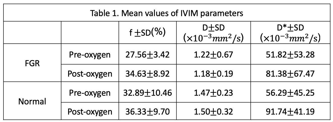
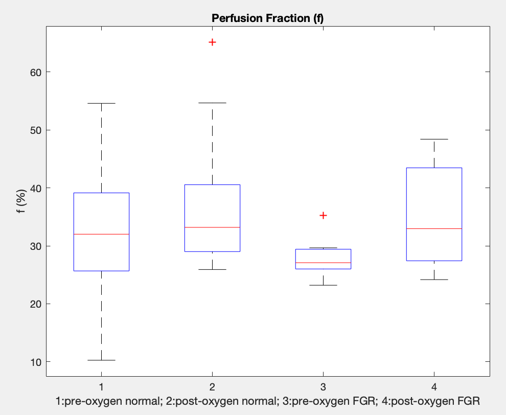
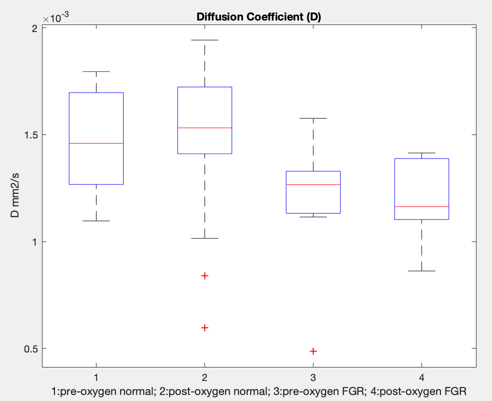
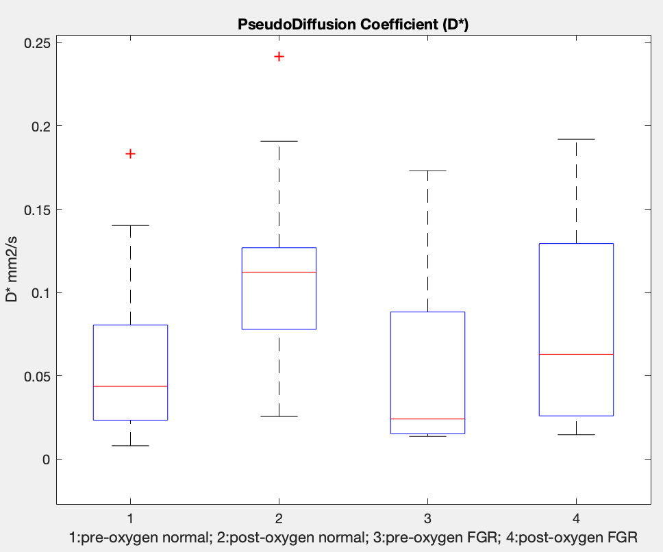
The mean values and standard deviation of IVIM parameters for both normal and FGR pregnancies before and after oxygenation inhalation were presented in Table1. It was found that before oxygenation inhalation, the IVIM parameter fpre was significantly lower in FGR group than that in normal group (P=0.002). In contrast, no significant differences were found for the parameters D (P=0.31) and D* (P=0.47) between these two groups, although they were relatively higher in normal groups. After oxygenation inhalation, fpost increased in both FGR (P=0.007) and normal groups (P=0.26), but significantly difference was only found in FGR group. Interestingly, fpost of FGR group reached a similar level compared to that of normal group (P=0.08). For Dpost, the values in FGR group was lower than that in normal groups but without significant difference (P=0.10). Compare with Dpre, Dpost was not significantly changed for FGR (P=0.69) and group (P=0.60), respectively. For D*post, it also increased in both FGR and normal groups compared to D*pre, but no significant difference were found (P=0.25, P=0.01). Also, between FGR and normal groups, no significant difference was found for D*post (P=0.28).
Conclusions
In this study, the effect of oxygenation therapy on FGR placenta perfusion has been explored by IVIM MRI. The parameter perfusion fraction f, could characterize the changes of placenta perfusion and indicated that FGR placenta perfusion could reach the normal level temporarily after oxygenation inhalation. The study showed that IVIM perfusion parameters could be served as an additive technique not only in identifying FGR patient from normal pregnancy but also useful in predicting or monitoring the hyperoxygenation therapy response in FGR pregnancies.Acknowledgements
No acknowledgement found.References
[1] Moore R J , Strachan B K , Tyler D J , et al. In utero perfusing fraction maps in normal and growth restricted pregnancy measured using IVIM echo-planar MRI[J]. Placenta, 2000, 21(7):726-732.
[2] Moore R J , Issa B , Tokarczuk P, et al. In utero perfusing fraction maps in normal and growth restricted pregnancy measured using IVIM echo-planar MRI[J]. Magnetic Resonance in Medicine, 2000, 43:295-302.
[3] Le B D, Breton E, Lallemand D, et al. MR imaging of intravoxel incoherent motions: application to diffusion and perfusion in neurologic disorders[J]. Radiology, 1986, 161(2):401.
[4] Bihan D L, Breton E, Lallemand D, et al. Separation of diffusion and perfusion in intravoxel incoherent motion MR imaging.[J]. Radiology, 1988, 168(2):497-505.
[5] Bonel H M , Stolz B , Diedrichsen L , et al. Diffusion-weighted MR imaging of the placenta in fetuses with placental insufficiency.[J]. Radiology, 2010, 257(3):810-9.
Figures