3595
Towards MR-based interrogation of the hypoxia-driven insulin resistance mechanism: Adipocytes size estimation.1Radiology, Washington University in St. Louis, School of Medicine, St. Louis, MO, United States
Synopsis
It has been suggested that adipocyte hypertrophy plays a key role in the pathogenesis of systemic insulin resistance and type 2 diabetes. A method for quantifying adipocyte size/hypertrophy in-vivo would be a major advance towards understanding the pathogenesis of type 2 diabetes. In-vivo quantification of adipocyte size might be achievable using short time regime diffusion MR, which carries information about size of the system. Herein, we explore the feasibility of measuring adipocyte size based on the diffusion of triglyceride within adipocytes and the principles of the short diffusion time regime.
Introduction
Obesity is assumed to be a primary cause of type 2 diabetes, but the pathogenesis of the disease is still not well understood. It has been suggested that adipose tissue hypoxia - a condition induced by adipocyte hypertrophy - initiates adipose tissue inflammation and leads to systemic insulin resistance1. A method for quantifying adipocyte size/hypertrophy in-vivo would be a major advance towards understanding the pathogenesis of type 2 diabetes, yet no such method exists. In-vivo quantification of adipocyte size might be achievable using short time regime diffusion MR, which carries information about size of the system and is defined as a regime in which the displacement of the particle is significantly smaller than the actual compartment size and b·D0<<1 2,3,4 (Eq. 1):
$$$D(t)=D_{0}\cdot(1-\frac{4}{3\sqrt{\pi}}\frac{1}{R}\sqrt{Δ\cdot D_{0}}) $$$
Where D(t) is the measured diffusivity of the system (calculated from a mono-exponential representation of the diffusion signal decay), D0 is the free diffusivity of the diffusing molecule of interest, and R is the radius of the system and Δ is the diffusion time. In the context of adipocyte size, the “system” is defined as a single spherical oil droplet that typically comprises >99% of the adipocyte volume (a common surrogate for adipocyte size), and the “diffusing molecules of interest” are the triglyceride molecules within the oil droplet.
Based on the short time regime theory, our objective is to develop a direct, non-invasive, and quantitative measure of adipocyte size using diffusion MR. We use Monte Carlo simulations, microcapillary phantoms and ex-vivo adipose tissue to explore this possibility.
Methods
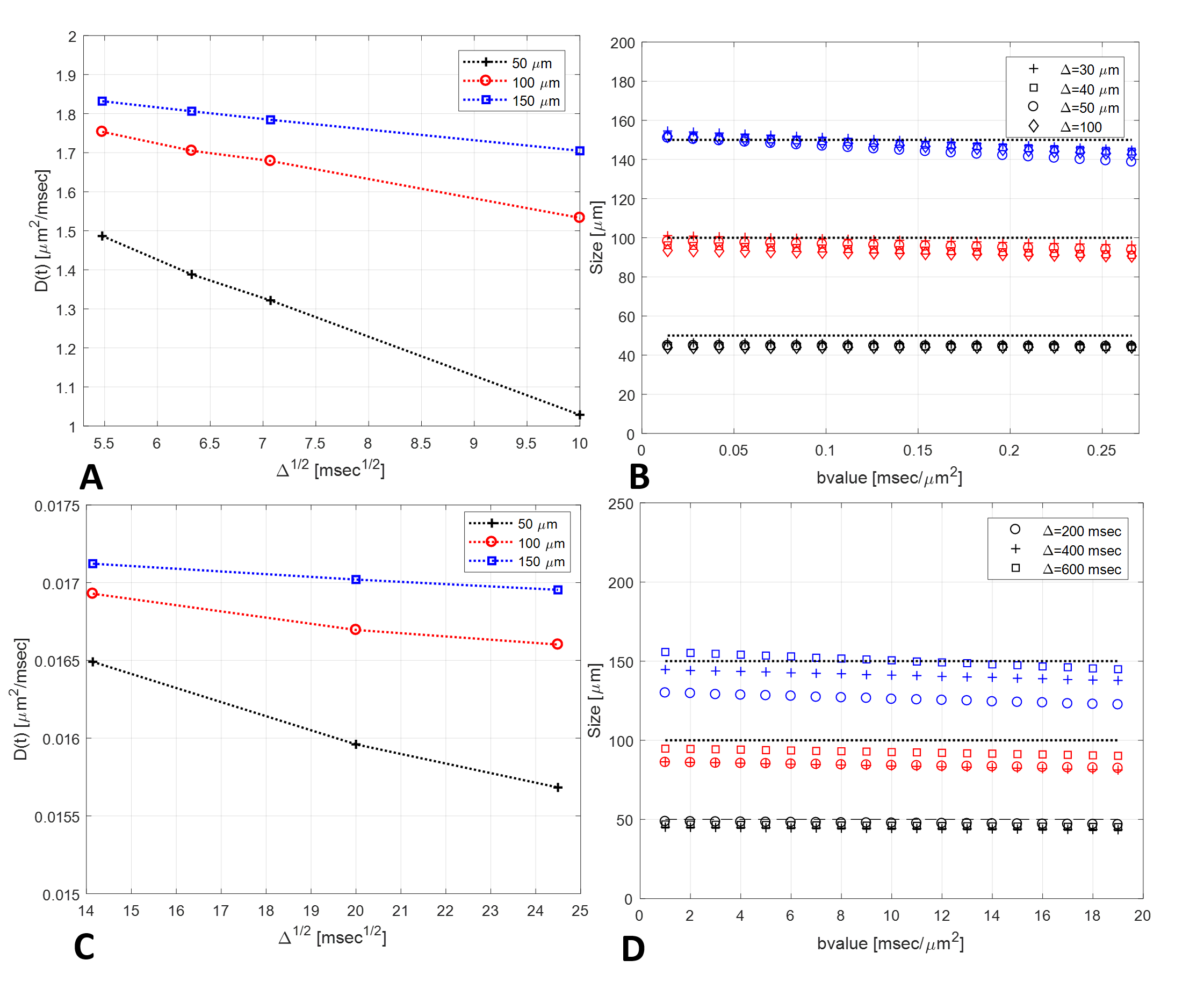
Simulations: Monte Carlo simulations of particle random-walk were performed on M (105-106) random particles with random starting positions inside of infinitely long cylinders with diameters of 50, 100 and 150 μm (roughly the size of adipocytes)4. Diffusion of water (D0=2 μm2/msec) and lipids (D0=0.0175 μm2/msec) inside the three different size cylinders was simulated at diffusion times ranging from 30 to 100 ms or 200 to 600 ms for water or lipids, respectively. These values fall into the short diffusion time regime.
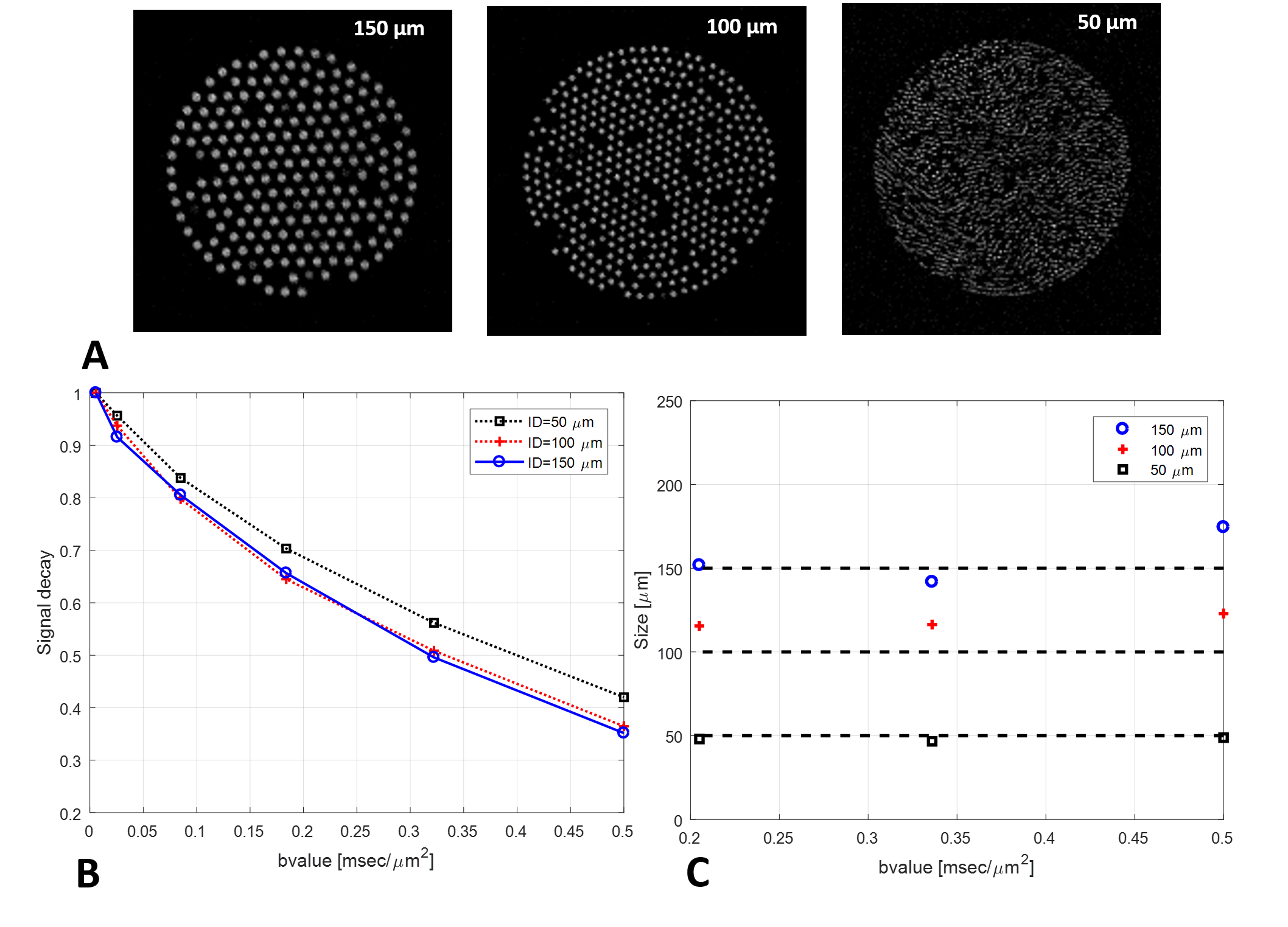
Experimental samples: Ground-truth phantoms constructed from water-filled microcapillaries having inner diameters of 50±5, 100±5 and 150±5 μm and formalin-fixed mice epididymal white adipose tissue (eWAT).
Short-time regime diffusion MR: Diffusion MR experiments were conducted at 11.7T MRI scanner using stimulated echo imaging sequence modified with diffusion encoding gradients. Experimental parameters for imaging of microcapillaries: TR/TE=8000/11 ms, ∆=30 ms (chosen based on simulations), 10 gradient steps, max b-value of 0.5 ms/μm2, NS=1. Experimental parameters for imaging of eWAT: TR/TE=3000/34.6 ms, ∆=400 ms (chosen based on simulations), 10 gradient steps, max b-value of 20 ms/μm2, NS=1.
Results and Discussion
Simulations: This simulation was used to interrogate the limitations of the model. Sizes estimated from the simulation data are in good agreement with the input sizes (Figure 1B,D). For example, in case of cylinders filled with lipids (Figure 1D), sizes of 45.6, 82.7, 139.7 μm are estimated for true input sizes of 50, 100 and 150 μm at ∆ of 400 ms and b-value of 14 ms/μm2. Importantly, the uncertainly of the diameter estimates increases with the true diameter. This can be explained by the inverse relationship between the term which carries the size information (rightmost term of Eq. 1) and the true radius of the system. Restated, as the radius increases, D(t) and D0 converge and the size information about the system diminishes. In practical terms, the results of this simulation imply that, to experimentally resolve large diameters, one must be aware of the relationship between the SNR, the true diameter, and the precision of the estimated diameter. A quantitative in-silico interrogation of this relationship is the subject of ongoing investigation.
Microcapillary phantoms: The calculated diameters of microcapillaries appear to be in good agreement with the ground-truth sizes obtained from manufacturer (dashed black lines in Figure 2C). For example, sizes of 48.0, 115.6 and 150.1 μm are estimated for true capillary sizes of 50, 100 and 150 μm at b-value of 0.2 ms/μm2. Rigorously quantification of the experimental uncertainty in the diameter measurements is ongoing.
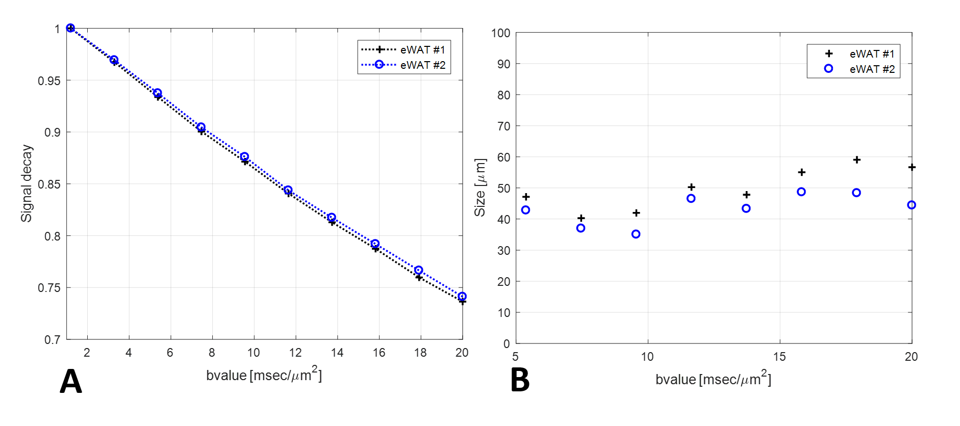
Ex vivo tissue: As a proof-of-principle, diffusion experiments were conducted on two eWAT samples from lean mice using a diffusion time of 400 ms. Here, the mean adipocyte sizes were found to be 49.8±6.8 μm and 43.2±5.0 μm, for the two samples respectively. These adipocyte size estimates are in good agreement with those measured by histology6.
Conclusion
Diffusion MR shows promise for quantifying adipocyte size in-vivo. However, the precision of the measurement is inversely related to the the true diameter of the system and directly related to the free diffusivity of the particle. Thus, one must carefully choose experimental parameters to optimize the fidelity of the model.Acknowledgements
No acknowledgement found.References
1. Trayhurn P. Hypoxia and adipose tissue function and dysfunction in obesity. Physiol Rev. 2013;93(1):1-21. 2. Sukstanskii AL, Ackerman JJH and Yablonskiy DA. Effects of barrier-induced nuclears magnetization inhomogeneities on diffusion-attenuated MR signal. Magn Reson Med. 2003; 50:735–742. 3. Sen PN. Time-dependent diffusion coefficient as a probe of geometry. Concepts in Magnetic Resonance Part A. 2004: 23A(1): 1–21 4. Sukstanskii AL and Yablonskiy DA. In vivo lung morphometry with hyperpolarized 3He diffusion MRI. Theoretical background. J Magn Reson. 2008; 190(2): 200–210. 5. Parlee SD, Lentz SI, Mori H and MacDougald OA. Quantifying size and number of adipocytes in adipose tissue. Methods Enzymol. 2014; 537: 93–122.Figures