3590
To Detect Metastatic Lymph Nodes Using Intravoxel Incoherent Motion Diffusion-weighted Imaging: A Longitudinal Study on Animal Model1Department of Medical Imaging, Nanfang Hospital, Southern Medical University, Guangzhou, China, 2Department of Medical Imaging, People's Hospital of Dehong Prefecture, Yunnan, China, 3Philips Healthcare, Guangzhou, China
Synopsis
Lymph node (LN) involvement is a dynamic process which may be reflected by intravoxel incoherent motion diffusion-weighted imaging (IVIM-DWI). However, there is no such studies so far. Therefore, the potential influence on the diagnosis performance for metastatic LN needs to be investigated. In this longitudinal study on animal models, metastatic LNs evolved in a different pattern from inflammatory LNs pathologically, and IVIM parameters differed between the two groups at particular time points. This indicates that IVIM-DWI can reflect the dynamic pathological changes of LN diseases, and its ability to detect metastatic LN may be affected by the course of disease.
Introduction
Accurate assessment of lymph node (LN) involvement preoperatively is important for cancer staging and treatment decision-making.1 LN metastasis is a dynamic process, as confirmed by pathological studies,2,3,4 but was scarcely studied longitudinally using non-invasive imaging methods. Intravoxel incoherent motion diffusion-weighted imaging (IVIM-DWI) can simultaneously quantify both tissue diffusion and perfusion,5 which theoretically allows for monitoring the process of tumor invasion into LNs. However, previously published related studies were cross-sectional,6-9 so the potential influence of disease evolution on the diagnosis performance for metastatic LNs remains unclear. The purpose of this study was to monitor the longitudinal changes of LNs metastases with IVIM-DWI on animal models, and to determine the relationship between the course of disease and the capability of IVIM-DWI in detecting metastatic LNs.Methods
Twenty New Zealand rabbits with 2.5 - 3.0 kg body weight were studied. Metastatic popliteal LNs were induced by inoculating VX2 cells suspension intramuscularly into either side of the thighs, while inflammatory nodes were induced by inoculating egg-yolk emulsion in the other side. Eight rabbits underwent IVIM-DWI on a 3T scanner (Achieva TX, Philips) with 14 b values (b = 0, 10, 20, 40, 60, 100, 140, 160, 200, 300, 500, 1000, 1500, 2000 s/mm2) at 2 hours prior to, and 14, 21, 28 days after developing disease models. Three rabbits randomly chosen from the remaining twelve rabbits were sacrificed at each time point to perform hematoxylin-eosin staining. Mann-Whitney U test were utilized for comparing the IVIM parameters (ADC, D, D*, f) between two groups.Results
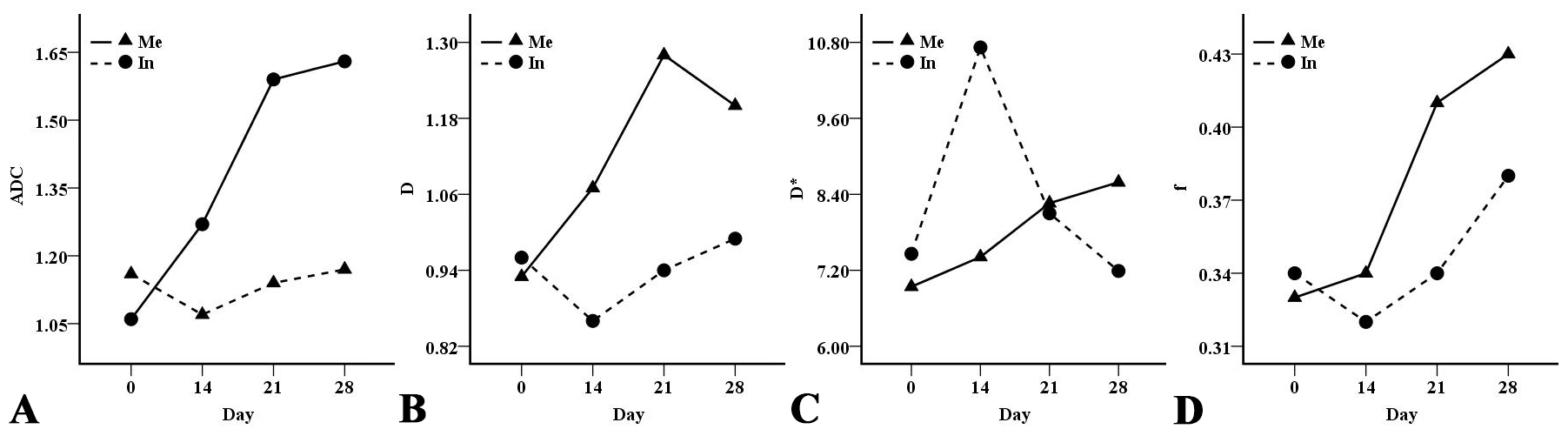
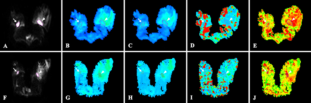
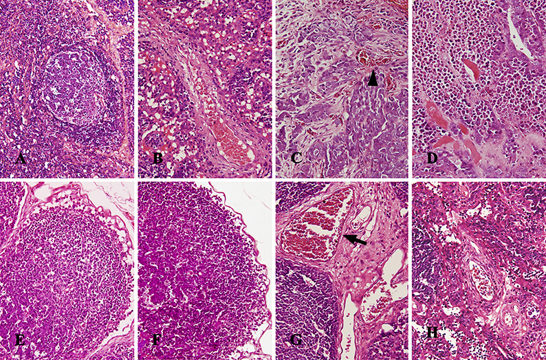
The longitudinal changes of IVIM parameters of two groups were shown in Figure 1. The patterns of dynamic change of ADC, D and D* were different between two groups. Both ADC and D in the metastatic group increased rapidly before day 21 and then slowed down slightly. The inflammatory group decreased in both ADC and D until day 14 and then increased. The mean value of ADC was significantly higher in metastatic nodes than in inflammatory nodes only at day 21 and day 28 (Day 21: 1.59 ± 0.36 × 10⁻³ mm2/s vs. 1.14 ± 0.29 × 10⁻³ mm2/s, P = 0.015; Day 28: 1.63 ± 0.30 × 10⁻³ mm2/s vs. 1.17 ± 0.15 × 10⁻³ mm2/s, P = 0.003) (Figures 1 and 2). Similar findings were observed in D (Day 21: 1.28 ± 0.43 × 10⁻³ mm2/s vs. 0.94 ± 0.27 × 10⁻³ mm2/s, P = 0.029; Day 28: 1.20 ± 0.17 × 10⁻³ mm2/s vs. 0.99 ± 0.15 × 10⁻³ mm2/s, P < 0.001) (Figures 1 and 2). The metastatic group increased in D* over time, while the inflammatory group peaked in day 14 and then decreased. The mean value of D* was significantly different between two groups merely at day 14 (7.41 ± 1.22 × 10⁻³ mm2/s vs. 10.72 ± 3.62 × 10⁻³ mm2/s, P = 0.001) (Figures 1 and 2). The patterns of dynamic change of f were similar in two groups and the mean value of f showed no significant difference between two groups at any time point. Histological studies showed that metastatic LNs were filled with increasing numbers of tumor cells and neovascularization over time, and focal necrosis gradually appeared (Figure 3). Inflammatory LNs were filled with diffuse lymphocytes and plasmacytes, associated with marked dilated vessels at day 14, and then these inflammatory changes gradually recessed (Figure 3).Discussion
In our study, metastatic LNs exhibited different patterns of dynamic change compared to inflammatory LNs, and IVIM parameters differed significantly between two groups merely at some particular time points. As shown by pathological examinations, cellularity, intracellular and extracellular space, hemodynamics and/or angiogenesis in both metastatic and inflammatory nodes changed as the disease progressed and recovered. Our results demonstrated that IVIM-DWI can reflect the dynamic process of both metastasis and inflammation in LNs. The stage of the diseased nodes can have an influence on IVIM parameters in differential diagnosis. A lower D* in metastatic nodes at day 14 was in accordance with less marked vascular changes, such as vasodilation, compared to the inflammatory nodes. Similarly, higher ADC and D in metastatic nodes at day 21 and day 28 indicated the relatively sparse cellularity, less limited extracellular space, and the existence of focal necrosis, compared to the high cellularity in inflammatory nodes. Notably, the present study was conducted on animal models, a study on patients with large population in the future is warranted to validate these findings.Conclusion
IVIM-DWI can reflect the dynamic pathological changes of LN diseases, and its ability to detect metastatic LN may be affected by the course of disease.Acknowledgements
No acknowledgement found.References
1. Qi L, Yan W, Chen K, et al. Discrimination of Malignant versus Benign Mediastinal Lymph Nodes Using Diffusion MRI with an IVIM Model. Eur Radiol. 2018; 28(3):1301-1309.
2. Qian CN, Berghuis B, Tsarfaty G, et al. Preparing the "soil": the primary tumor induces vasculature reorganization in the sentinel lymph node before the arrival of metastatic cancer cells. Cancer Res. 2006; 66(21):10365-10376.
3. Sleeman JP. The lymph node pre-metastatic niche. J Mol Med (Berl). 2015; 93(11):1173-1184.
4. Karaman S, Detmar M. Mechanisms of lymphatic metastasis. J Clin Invest. 2014; 124(3):922-928
5. Le Bihan D. What can we see with IVIM MRI? Neuroimage. 2017; doi: 10.1016/j.neuroimage.2017.12.062.
6. Liang L, Luo X, Lian Z, et al. Lymph node metastasis in head and neck squamous carcinoma: Efficacy of intravoxel incoherent motion magnetic resonance imaging for the differential diagnosis. Eur J Radiol. 2017; 90:159-165.
7. Qiu L, Liu XL, Liu SR, et al. Role of quantitative intravoxel incoherent motion parameters in the preoperative diagnosis of nodal metastasis in patients with rectal carcinoma. J Magn Reson Imaging. 2016; 44(4):1031-1039.
8. Wu Q, Zheng D, Shi L, et al. Differentiating metastatic from nonmetastatic lymph nodes in cervical cancer patients using monoexponential, biexponential, and stretched exponential diffusion-weighted MR imaging. Eur Radiol. 2017; 27(12):5272-5279.
9. Zhu Y, Li X, Wang F, et al. Intravoxel incoherent motion diffusion-weighted magnetic resonance imaging in characterization of axillary lymph nodes: Preliminary animal experience. Magn Reson Imaging. 2018; 52:46-52.
Figures