3581
Diffusion Kurtosis Imaging of the Brain with Free Water Elimination1Radiology, Miller School of Medicine, University of Miami, Miami, FL, United States
Synopsis
In diffusion weighted imaging (DWI) of the brain, a single voxel may contain gray matter (GM), white matter (WM), as well as free water embedded in GM, WM, and cerebrospinal fluid (CSF), each with different diffusion profiles. Free water elimination (FWE) is a method used to separate the free water components from tissue water. In this work, we extend FWE by fitting with a diffusion kurtosis imaging (DKI) model as it is a better descriptor of the non-Gaussian diffusion that occurs in tissue water.
Introduction
Free water contamination refers to the presence
of fast and isotropically diffusing water mixed with anisotropically
diffusing tissue water in a voxel. The
contribution to this free water can come from the cerebrospinal fluid (CSF) partial voluming or from excessive
amounts of extra-cellular water content.1,2 In diffusion tensor
imaging (DTI) studies, where a single tensor is used to describe the water diffusion
profile of a voxel, the presence of free water biases the measured
diffusion metrics.1,3 Free water elimination (FWE) has been proposed
as a dual compartment fitting method to separate the free water fraction from the anisotropic water diffusing in tissue, which provides a corrected DWI-signal representing the
restricted water diffusion within biological cells.1,4,5 However, biological
restrictions in the tissue microstructure cause the water diffusion to be
non-Gaussian in its distribution. Using a diffusion kurtosis imaging (DKI)
model with FWE is therefore better suited to describe the signal than DTI which
assumes Gaussianity.5,6,7 Additionally, DKI can be used with high
b-values making it more sensitive to regions with large diffusion restrictions.
Thus, the aim of this work is to extend the commonly used FWE-DTI method with
kurtosis fitting using a clinically feasible two-shell acquisition and
demonstrate its usability in extracting diffusivity and kurtosis metric maps.Methods
The FWE-DTI model is described by a simple bi-exponential expansion of DTI1,4
$$$S_{i}=S_{0}\left[\left(1-f\right)exp\left(-b_{i}g_i^T\,D_{tissue}g_{i}\right)+f\,exp\left(-bD_{iso}\right)\right]$$$. (1)
Here, the measured diffusion-weighted signal $$$S_{i}$$$ is the combination of free water, with a volume fraction $$$f$$$ and
isotropic diffusion $$$D_{iso}$$$ set at a constant value of $$$3\times10^{-3}$$$mm2/s,
and tissue water, with a volume fraction $$$\left(1-f\right)$$$
and diffusion tensor $$$D_{tissue}$$$ characterized by $$$6$$$ independent elements $$$\theta_{D}=\left\{D_{ij}\right\}_{i\leq\,j\leq\,3}$$$. $$$S_{0}$$$
is the signal when no diffusion sensitization gradient is applied and $$$g_{i}$$$ is the $$$i$$$-th component of the normalized
diffusion weighting gradient direction vector $$$\bf\,g$$$ with $$$i=1,...,m$$$ for $$$m$$$ gradient directions.
We expand this formulation using the DKI model6
$$$S_{i}=S_{0}\left[\left(1-f\right)exp\left[-b_{i}g_i^T\,D\,g_{i}\,+\,\frac{b_i^2}{6}\left(\sum_{i=1}^3\frac{D_{ii}}{3}\right)^2\sum_{i,j,k,l=1}^3g_{i}g_{j}g_{k}g_{l}W_{ijkl}\right]+f\,exp\left(-bD_{iso}\right)\right]$$$, (2)
where $$$W_{ijkl}$$$ represents the $$$ijkl$$$-th element of the fully symmetric fourth-order diffusion kurtosis tensor $$$W$$$, which can be characterized by 15 independent elements $$$\theta_{K}=\left\{W_{ijkl}\right\}_{i\leq\,j\leq\,k\leq\,l\leq3} $$$. Using the bi-exponential expression with DKI, the fitting problem becomes difficult and ill-conditioned as we now have to find 21 parameters $$$\theta=\left[\theta_{D},\theta_{K}\right]$$$. We use a two-step approach, with a weighted least squares (WLS) initial estimation followed by a non-linear least squares (NLS) step to refine the solution as proposed by Hoy et al.4,8 The goal is to find the $$$\left(f,\theta\right)$$$ pair that minimizes the WLS objective function
$$$F_{WLS}=\frac{1}{2}\sum_{i=1}^m\omega_i^2\left( y_{i}-\sum_{j=1}^{22}A_{ij}\gamma_{j}\right)^2$$$. (3)
The weights $$$\omega_i$$$ are set equal to the measured signal $$$s_i$$$, $$$A$$$ is the $$$\left(m\times22\right)$$$ diffusion encoding matrix, and $$$\gamma$$$ is the solution parameter matrix estimated by
$$$\gamma=\left( A^T S^2 A\right)^{-1}A^TS^2y$$$, (4)
where $$$S$$$ is a diagonal matrix of the measured diffusion signal, and $$$y$$$ is the natural log of the free water adjusted signal given by
$$$y_{ik}=ln\left\{\frac{s_i-s_0f_k\,exp\left(-b\,D_{iso}\right)}{\left(1-f_k\right)}\right\}$$$, (5)
with $$$k=1,…,n$$$ and $$$n$$$ is the number of $$$f$$$-values fitted simultaneously. Our method is implemented in Python using the Dipy libraries. The initial WLS solution is found by searching for the solution over intervals of $$$f=\left\{0,0.1,0.2,…1\right\}$$$. A second and third iteration are used to improve the solution over smaller intervals sizes of 0.01 and 0.001 respectively. The solution is further refined during NLS fitting using a modified Levenberg-Marquard algorithm to minimize the NLS objective function
$$$F_{NLS}=\frac{1}{2}\sum_{i=1}^m\left[s_i-S_0\,f\,exp\left(-b_i\,D_{iso}\right)-\left(1-f\right)exp\left(\sum_{j=1}^{22}A_{ij}\gamma_{j}\right)\right]^2$$$
Results and Discussion
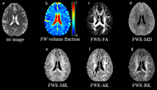
We applied our FWE-DKI model on a dataset acquired
at 3 T from the brain of a human volunteer (female, age 31) using two nonzero
b-values (b=1000/2000 s/mm2; 30
direction and 2 averages per shell) and 6 b0 images. Fig 1 shows the parametric
maps obtained with our FWE-DKI. We obtain the free water volume fraction (Fig 1b) together with commonly obtained
diffusivity metric maps FA and MD (Fig 1c-1d). In addition, we can obtain FWE mean, axial, and radial kurtosis maps (Fig 1e-1g).Conclusion
We have successfully extended the FWE model to DKI and implemented the method using a combination of WLS and NLS fitting to solve the ill-conditioned problem. This method does not require any parameter approximation, can be applied to a simple dual-shell acquisition, and produces FWE kurtosis maps.Acknowledgements
Funding from NIH grant, R01 NS094043References
1. Pasternak O, et al. Free water elimination and mapping from diffusion MRI. Magn Reson Med 2009;62(3):717-730.
2. Lyall AE, et al. Greater extracellular free-water in first-episode psychosis predicts better neurocognitive functioning. Mol Psychiatry 2018;23(3):701–707.
3. Pasternak O, et al. Estimation of extracellular volume from regularized multi-Shell diffusion MRI. Med Image Comput Comput Assist Interv 2012;15(Pt 2):305-312
4. Hoy AR, et al. Optimization of a free water elimination two-compartment model for diffusion tensor imaging. NeuroImage 2014;103:323-333.
5. Collier, et al. Diffusion Kurtosis Imaging With Free Water Elimination: A Bayesian Estimation Approach. Magn Resn Med 2018;80(2):802-813.
6. Jensen JH, et al. Diffusional kurtosis imaging: the quantification of non-gaussian water diffusion by means of magnetic resonance imaging. Magn Reson Med 2005;53(6):1432-1440.
7. Jensen JH and Helpern JA (2010). MRI quantification of
non-Gaussian water diffusion by kurtosis analysis. NMR in
Biomedicine 2010;23(7):698-710
8. Henriques RN, et al. bioRxiv 2017. doi: http://dx.doi.org/10.1101/108795.
Figures