3579
Resolving crossing fibers with the inclusion of intra-axonal diffusion modeling1Biomedical Engineering, Washington University in St. Louis, st louis, MO, United States, 2Radiology, Washington University School of Medicine, St. Louis, MO, United States, University City, MO, United States, 3Chemistry, Washington University in St. Louis, st louis, MO, United States, 4Radiology, Washington University School of Medicine, St. Louis, MO, United States, Saint Louis, MO, United States
Synopsis
A new diffusion histology imaging (DHI) model is proposed to model crossing fibers considering both intra- and extra-axonal water diffusion, along with extra-axonal isotropic diffusion within an image voxel. Both Monte-Carlo simulation and in vivo MRI data from one healthy volunteer brain were examined to assess whether DHI can resolve crossing fibers while quantifying axonal injury, demyelination, and inflammation.
Introduction
Existing diffusion MRI methods focuses on accurately resolving crossing fibers while failing to assess fiber axial and radial diffusivity. None of existing methods is able to quantify axial and radial diffusivity of individual crossing fibers. We previously developed diffusion basis spectrum imaging (DBSI1) to resolve crossing fibers while also accurately assess axial and radial diffusivity of individual fibers. In this report, we will present a further improvement of DBSI, i.e., diffusion histology imaging (DHI), to include quantifying intra-axonal diffusivity to more sensitively assess axonal injury and better assess axonal density. We demonstrate the utility of DHI employing a Monte-Carlo simulation and diffusion-weighted MRI data from a healthy control volunteer. DHI accurately estimated fiber crossing angles and axial/radial diffusivity of individual crossing fibers even under low SNR and relatively low diffusion gradient strength.Materials and Methods
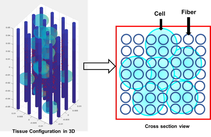
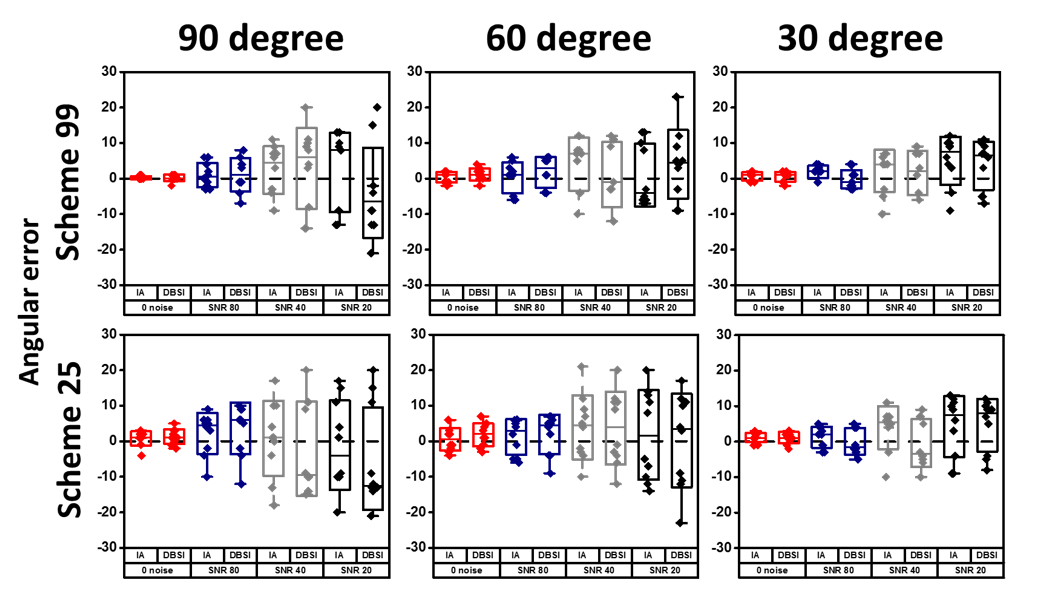
DHI: The new DHI models water diffusion in multiple micro-compartments of white matter tracts including anisotropic intra-axonal diffusion, anisotropic extra-axonal diffusion, and isotropic extra-axonal diffusion within an image voxel. Monte-Carlo simulation: The simulation was performed by tracking the path of random walk of 1 × 10⁶ randomly distributed water molecules in a coherent nerve model (Fig. 1). The size of the axon and cells were adapted according to literature reports2.The number of water molecules inside cell is fixed at 15% of the total number. Diffusivity for free water was set to be 3 μm2/ms (assuming 37 °C). MRI acquisition parameters are TE=37 ms, Δ=18 ms, δ=6 ms, maximum b-value 2200 s/mm2 corresponding to our previous in vivo mouse optic nerve diffusion MRI experiments. Two coherent fiber bundles are allowed to cross with each other at 30, 60, 90 degrees. Rician noise was added to the diffusion MRI signal at SNR = 20, 40, 80.The simulation was repeated for 10 times at each SNR and inter-axonal gaps. Human subject: Procedures involving human subjects were all approved by the Institutional Review Board of Washington University. All subjects provided informed consent before their participation in the study. In-Vivo DW-MRI: All subjects underwent diffusion-weighted MRI at 3.0T using a multi-b value diffusion-weighting scheme (Trio; Siemens, Erlangen, Germany). Diffusion-weighted images (DWIs) were collected with a 99-direction multi-b-value diffusion scheme using a single-shot spin-echo echo-planar imaging sequence with the following key parameters: voxel size = 2×2×2 mm3; Maximum b-value = 1500 s/mm2; acquisition time = 15 minutes. DBSI and DHI were computed using the in-house software developed using Matlab.Results and Discussion
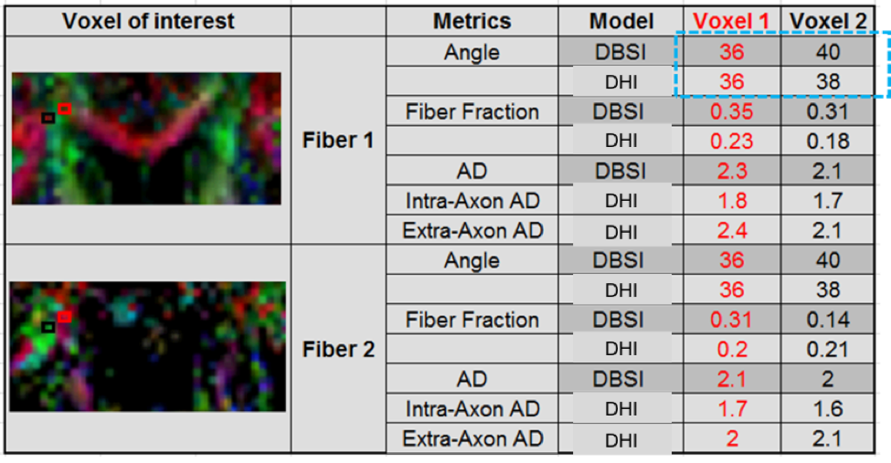
The crossing fiber angle estimation was compared to the in silico true angles. The estimated errors were comparable between DHI and DBSI at all SNR levels (Fig. 2). In vivo diffusion-weighted MRI data from a healthy volunteer were analyzed by DBSI and DHI at the intersection of the corpus callosum and corona radiata. The fiber crossing angle estimated was comparable between DBSI and DHI (Fig. 4). DHI and DBSI performed comparably in estimating crossing angles both in vivo and in silico.Conclusion
DHI resolves crossing fibers accurately estimating fiber crossing angles. Results have shown that DHI provides accurate and precise estimates of fiber orientations as does DBSI. Both DBSI and DHI resolve fibers crossing at relatively low crossing angles even under the influence of low SNR. In silico DHI and DBSI estimation was also experimentally validated using in vivo diffusion MRI data from a healthy volunteer.Acknowledgements
This work was supported in part by NIH R01-NS047592, P01-NS059560, U01-EY025500, National Multiple Sclerosis Society (NMSS) RG 5258-A-5, RG 1701-26617.References
1. Wang Y, Wang Q, Song SK et al. Quantification of increased cellularity during inflammatory demyelination. Brain. 2011; 134: 3590–601.
2. Chiang CW, Wang Y, Sun P et al. Quantifying white matter tract diffusion parameters in the presence of increased extra-fiber cellularity and vasogenic edema. Neuroimage. 2014;
Figures