3575
An in vivo investigation on quantitative metrics of diffusion kurtosis tensor: the effect of diffusion gradient parameters in the clinical setting1Institute of Biomedical Engineering and Nanomedicine, National Health Research Institutes, Miaoli County, Taiwan, 2Institute of Neuroscience and Medicine 4, INM-4, Forschungszentrum Jülich, Jülich, Germany, 3Department of Computer Science, National Chiao Tung University, Hsinchu, Taiwan, 4Institute of Neuroscience and Medicine 11, INM-11, JARA, Forschungszentrum Jülich, Jülich, Germany, 5JARA - BRAIN - Translational Medicine, Aachen, Germany, 6Department of Neurology, RWTH Aachen University, Aachen, Germany, 7Institute of Medical Device and Imaging, National Taiwan University College of Medicine, Taipei, Taiwan
Synopsis
Diffusion kurtosis imaging (DKI) is an emerging technique that provides additional information to delineate tissue microstructures by quantifying the non-Gaussian water molecular diffusion. Although the capability of DKI has been demonstrated, the effects of diffusion gradient parameters on its quantitative metrics, particularly in the clinical setting, have not been fully understood yet. This study aims to investigate the effect of diffusion gradient parameters on diffusion kurtosis tensor calculation and its quantitative metrics. In vivo results show that diffusion gradient duration has incremental influence on DKI quantitative metrics in the clinical setting. Further investigation with more subjects would help to statistically solidify our findings.
PURPOSE
Diffusion kurtosis imaging (DKI), a recently developed diffusion MRI technique, provides additional, relevant information on microstructural changes than diffusion tensor imaging (DTI) does in brain diseases1-3. However, the effects of diffusion gradient characteristics (e.g. strength, duration and separation) on its quantitative metrics, particularly in the clinical settings, have not been fully understood yet. Previously, Minati et al.4 performed an in vivo study on rat thalamus and found that increasing gradient separation time (Δ) reduced diffusion kurtosis, whereas increasing gradient pulse duration (δ) elevated diffusion coefficient derived from DKI model. In a simulation study, Jensen et al.5 showed that the prolonged δ deviated apparent diffusion kurtosis at most a few percent using an analytic expression with model parameters similar as clinically used ones. Both studies analysed diffusion kurtosis by fitting DKI model on the 1D profile of diffusion signals along a single diffusion encoding direction, which does not fully describe the diffusion signal in anisotropic systems. To deconstruct the DKI protocol in the clinical setting, this study aims to investigate the effect of diffusion gradient timing parameters on diffusion kurtosis tensor calculation and its quantitative metrics. We expect to elucidate the relationship between diffusion gradient characteristics and their influence on diffusion kurtosis metrics.METHODS
RESULTS
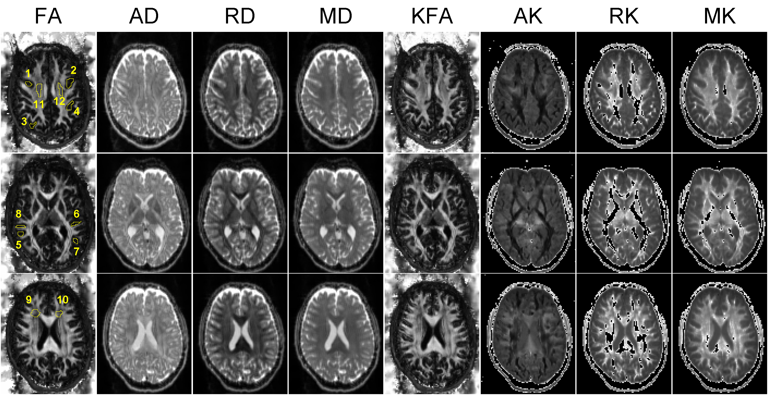
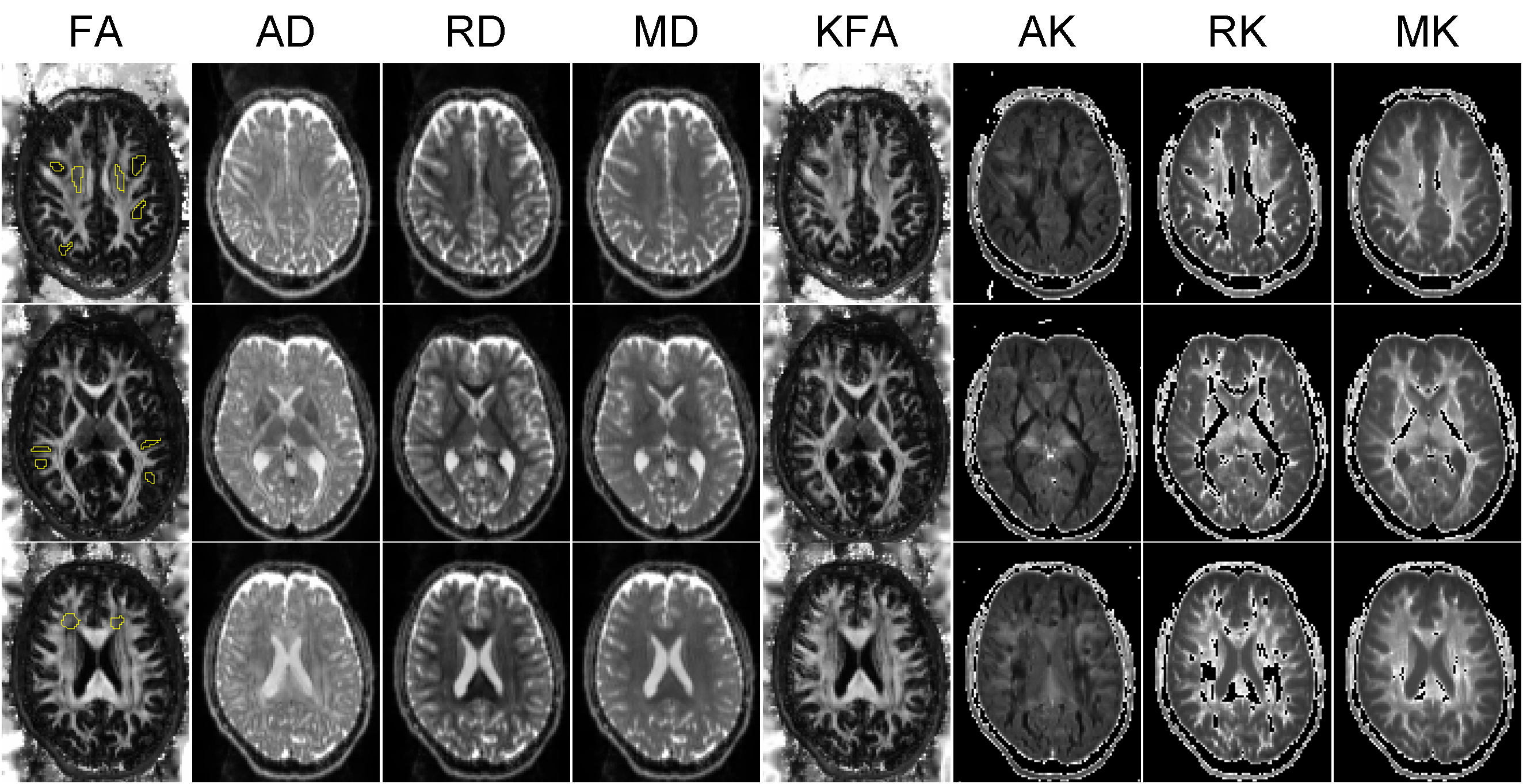
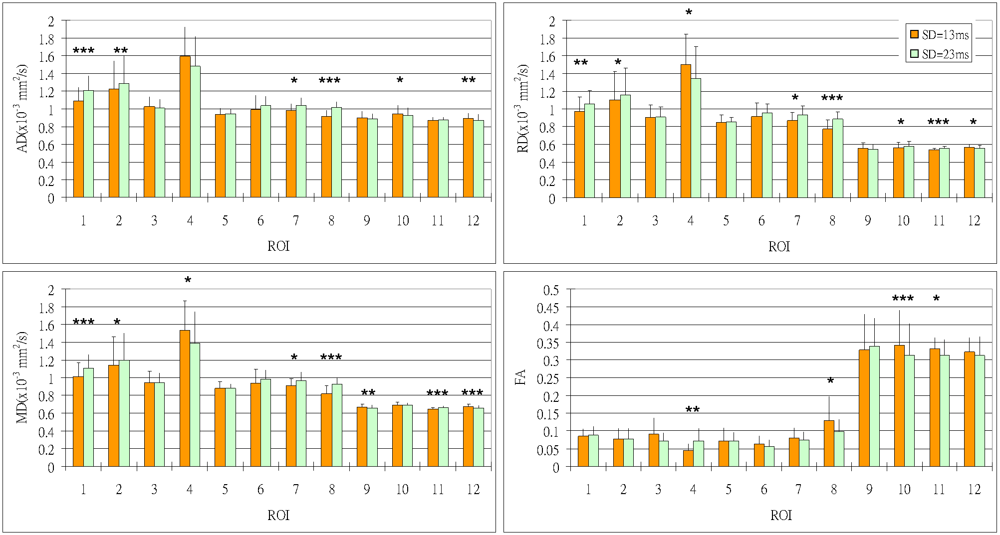
Figures 1 and 2 show the DTI/DKI metrics by using δ of 13 and 23 ms, respectively. There are no significant visual differences between the parametric maps using different δ. We performed the ROI-based statistical comparisons between the maps derived from data sets with δ of 13 and 23 ms, shown in Figures 3 and 4, respectively. As shown in both figures, the use of different δ deviates the quantitative measures of diffusion characteristics in some grey and white matter regions. We also observed that the regions with significant differences, among MD, AD and RD are relatively similar, whereas those among MK, AK and RK are generally different. This finding may imply that these three kurtosis measures can have their respective microstructural underpinnings and sensitivities to different kinds of tissue structures. The change of δ has similar impact on both FA and KFA, especially in region 8, 10, and 11, where FA and KFA significantly decrease with increasing δ.DISCUSSION AND CONCLUSIONS
Our preliminary results of in vivo human brain show significant regional differences of DTI/DKI quantitative metrics in some specific ROIs between the uses of different δ. We also observed different patterns between diffusivity and kurtosis metrics, whereas a similar tendency was found between FA and KFA. Our results are not fully consistent with previous simulation and in vivo animal studies4,5. Several factors may affect the dependency of diffusion kurtosis with δ, such as diffusion encoding scheme, signal-to-noise ratio and partial volume effect. Also, the complexity of tissue microstructures may deviate the sensitivity of diffusion kurtosis since a previous study has demonstrated the strong effect of fiber crossings and their crossing angles on diffusion kurtosis metrics11. In conclusion, our preliminary results show that one of the diffusion gradient parameters, i.e. δ, has incremental influence on quantitative metrics derived from DKI data in the clinical setting. To solidify our observations, the sample size has to be increased and an investigation on diffusion gradient separation time has to be conducted.Acknowledgements
We thank for the funding supports from National Health Research Institutes (BN-107-PP-06) and Taiwan Ministry of Science and Technology (107-2911-I-400-502 and 107-2221-E-400-001).References
- Chen, X.-r., Zeng, J.-y., Shen, Z.-W., Kong, L.-m. & Zheng, W.-b. Diffusion Kurtosis Imaging Detects Microstructural Changes in the Brain after Acute Alcohol Intoxication in Rats. BioMed Research International 2017, 6 (2017).
- Spampinato, M.V. et al. Diffusional Kurtosis Imaging and Motor Outcome in Acute Ischemic Stroke. AJNR. American journal of neuroradiology 38, 1328-1334 (2017).
- Zhang, G. et al. Diffusion Kurtosis Imaging of Substantia Nigra Is a Sensitive Method for Early Diagnosis and Disease Evaluation in Parkinson's Disease. Parkinson’s Disease 2015, 5 (2015).
- Minati, L. et al. Effect of diffusion-sensitizing gradient timings on the exponential, biexponential and diffusional kurtosis model parameters: in-vivo measurements in the rat thalamus. Vol. 23 115-121 (2010).
- Jensen, J.H. & Helpern, J.A. Effect of gradient pulse duration on MRI estimation of the diffusional kurtosis for a two-compartment exchange model. Journal of Magnetic Resonance 210, 233-237 (2011).
- Manjón, J.V. et al. Diffusion Weighted Image Denoising Using Overcomplete Local PCA. PLOS ONE 8, e73021 (2013).
- Jensen, J.H. & Helpern, J.A. MRI quantification of non‐Gaussian water diffusion by kurtosis analysis. NMR in Biomedicine 23, 698-710 (2010).
- Tabesh, A., Jensen, J.H., Ardekani, B.A. & Helpern, J.A. Estimation of tensors and tensor‐derived measures in diffusional kurtosis imaging. Magnetic Resonance in Medicine 65, 823-836 (2011).
- Ben-Israel, A. & Greville, T.N. Generalized inverses: theory and applications, (Springer Science & Business Media, 2003).
- Glenn, G.R., Helpern, J.A., Tabesh, A. & Jensen, J.H. Quantitative assessment of diffusional kurtosis anisotropy. NMR in Biomedicine 28, 448-459 (2015).
- Ankele, M. & Schultz, T. Quantifying Microstructure in Fiber Crossings with Diffusional Kurtosis. 150-157 (Springer International Publishing, Cham).
Figures