3559
Machine learning based estimation of axonal properties in the presence of beading1UNIRS, CEA/Joliot/Neurospin, GIf-sur-Yvette, France, 2Research Centre Jülich, Institute of Neuroscience and Medicine, Jülich, Germany, Juelich, Germany, 3UNATI, CEA/Joliot/Neurospin, GIf-sur-Yvette, France
Synopsis
In this work, we investigate the potential of machine learning techniques to make one step forward by quantitatively estimating beading amplitude, a specific marker of pathological beading using frequency-dependent changes in diffusion measurements. Classification and regression are performed using Extremely Randomized Trees from OGSE signals corresponding to 6 distinct frequencies and synthesized from numerical simulations in realistic white matter phantoms depicting beaded axons.
Introduction
Axonal beading is a morphological change indicative of neuronal injury which might be implicated in numerous human degenerative disorders of the CNS1 and is observed after strokes due to ischemia-induced swelling2. Recent simulation studies2-4 suggest that diffusion-weighted MRI is sensitive to beading which causes a substantial decrease of the apparent diffusion coefficient. In particular, the use of Oscillating-Gradient Spin-Echo (OGSE) sequences showed a strong dependence of axial and radial dispersive diffusivity to beading in both intra- and extra-cellular spaces3,4. In this work, we investigate the potential of machine learning techniques5 to make one step forward by quantitatively estimating beading amplitude (BA), a specific marker of pathological beading2,3, using frequency-dependent changes in diffusion measurements. Classification and regression are performed using Extremely Randomized Trees (ExtraTrees) from OGSE signals corresponding to 6 distinct frequencies and synthesized from numerical simulations in realistic white matter phantoms depicting beaded axons.Material & Methods
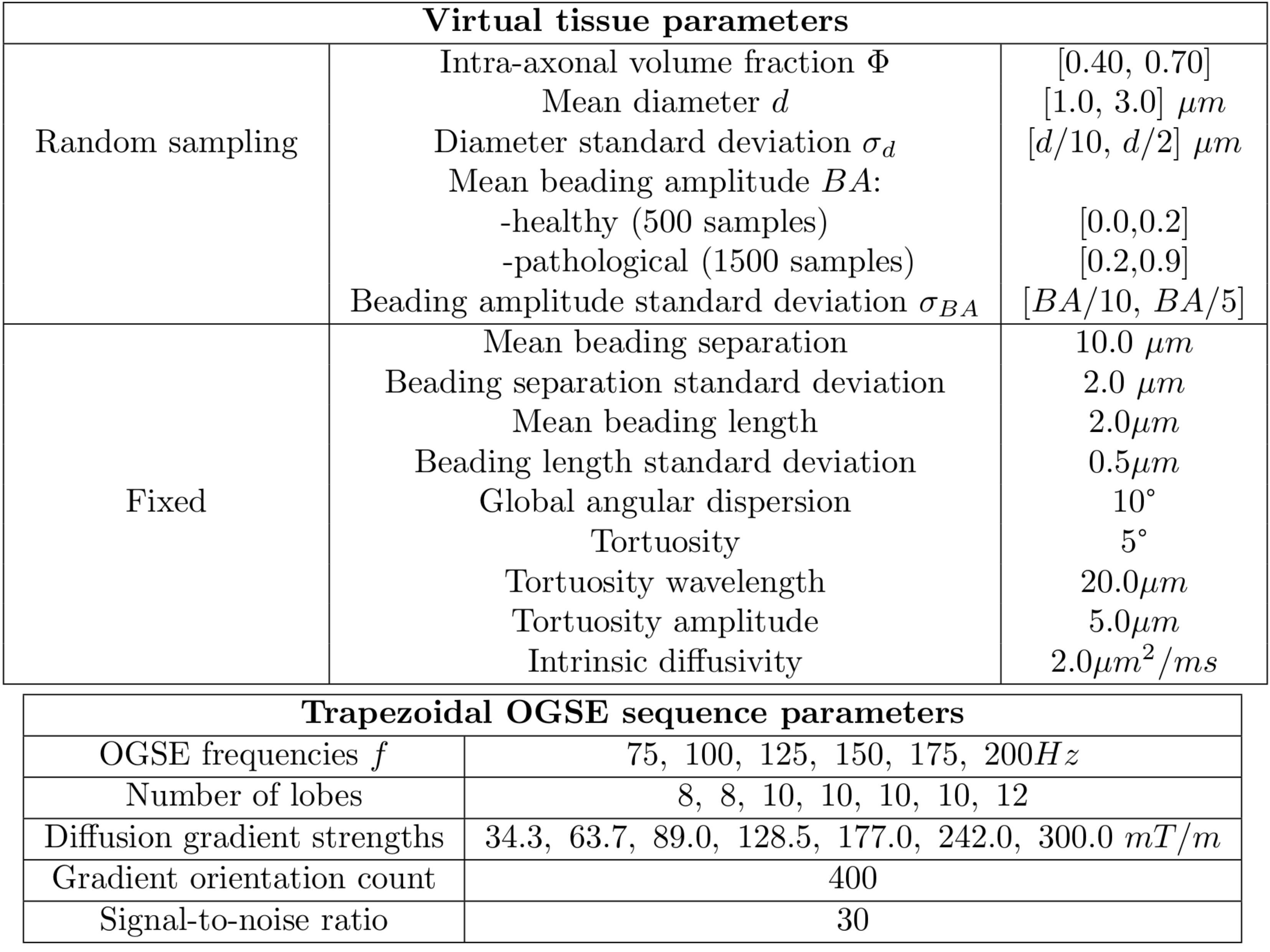
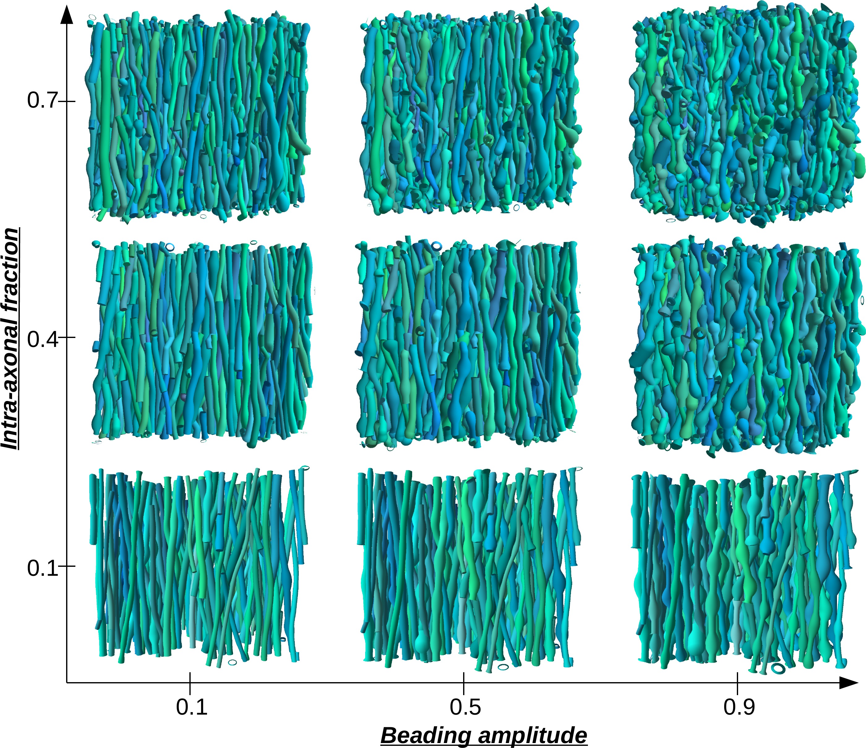
2000 virtual white matter tissues each containing 2000 axonal fibers with randomly sampled mean axonal diameters, packing densities and mean BA (fig.1) were created, including 500 samples corresponding to healthy tissues ($$$BA < 0.2$$$)2,3 and 1500 samples to pathological tissues ($$$BA \in [0.2,0.9]$$$). A novel GPU-based phantom simulator modeling axonal fibers from sets of overlapping spheres and enabling to model global angular dispersion, axonal tortuosity and beading at high packing densities was employed to generate the tissues (fig.2).
Monte-Carlo simulations were subsequently performed on each virtual tissue using 105 spins6,7 with a random walk step equal to 2% of the mean axonal diameter for each tissue. Equal-step-length random leap8 was employed for particle-membrane interaction with impermeable membranes. OGSE diffusion-weighted signals at 6 distinct frequencies from 75Hz to 200Hz and constant $$$b$$$-value of $$$450 s/mm^2$$$ were synthesized according to the protocol given in fig.1, adding noise to reach SNR=30 clinically achievable with a Connectome gradient set.
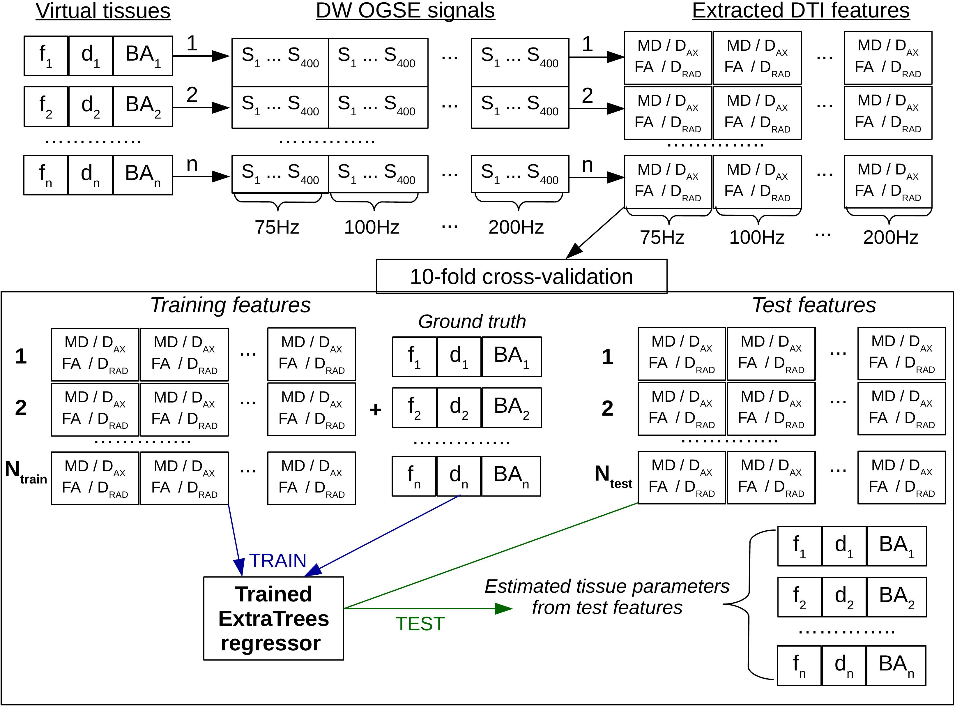
A feature vector was computed from each synthesized OGSE signal using a DTI analysis, comprising the mean diffusivity, fractional anisotropy, axial and radial diffusivities for the 6 OGSE frequencies resulting in 24 features per tissue.
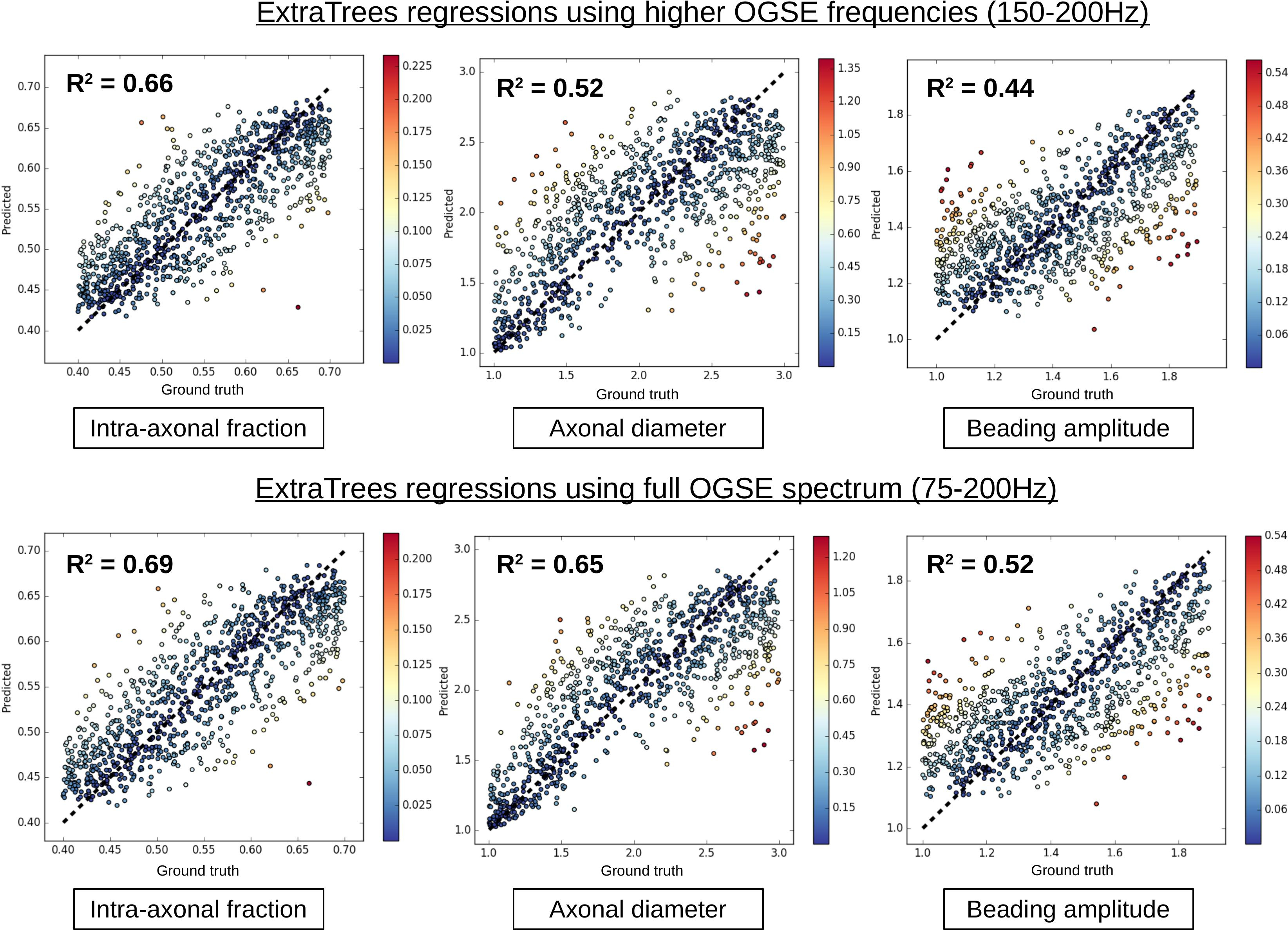
An ExtraTrees classifier from Scikit-Learn9 was trained on a subsample of the dictionary of generated signals containing 500 healthy and 500 pathological samples to discriminate between healthy and pathological tissues (fig.3). An ExtraTrees regressor was trained on the whole dictionary (500 healthy and 1500 pathological samples) to learn the mapping between the extracted features and the underlying microstructure parameters of interest : intra-axonal volume fraction, axon diameter and BA. Both classifier and regressor used 100 trees and a maximum depth of 20 (other parameters were set to default values). All performance metrics were computed using a 10-fold cross-validation strategy to reduce bias due to the choice of training data.
Results and discussion
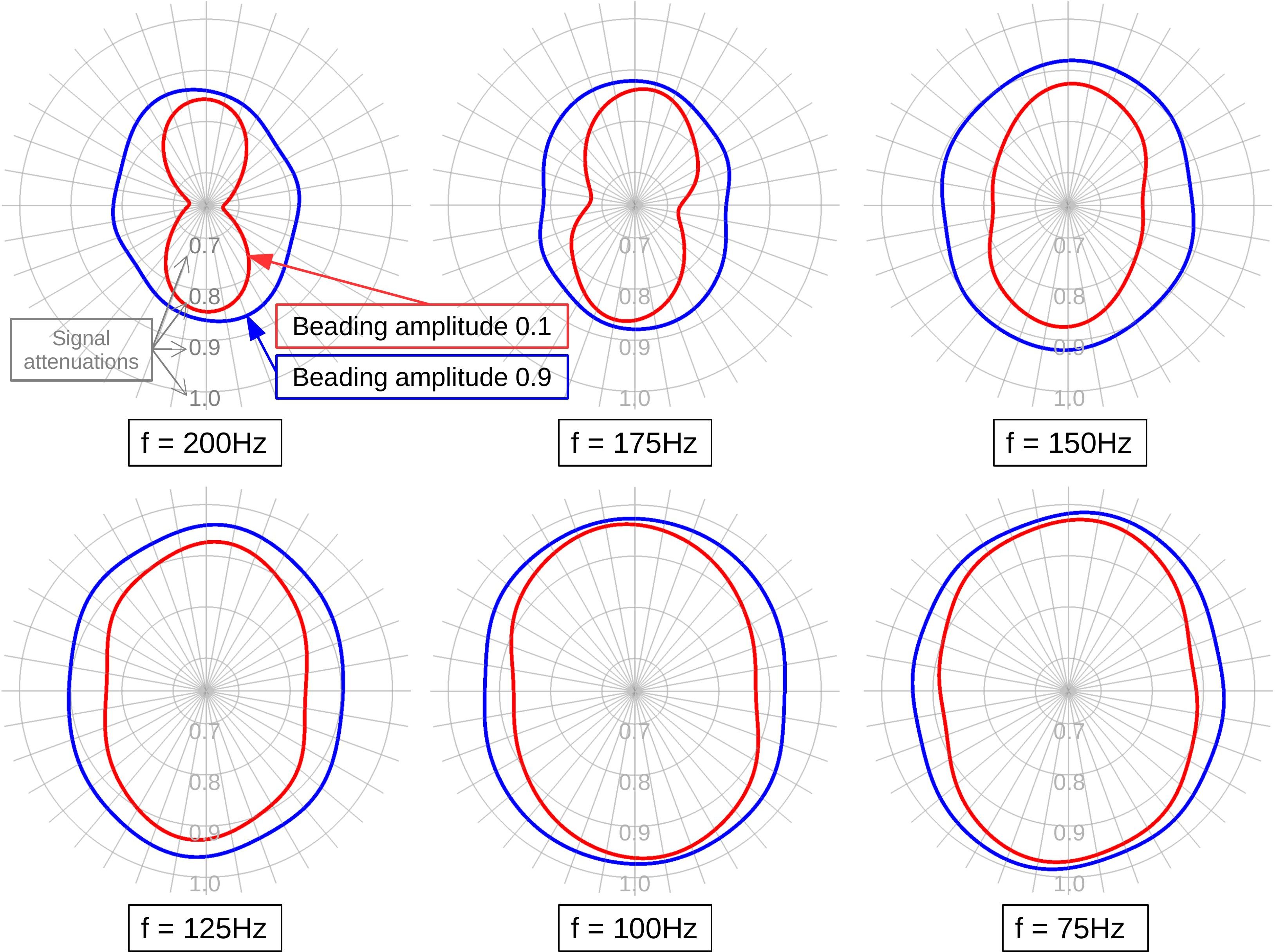
Fig.2 shows example tissues demonstrating the ability of the employed phantom generator to control the intra-axonal fraction and BA of each constructed tissue. The impact of BA on the diffusion-weighted signal at each OGSE frequency is illustrated in fig.4, which indicates that differences in BA are more prominent at higher OGSE frequencies (150-200Hz).
However, using the full frequency spectrum instead of only higher frequencies improves the discrimination between healthy and pathological tissues. Classification accuracies of 75.2/70.4% are obtained with the full spectrum/higher frequencies only, with precision and recall scores of 77.3/74.3% and 91.4/87.8% respectively, showing that the classifier trained with the full spectrum has both a low rate of false positive and false negatives. Regressions of BA and other microstructure parameters from the OGSE signals are also improved using the full OGSE frequency spectrum: $$$R^2$$$ scores of 0.69/0.66, 0.65/0.52 and 0.53/0.44 are thus obtained for intra-axonal fraction, axonal diameter and BA respectively for the global and partial frequency spectra, showing substantial correlations between predictions and ground truth at the employed noise level (SNR=30), which could be further improved using a bigger training dataset. Mean absolute errors (MAE) of 0.039/0.04, 0.26/0.32 $$$\mu m$$$ and 0.13/0.15 were obtained for intra-axonal fraction, axon diameter and BA respectively. The diameter MAE is notably low for a SNR of 30 considering the range of estimated diameters (1-3 $$$\mu m$$$).
Conclusion
This work introduces a machine learning framework to infer beading amplitudes, intra-axonal fraction and axonal diameter. Axonal beading has an important effect in biasing parameter estimation and each employed OGSE frequency brings supplementary information enabling to better decode the underlying tissue geometry. Future work will consist in applying the proposed framework to clinical data with the hope of improving the diagnosis of white matter injuries.Acknowledgements
This project has received funding from the European Union’s Horizon 2020 Research and Innovation Programme under Grant Agreement No. 785907 (HBP SGA2).References
1. Roediger B. et al. Oxidative stress induces axonal beading in cultured human brain tissue. Neurobiology of disease 13.3: 222-229, 2003.
2. Budde, MD. et al. Neurite beading is sufficient to decrease the apparent diffusion coefficient after ischemic stroke. Proceedings of the National Academy of Sciences 107.32: 14472-14477, 2010.
3. Palombo M et al. Can we detect the effect of spines and leaflets on the diffusion of brain intracellular metabolites? NeuroImage, 2017.
4. Ginsburger K et al. Improving the realism of white matter numerical phantoms: A step toward a better understanding of the influence of structural disorders in diffusion MRI. Frontiers in Physics,6:12, 2018.
5. Nedjati-Gilani GL. et al. Machine learning based compartment models with permeability for white matter microstructure imaging. NeuroImage 150: 119-135, 2017.
6. Hall, M et al. Realistic voxel sizes and reduced signal variation in Monte-Carlo simulation for diffusion MR data synthesis. arXiv preprint arXiv:1701.03634, 2017.
7. Fieremans, E et al. Physical and numerical phantoms for the validation of brain microstructural MRI: A cookbook. NeuroImage, 2018.
8. Xing, H et al. Investigation of different boundary treatment methods in Monte‐Carlo simulations of diffusion NMR. Magnetic resonance in medicine 70.4: 1167-1172, 2013.
9. Pedregosa, Fabian, et al. Scikit-learn: Machine learning in Python. Journal of machine learning research 12:2825-2830, 2011.
Figures