3556
Time dependence and stability of diffusion tensor metrics in a hydrophilic, electrospun, water-perfused, hollow fiber phantom at 3T1Developmental Imaging and Biophysics, University College London, London, United Kingdom, 2Medical Radiation Physics, National Physical Laboratory, London, United Kingdom, 3University of Manchester, Manchester, United Kingdom, 4Neurology Applications Development, Siemens Healthcare GmbH, Erlangen, Germany
Synopsis
We assess the dependence of Diffusion Tensor Imaging metrics on diffusion time using a well-characterised hydrophilic phantom comprised of parallel hollow fibers with radii comparable to axons in healthy human white matter.
Purpose
To assess the dependence of Diffusion Tensor Imaging metrics on diffusion time using a well-characterised hydrophilic phantom comprised of parallel hollow fibers with radii comparable to axons in healthy human white matter.Introduction
Diffusion-weighted Imaging (DWI) is known to reveal information about the diffusion micro-environment, but despite considerable research effort many promising techniques are yet to gain traction in the clinic. Often, the difficulty is in assessing and demonstrating reproducibility. Phantoms which mimic the tissue environment are an important tool in analysing reproducibility. Well-characterised physical models allow DWI-based analyses to be compared to a known ground truth. Neural white matter is a key application for diffusion imaging. There has been considerable interest in fiber-based phantoms (e.g. [1],[2]). Producing fibers with radii representative of white matter axons is challenging, however, and both of these examples consist of close-packed solid fibers. The use of solid fibers is an obvious discrepancy between phantom and tissue structure, however, and this has driven interest in hollow-fiber phantoms. Recently, [3] employed electro-spinning to construct a hollow-fiber phantom but this phantom was hydrophobic, necessitating perfusion with a non-aqueous fluid such as cyclohexane. Here, we consider diffusion measurements in a hydrophilic electrospun phantom consisting of parallel hollow fibers perfused with water [4]. The hollow-fiber structure is well-characterised and contains structure with size and geometry comparable to healthy human white matter. Complex configurations or barriers to diffusion are suggestive of an environment in which diffusion may exhibit a time-dependence [5], which may be an important consideration for study design. This work investigates whether diffusion time is an important effect in diffusion tensor analysis of DWI data of diffusion in the phantom. We investigate the behaviour of DTI metrics across a range of diffusion times to assess their stability.Methods
Data were acquired on a 3T MAGNETOM Prisma scanner (Siemens Healthcare, Erlangen, Germany) using a 64-channel head coil. The phantom was immersed in a water bath for acquisition. Diffusion data were acquired with a prototype sequence using echo-planar imaging and a stimulated-echo (STEAM) preparation with pulse duration 10ms and 7 diffusion times (DL) of 55, 80, 100, 150 ,200, 250, 300ms, each acquiring non-collinear 24 directions at b-values of 0, 800, 1000, 1200, 1400, 1800 and 2000 s/mm2. Voxel size was 2x2 mm with a slice thickness of 3 mm, and a TE and TR of 50 ms and 1500 ms respectively. Tensor fitting was performed per diffusion time using Tractor [http://www.tractor-mri.org.uk/] and fsl [https://fsl.fmrib.ox.ac.uk/fsl/fslwiki/] using a weighted least squares fit. Regions of interest were drawn on the MD images reconstructed at the shortest diffusion time. MD, FA, and principle eigenvectors were analysed in each voxel of interest. Angular dispersion of eigenvectors was calculated relative to the spherical mean of all directions in the ROI using the dot product.Results
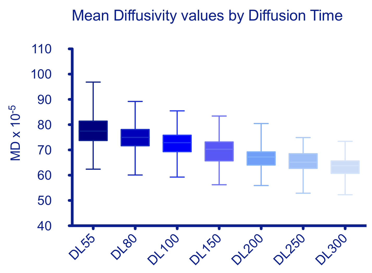
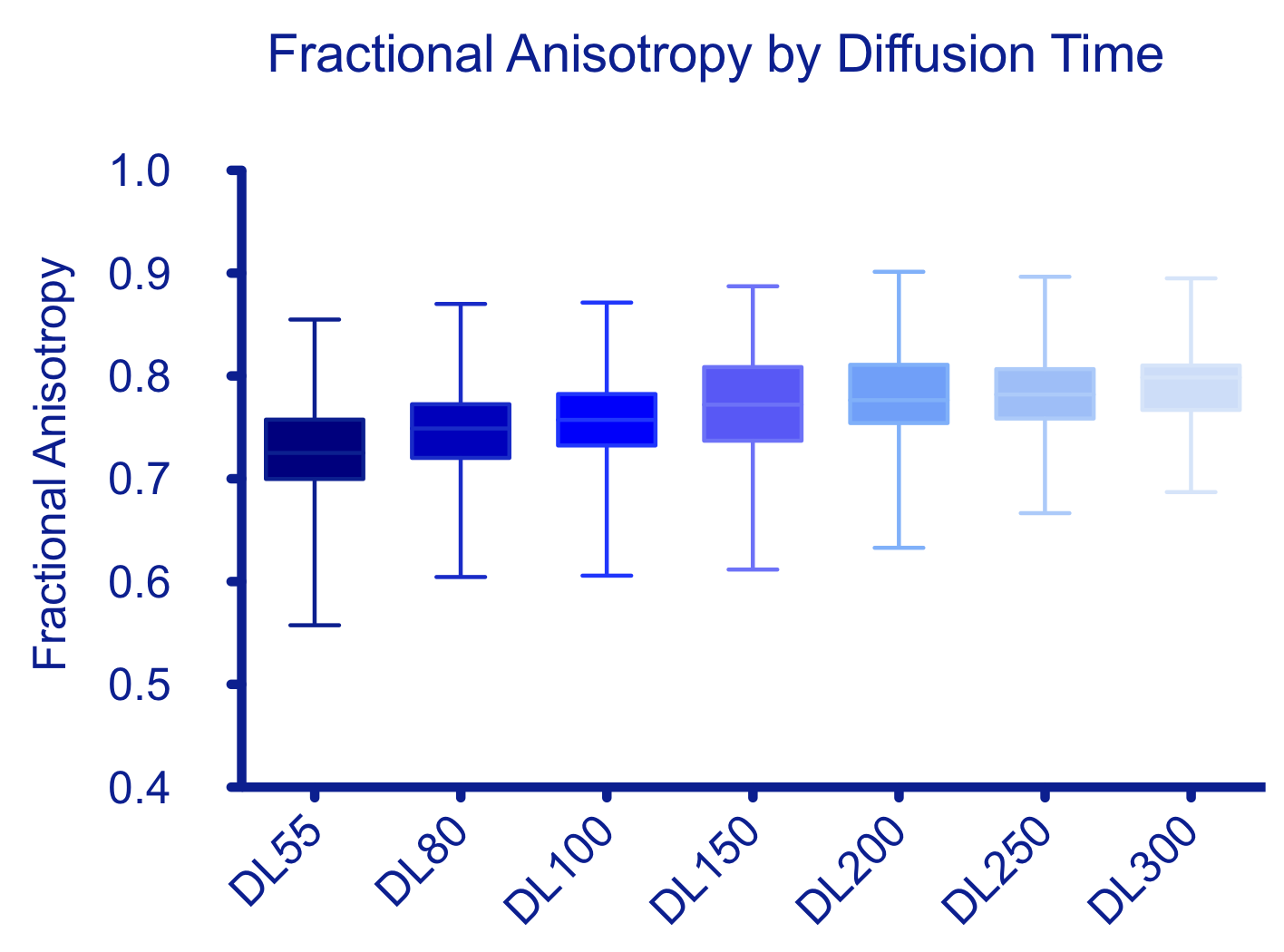
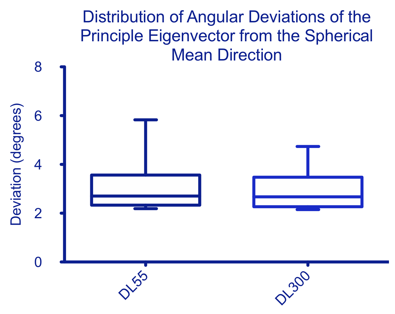
Mean diffusivity and fractional anisotropy values are shown in Figs. 1 and 2. The mean MD decreases from 77.74 x 10⁻⁵mm2/s at DL=55ms to 63.09 x 10⁻⁵mm2/s at 300 ms. Mean FA increases from 0.720 at DL=55 ms to 0.795 at DL=300ms. The differences between neighboring values are not significant but the differences across the complete range are (MD: p<0.0001, FA: p=<0.0001). In the remaining experiment we report results for the shortest and longest diffusion times to save space. Angular deviations from the spherical mean at the shortest and longest diffusion times are shown in Fig-3. We observe a mean angular deviation of 2.31 degrees (std dev 0.80 degrees) at DL=55 ms and of 2.38 degrees (std dev 0.76 degrees) at 300 ms. The difference between timepoints had a p-value of 0.74.Discussion and Conclusions
We observe a consistent trend in MD and FA as a function
of diffusion time: a decrease in MD and an increase in FA. There is significant
change in FA and MD across the range of diffusion times considered. This
indicates that the diffusion tensor measured in this phantom is not completely
stable with respect to diffusion time, and that diffusion time must be
explicitly considered when comparing acquisitions between sites and scanners
using phantoms of this kind. This demonstrates the importance of controlled
diffusion time in diffusion MRI reproducibility.
We observe no significant differences in the distribution
of angular deviations of principle eigenvectors from the mean direction.
Together this suggests an overall reduction in radial diffusivity with
diffusion time, altering the shape but not orientation of the tensors. Acknowledgements
MGH and AM are supported in part by a grant from Great Ormond St Hospital’s Biomedical Research Centre. AM is also supported by a grant from the National Physical Laboratory.References
[1] Poupon C, Rieul B, Kezele I, Perrin M, Poupon F, and Mangin JF New diffusion phantoms dedicated to the study and validation of high-angular-resolution diffusion imaging (HARDI) models. Magn Reson Med 60 (2008) 1276–1283
[2] Fieremans E, De Deene Y, Delputte S, Özdemir MS, D’Asseler Y, Vlassenbroeck J, Deblaere K, Achten E, and Lemahieu I. Simulation and experimental verification of the diffusion in an anisotropic fiber phantom. J. Magn. Reson. 190(2) (2008) 189–199
[3] Grech-Sollars M, Zhou F-L, Waldman AD, Parker GJM, and Hubbard Cristinacce PL, Stability and reproducibility of co-electrospun brain-mimicking phantoms for quality assurance of diffusion MRI sequences, NeuroImage, 181 (2018) 395-402
[4] Zhou F-L, Li Z, Gough JE, Hubbard Cristinacce PL, and Parker GJM Axon mimicking hydrophilic hollow polycaprolactone microfibres for diffusion magnetic resonance imaging Mater Des. 137 (2018) 394–403
[5] Novikov DS, Fieremans E, Jensen JH, Helpern JA. Random walk with barriers. Nat Phys. 7(6) (2011) 508-514.
Figures