3552
Reliability of microstructural quantification with diffusion MRI in brain tumors.1Padova Neuroscience Center, University of Padova, Padova, Italy, 2Department of Information Engineering, University of Padova, Padova, Italy, 3Department of Neuroscience, University of Padova, Padova, Italy
Synopsis
Most existing biophysical models used to quantify diffusion microstructural information are designed for the healthy brain. When pathological processes occur, the diffusion signal of cancerous regions is altered, possibly biasing the fitting results of these models and making their parameter estimates unreliable. In this work, we investigate the precision and accuracy of estimates provided by several well known diffusion models, by evaluating their goodness-of-fit with residual sum of squares map and the parameter reliability with the bootstrap technique.
Introduction
Diffusion magnetic resonance imaging (dMRI) has, throughout recent years, seen the ever increasing utilization of biophysical modeling approaches in order to overcome the specificity limitations of more conventional measures such as Diffusion Tensor Imaging1 (DTI). Such methodologies are usually structured to fit the diffusion signal inside healthy brain tissue, but they have also been employed to characterize a wide range of diseases2,3. However, the microstructural alterations which occur during pathological processes could affect the features of the diffusion signal inside several brain regions. Therefore, every biophysical model may introduce significant bias in the identification process and/or produce parameter estimates which are dependent on the noise pattern which affects dMRI images. In this work, we investigate if brain tumors introduce these issues for several well known biophysical models.Methods
The dataset was composed by 12 patients diagnosed with brain tumors. We considered the original version of NODDI4, its linearization within the AMICO framework5, and the two available implementations of the Spherical Mean Technique (SMT), i.e the tensor6 and the bi-compartment model7. Diffusion weighted images were acquired on a Siemens Biograph mmR scanner at 3T, with a two-shell diffusion protocol (10 b0 images, 30 diffusion directions at b=710 and 60 diffusion directions at b=2855). Each diffusion volume was acquired in both AP and PA phase encoding directions. The available data underwent a preprocessing pipeline based on MRtrix which included PCA based denoising8 and correction for readout distortion9, eddy current and subject motion10. The computation of the Residual Sum of Squares (RSS) maps was chosen as goodness-of-fit metric, while the uncertainty of parameter estimates was quantified by means of the bootstrap technique11. In NODDI’s case, for each iteration the model residuals were shuffled and re-added to the model prediction to artificially generate a new signal; in SMT’s case, bootstrapping consisted in sampling with replacement the available diffusion directions, separately for the two diffusion shells.Results and discussion
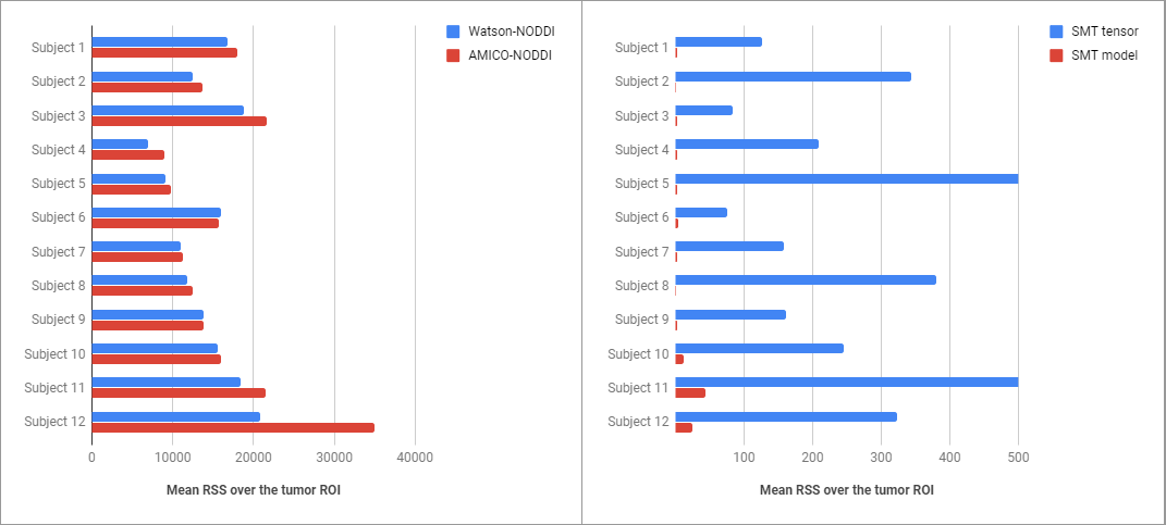
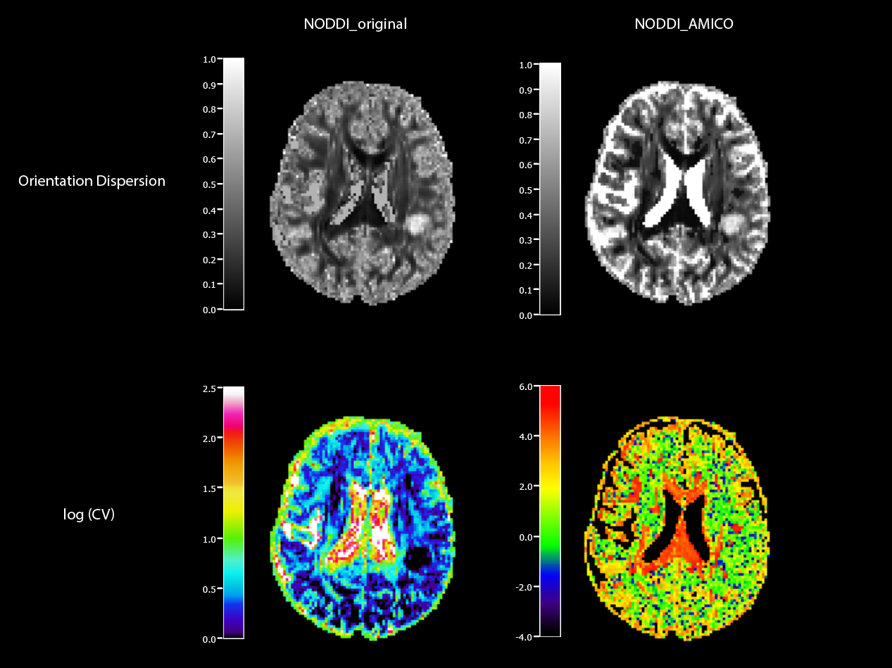
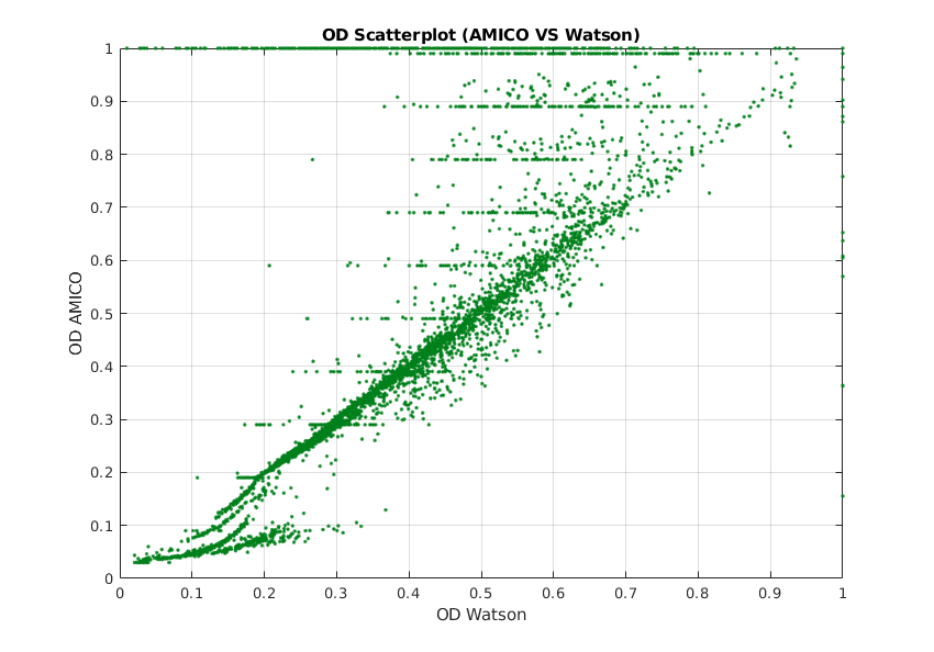
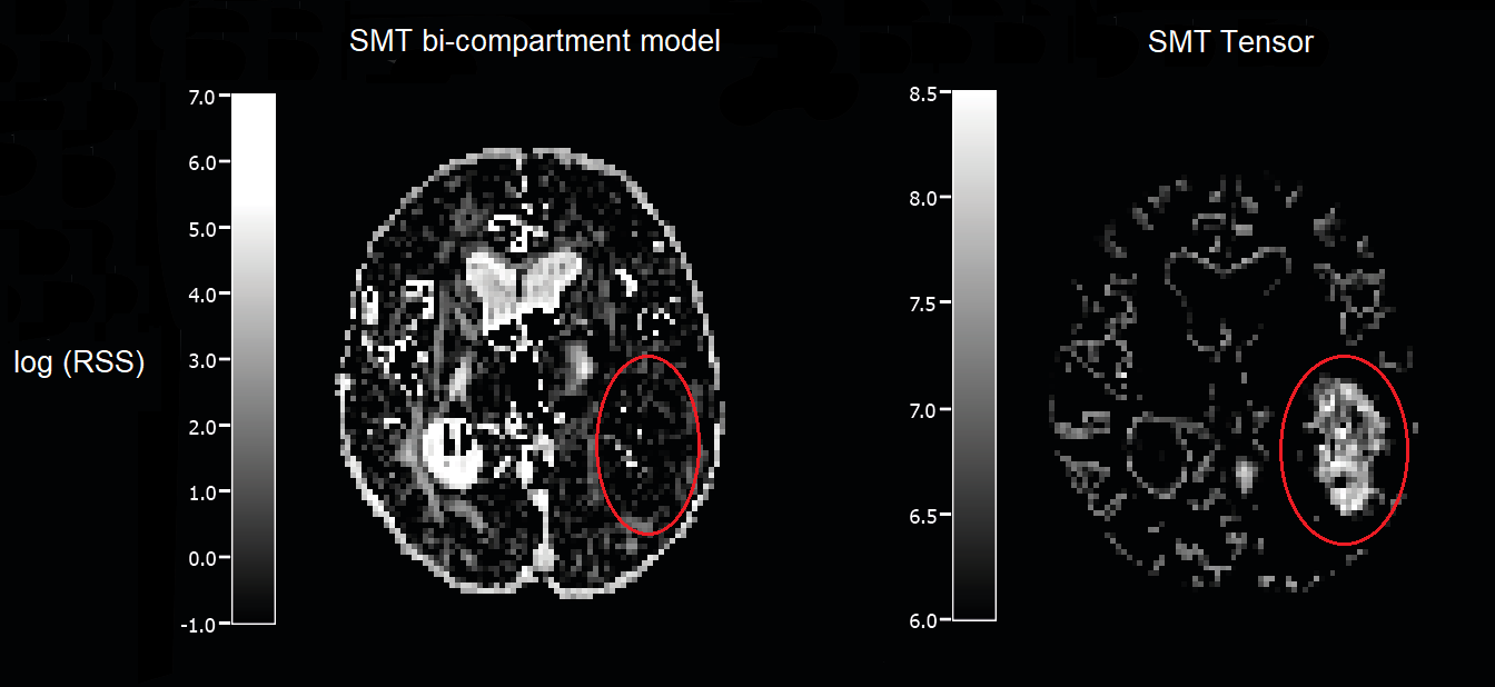
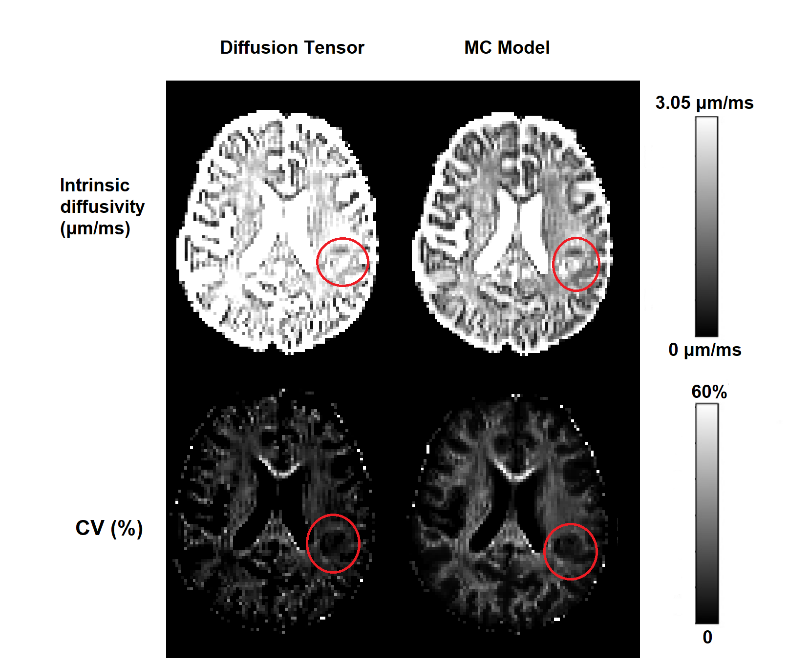
NODDI-AMICO is found to consistently score higher RSS values both across normal appearing brain tissue and in the tumour interested region with respect to the original NODDI. For 10 out of 12 subjects, the relative increase is small (<5%), while in the remaining cases, it is moderate to high (16%/ 67%). This result was partially expected, as the linearization of a model usually provides a tradeoff between identification time and fitting goodness. Estimated parameters’ Coefficients of Variation (CVs) do not appear to vary between cancerous and normal appearing tissue for both formulations, providing comparable values except for the Orientation Dispersion Index (ODI), for which AMICO has higher uncertainty in white matter surrounding the ventricles, where most CVs are in the range 70%-120% (Figure 2). Interestingly, NODDI model fits are worse in zones with high water presence, e.g. ventricles and cerebrospinal fluid filled regions. We presume this is due to difficulties concerning model identification when the isovolumetric volume fraction approaches the unitary value. This behaviour is not found in AMICO-NODDI, where the employed regularization term may in this sense have helped. Visualizing the scatterplot of both models joint parameter values reveals discretization issues for both ODI and the intracellular volume fraction (Figure 3). The comparison between the two available versions of SMT, i.e the tensor and the bi-compartment model, revealed that the former struggles to fit the data in voxels which mark the transition between different brain tissues, and generally presents higher model residuals than the latter. Moreover, tumour related regions showed a considerably high RSS value for the tensor SMT (>1000% increase over the bi-compartment model), suggesting that this model may not be adequate to describe cancerous tissues (Figure 4). Computing the CVs of the parameter estimates shows that, interestingly, in some tumor-related regions the accuracy of intrinsic and transverse diffusivities is higher with respect to normal appearing white matter for both SMT formulations (Figure 5).Conclusion
while the original NODDI implementation, AMICO-NODDI and the bi-compartment SMT seem to produce reliable parameter estimates and provide a good fit even in cancerous tissue, the tensor SMT features severely high RSS, suggesting this model may be inadequate in this case. The computation of the RSS maps and of the coefficients of variation are simple yet powerful instruments to detect possible unreliabilities in the employed microstructural models, and should carefully be considered when applying existing methodologies in an environment which differs from healthy brain situations.Acknowledgements
We acknowledge that the multi-band EPI sequence was made available from University of Minnesota through the C2P Siemens sharing mechanism.References
1. Basser, Peter J., James Mattiello, and Denis LeBihan. "MR diffusion tensor spectroscopy and imaging." Biophysical journal66.1 (1994): 259-267.
2. Maximov, Ivan I., Aram S. Tonoyan, and Igor N. Pronin. "Differentiation of glioma malignancy grade using diffusion MRI." Physica Medica 40 (2017): 24-32.
3. Spanò, Barbara, et al. "Disruption of neurite morphology parallels MS progression." Neurology-Neuroimmunology Neuroinflammation 5.6 (2018): e502.
4. Zhang, Hui, et al. "NODDI: practical in vivo neurite orientation dispersion and density imaging of the human brain." Neuroimage 61.4 (2012): 1000-1016.
5. Daducci, Alessandro, et al. "Accelerated microstructure imaging via convex optimization (AMICO) from diffusion MRI data." NeuroImage 105 (2015): 32-44.
6. Kaden, Enrico, Frithjof Kruggel, and Daniel C. Alexander. "Quantitative mapping of the per‐axon diffusion coefficients in brain white matter." Magnetic resonance in medicine 75.4 (2016): 1752-1763.
7. Kaden, Enrico, et al. "Multi-compartment microscopic diffusion imaging." NeuroImage 139 (2016): 346-359.
8. Veraart J.; Novikov D.S.; Christiaens D.; Ades-aron B.; Sijbers J., Fieremans E.; Denoising of diffusion MRI using random matrix theory, NeuroImage, vol.142, 2016, p.394-406.6
9. Andersson J.L.R., Skare S., Ashburner J.; How to correct susceptibility distortions in spin-echo echo-planar images: application to diffusion tensor imaging, NeuroImage, 2003, 20(2):870-888.
10. Jesper L. R. Andersson, Stamatios N. Sotiropoulos., An integrated approach to correction for off-resonance effects and subject movement in diffusion MR imaging, NeuroImage, vol. 125, 2016, p.1063-1078.
11. Efron, Bradley and Robert J Tibshirani (1994). An introduction to the bootstrap. CRC press.
Figures