3551
Microstructural Parameter Estimation of Skeletal Muscle Using Random Forest Model of dMRI1Mechanical Science and Engineering, University of Illinois at Urbana Champaign, Urbana, IL, United States, 2Biomedical Engineering, Illinois Institute of Technology, Chicago, IL, United States, 3Beckman Institute for Advanced Science and Technology, Univeristy of Illinois at Urbana Champaign, Urbana, IL, United States, 4Radiology, Carle Foundation Hospital, Urbana, IL, United States
Synopsis
Results are presented for a random forest model to estimate skeletal muscle microstructure parameters from
Introduction
Employing diffusion MRI (dMRI) to estimate microstructure of skeletal muscle in vivo requires an accurate model of dMRI’s relationship to the muscle microstructure. Current models often reduce the number of microstructural parameters to allow for more stable fitting, for example by discarding the extracellular matrix. However, extracellular properties play an important role in how force is transmitted from the individual myocyte to the tendon during activation.1,2 Machine learning (ML) techniques allow estimation of structural parameters by training models with numerically simulated dMRI data and have shown success in estimating myelin permeability in white matter.3 Combining our previous work on dMRI simulations4 with ML techniques may enable estimations of microstructural properties with accuracy not available from current models. Here preliminary results are presented for the development of a random forest model for skeletal muscle microstructure estimation from dMRI measurements.Methods
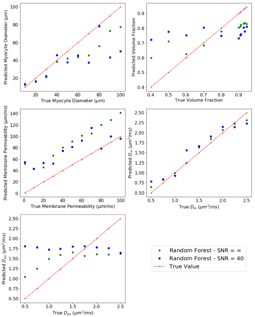
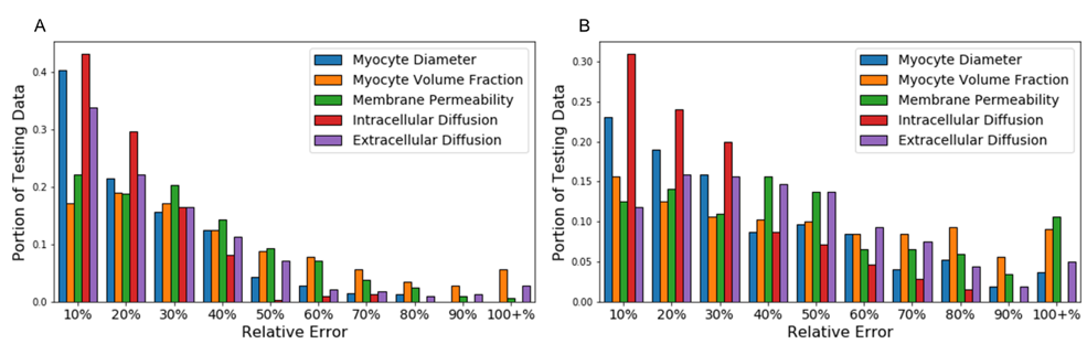
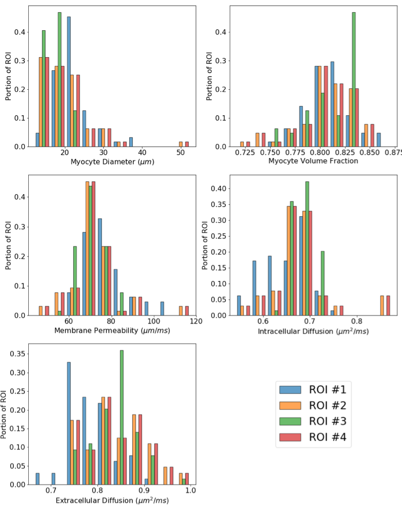
Using a numerical model based on a Lattice Boltzmann Method solution of the Bloch-Torrey equation,4 dMRI signals were computed on the San Diego Supercomputing Center’s Comet cluster5 over a domain of periodically packed hexagonal cells surrounded by a permeable membrane and embedded in an extracellular matrix. The domain was parameterized by intra- and extracellular diffusion, cell diameter, membrane permeability, and volume fraction which were sampled with a Sobol sequence6 to define 1600 parameter combinations (Table 1). PGSE sequences were simulated for 5 b-values (500, 750, 1000, 1500, and 2000 s/mm2). At each b-value 5 diffusion times (Δ) were simulated (10, 25, 50, 75, and 100 ms) with a gradient timing of 7.5 ms and the gradient strength adjusted to maintain a constant b-value. Six gradient directions were used to estimate a diffusion tensor from which fractional anisotropy (FA), apparent diffusion coefficient (ADC) and the tensor’s eigenvalues (λ1\λ2\λ3) were extracted, leading to 125 features for the model. This dataset was used to train a random forest (RF) model using the open source scikit-learn package in python.7 For testing data, simulations were performed with a single microstructural parameter varied and all other parameters held constant. Additionally, a data set using a Sobol sequence to generate 320 parameter combinations was constructed. Both testing sets were compared with the RF model for noise-free signal and with added Gaussian noise (SNR=40). Finally, the RF model was used to estimate microstructural parameters from an ex-vivo porcine myocardium, used as a substitute for skeletal muscle due to its similar microstructure, and imaged with a Siemens 3T TRIO scanner using a monopolar diffusion-weighted sequence with b-values of 800, 1500 and 2500 s/mm2, TR/TE/Δ/δ = 2930/94/44.35/22.93 ms, 2x2x2 mm resolution and 64 gradient directions. Because only three b-values at a single diffusion time were acquired, the model was retrained from the training data for only these three sequences (requiring additional simulations for b=2500 s/mm2).Results and Discussion
The RF model is compared with the two testing data sets in Figures 1 and 2. Figure 1 compares how well the RF model tracks changes in particular microstructural parameters. Figure 2 shows how the RF model compares with the Sobol sequence test data. Figure 3 shows the RF model’s microstructural estimations for the ex-vivo porcine phantom. The RF model was trained with 100 trees as it was found that more than 100 trees did not increase its ability to explain parameter variation (Table 2). The RF model is most accurate for estimating cell diameter and intracellular diffusion, which are also the parameters current models are best able to estimate. The model struggles to estimate volume fraction, likely due to low contrast between the intra- and extracellular diffusion. This RF model predicts parameters from 25 different dMRI sequences, however, using only three b-values at one diffusion time, the model can estimate tissue parameters from an experimental phantom demonstrating that the model can generalize beyond numerical data, an important first step. Future work is required to validate these predictions with histology and to reduce the number of sequences used to generate features.Conclusion
We have developed a random forest model to predict microstructural parameters of skeletal muscle from dMRI signal. The model has reasonable accuracy compared with numerically simulated data and provides physiologically reasonable values when applied to ex-vivo porcine heart. These results show that RF models are capable of predicting microstructural variations and that the model may be generalizable beyond the numerical data used to train it. More work is needed to accurately extract extracellular structural parameters, but random forest models appear to be a promising method of microstructure estimation in skeletal muscle.Acknowledgements
Funding for this work was provided by NSF Grant CMMI-1437113 and NSF Graduate Research Fellowship for NMN. This work used the Extreme Science and Engineering Discovery Environment (XSEDE), which is supported by National Science Foundation grant number ACI-1548562 and provided access to the SDSC Comet Cluster under allocation #TG-MCB180044.References
[1] Gillies, A. R., & Lieber, R. L. (2011). Structure and function of the skeletal muscle extracellular matrix. Muscle & nerve, 44(3), 318-331.
[2] Purslow, P. P. (2002). The structure and functional significance of variations in the connective tissue within muscle. Comparative Biochemistry and Physiology - A Molecular and Integrative Physiology, 133(4), 947–966.
[3] Nedjati-Gilani, G. L., Schneider, T., Hall, M. G., et al. (2017). Machine learning based compartment models with permeability for white matter microstructure imaging. NeuroImage, 150, 119-135.
[4] Naughton, N. M., Tennyson, C. G., & Georgiadis, J. G., Lattice Boltzmann method for simulation of diffusion magnetic resonance imaging physics in heterogeneous tissue models. Journal of Computational Physics, (submitted 2018)
[5] Towns, J., Cockerill, T., Dahan, M., Foster, I., et al. (2014). XSEDE: accelerating scientific discovery. Computing in Science & Engineering, 16(5), 62-74.
[6] Herman, J. and Usher, W. (2017) SALib: An open-source Python library for sensitivity analysis. Journal of Open Source Software, 2(9).
[7] Pedregosa, F., Varoquaux, G., Gramfort, A., et al. (2011). Scikit-learn: Machine learning in Python. Journal of machine learning research, 12(Oct), 2825-2830.
Figures