3546
Diffusion Time Dependence in Crossing Fiber Area in in vivo Human White Matter1Radiology, Juntendo University School of Medicine, Tokyo, Japan, 2Radiology, The University of Tokyo Graduate School of Medicine, Tokyo, Japan, 3Radiology, National Institute of Radiological Sciences, Chiba, Japan, 4Siemens Japan K.K, Tokyo, Japan, 5Siemens Healthcare GmbH, Erlangen, Germany
Synopsis
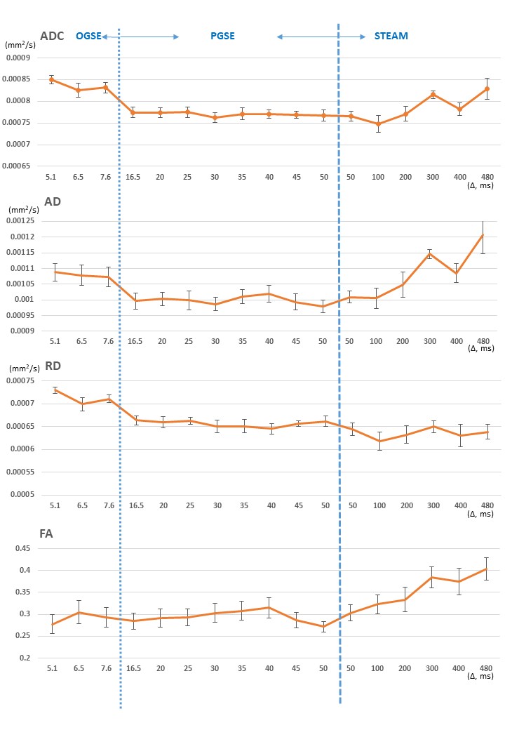
We investigated the diffusion time dependency of diffusion metrics in the white matter crossing fiber areas with different diffusion times using OGSE, PGSE and STEAM-DTI in in vivo human white matter at 3T. Our results show that ADC and AD decreased with increasing diffusion time at Δless than 100ms; after that ADC increased with increasing diffusion time. RD decreased and FA increased with increasing diffusion time. Moreover, the changes of white matter fODF in the white matter crossing fiber area at different diffusion times are shown.
Introduction
It is well known that quantitative diffusion metrics change with different diffusion time Δdue to tissue microstructure1-6. Observations of time-dependent diffusion parameters have been reported in in vivo brain of human subjects at times ranging from 40 to 800 ms using STimulated Echo Acquisition Mode (STEAM)-DTI1. Moreover, oscillating gradient spin echo (OGSE) diffusion-weighted sequences are able to probe shorter diffusion times compared to the clinically widely used pulsed gradient spin echo (PGSE), and are capable of demonstrating time-dependent diffusion4-6. In humans, Baron et al. combined OGSE (25 and 50 Hz) and PGSE methods (t = 20 and 40 ms) for a total diffusion time range from 4 to 40 ms, and have shown time-dependent diffusion5. Therefore, combination of OGSE, PGSE and STEAM-DTI can lead to the estimation of tissue microstructure at a wider range of diffusion times. Moreover, none of the previous reports above investigated the diffusion time dependence in the white matter crossing fiber area. The purpose of this study was to investigate the diffusion time dependency of diffusion metrics in the white matter crossing fiber area with different diffusion time using OGSE, PGSE and STEAM-DTI.Methods
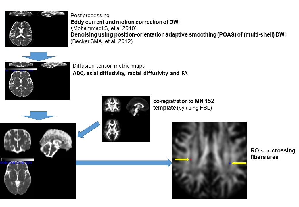
Six normal volunteers (mean 66 y.o., 3 women and 3 men) were scanned on a 3T magnetic resonance (MR) scanner (MAGNETOM Prisma, Siemens Healthcare, Erlangen, Germany) with a 64-channel head/neck coil. Imaging parameters for each diffusion MR imaging (employing a prototype sequence) were as follows: repetition time/echo time, 8100ms/130ms; section thickness, 5 mm; 30 slices; field of view, 200 x 200 mm2; matrix, 164 x 164; imaging time, approximately 2 min for each; b value of 1000 s/mm2 with a b=0 image and diffusion encoding in 6 directions for every b-value; effective Δ= 5.1, 6.5 and 7.6 ms (corresponding frequency =40Hz, 30Hz, 25Hz , respectively) on OGSE6, 16.5, 20, 25, 30, 35, 40, 45, and 50 ms on PGSE and 50, 100, 200, 300, 400, and 480 ms on STEAM-DTI. After eddy current and motion correction of DWI data, diffusion metrics maps were generated. The following diffusion metrics are considered; apparent diffusion coefficient (ADC), axial diffusivity (AD), radial diffusivity (RD) and fractional anisotropy (FA). We used the FMRIB Software Library linear image registration tool to register all diffusion metrics maps to the MNI152 template. We used Johns Hopkins University (JHU) ICBM-DTI-81 white-matter (WM) labels atlas for specifying fiber crossing point. These procedures are summarized in Figure 1. Moreover, white matter fiber orientation distribution function (fODF) based on an estimate of the signal expected for a single-fiber white matter population was estimated by using MRtrix3 (http://www.mrtrix.org/ ).Results
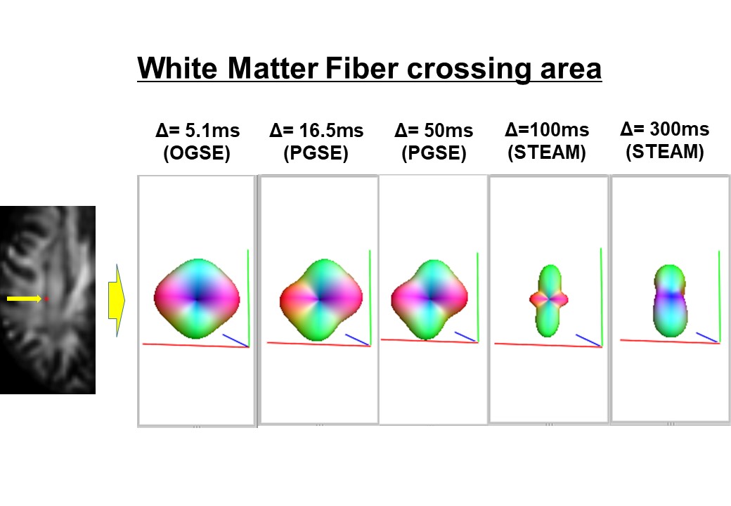
The results of diffusion metrics in the white matter crossing fiber area are shown in the graphs. Briefly, ADC and AD decreased with the increase of diffusion time for Δ less than 100ms; after that both parameters increased with the increase of diffusion time. RD decreased and FA increased with the increase of diffusion time. Moreover, the changes of white matter fODF in the white matter crossing fiber area at different diffusion times are shown in Figure 2.Discussion and Conclusion
The results show the diffusion time dependency of the diffusion metrics including ADC, AD RD and FA in the white matter crossing fiber area. Possible reasons for increasing ADC and AD atΔlonger than 100ms include: the effect of the water exchange between intra- and extra-axonal spaces, the dominant white matter fiber signal, and a diffusion time dependency of the extra-cellular component protons at long diffusion times. Moreover, the diffusion time dependency of fODF in the white matter crossing fiber area was observed. It is interesting that the fODF exhibits a smaller contribution of non-dominant fiber orientation at the longer diffusion time (Δof more than 100ms). Therefore, diffusion time itself affects the fODF and this may lead to an effect on white matter tractographyrepresentations.
In conclusion, diffusion time dependence is confirmed on diffusion tensor metrics in vivo in the white matter crossing fiber area and the change of fODF. We should pay attention to the diffusion times for interpretation of the diffusion metrics and fiber orientation for clinical use.
Acknowledgements
This work was supported by JSPS KAKENHI Grant Number 16K10328 and 18H02772.References
1. Fieremans E, et al. In vivo observation and biophysical interpretation of time-dependent diffusion in human white matter. Neuroimage. 2016 Apr 1;129:414-427.
2. Novikov, D. S., et al. Revealing mesoscopic structural universality with diffusion. Proceedings of the National Academy of Sciences, 2014;111(14), 5088-5093.
3. Novikov, D. S., & Kiselev, V. G. Effective medium theory of a diffusion-weighted signal. NMR in Biomedicine, 2010 23(7), 682-697.
4. Andica C, et al. Spatial Restriction within Intracranial Epidermoid Cysts Observed Using Short Diffusion-time Diffusion-weighted Imaging. Magn Reson Med Sci. 2018 Jul 10;17(3):269-272.
5. Baron CA, Beaulieu C. Oscillating gradient spin-echo (OGSE) diffusion tensor imaging of the human brain. Magn Reson Med. 2014 Sep;72(3):726-36.
6. Van AT, et al. In vivo investigation of restricted diffusion in the human brain with optimized oscillating diffusion gradient encoding. Magn Reson Med. 2014 Jan;71(1):83-94.
Figures