3544
What perivascular spaces do to diffusion MRI?1Medical Physics, University Medical Centre Freiburg, Freiburg, Germany, 2Spemann Graduate School of Biology and Medicine (SGBM), University of Freiburg, Freiburg, Germany, 3Faculty of Biology, University of Freiburg, Freiburg, Germany, 4University Medical Centre Freiburg, Freiburg, Germany
Synopsis
PVS is gaining importance in a many research fields. In using both in vivo and formalin fixed ex vivo methods, we sought to better understand what parameters in dMRI may be used to understood be as PVS in brain tissue.
Introduction
The perivascular spaces surround the vasculature of the brain, which is an integral part of the glymphatic system responsible for the removal of waste accumulated in the brain1. This role and further discoveries of its relationships to numerous neurological pathologies have invoked much interest1. In particular, diffusion MRI (dMRI) is sensitive to the presence of PVS. It is expected to appear as a non-negligible ‘fast-diffusion’ water fraction in solid white matter (WM)2. Presence of such water component does not fit into common white matter mircostructure models3 which may be seen as a confounding factor4, or as a useful feature. Here we propose a qualitative analysis of PVS ex and in-vivo and argue that PVS should not be neglected in structural, as well as, dMRI analysis.Methods
Ex-vivo brain preparation
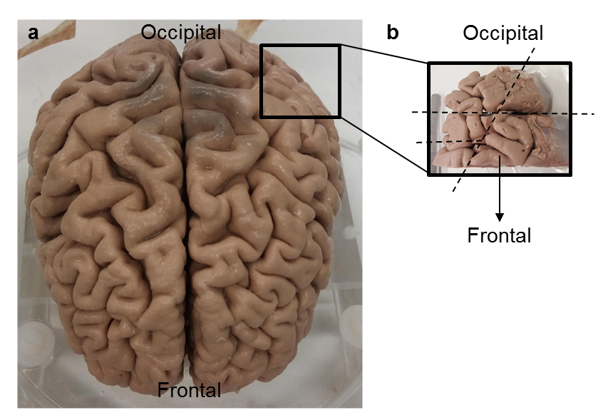
The post mortem brain (Figure 1a) was obtained from the Pathology Department, University Medical Centre Freiburg. It was female, over 80 years old with no known neurological medical histology or pathology. The body was fixed with formalin from the femoral artery and the brain was extracted upon complete perfusion. Figure 1b shows the region of the brain (Figure 1a) in which brain pieces were sectioned out for high-resolution scanning. Dimensions of the pieces are 2.3x1.5x5cm3. Brain pieces were incubated in 0.005% NaN3 in PBS for 24 hours prior to scanning.
MRI acquisition
Ex vivo scans were made with 9.4T Bruker scanner with a bore diameter of 200 mm. Scanning parameters were: TE = 40.22 ms, isotropic resolution of 0.18 mm, b value = 8000 m/s2. In vivo scans of a healthy human subject were made with 3T Siemens Prisma-Fit scanner with bore diameter of 60cm. Scanning parameters for in vivo volunteer were: For T2-weighted compressed sensing, TE = 175, Flip Angle =120, RepTime = 2000, isotropic resolution = 0.6x0.6x0.6mm3. T1 map was obtained with a resolution of 1x1x1 mm3, inversion time (TI) = 1100ms. At this time, the tissue compartments and the CSF have opposite phase. The free water fraction was one of the microstructure parameters evaluated using diffusion weighted imaging8 with TE = 76, RepTime = 8000, isotropic resolution = 1x1x1 mm3, q-ball (a hexagonal q space imaging scheme with 40 weightings) up to b-value = 2000 m/s2.
Visualisation
Visualisation of the scans was performed using NORA, a medical imaging platform [http://www.nora-imaging.com/].
Results
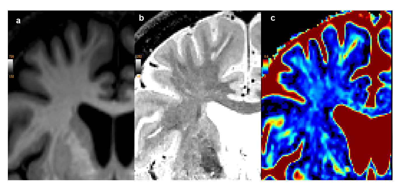
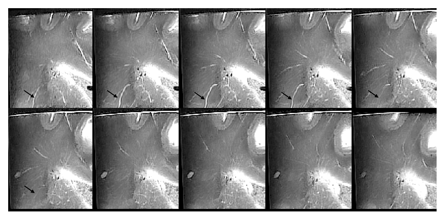
Figure 2a shows a presence of such water in T1-weighted images. Figure 2b shows the in vivo anatomical T2-weighted image. Figure 2c shows the corresponding fast-diffusion water fraction which indicated widespread distribution of fast-diffusion water in vivo. In Figure 3, ex vivo formalin fixed brain piece in T2- weighted high-resolution images visualises a number of PVS, however the total PVS volume is lower than that of on vivo fast-diffusion water.Discussion
With the evidence provided by the high-resolution MRI, we hypothesise that the fast diffusing water observer using dMRI reflects the density of PVS. The high resolution ex vivo imaging reveals PVS not visible in vivo (cf. Figure 2 and 3). An estimate of the fraction of revealed PVS can be made using the approximate scaling in the vascular system that can be described as a self-similar tree obeying the Murray’s law according to which the third power of the vessel diameter conserves at bifurcations. Applied to the whole arterial system this gives about 22 generations5, while the resolution increase from 1 mm to 0.18 mm should make about 7 generations of PVS visible. This implies that about 1/ 3 of the total PVS volume, that is a few per cent should become directly observable in the high resolution MRI. The approximate nature of this estimate, the drastic change in the tissue structure upon death and fixation and image imperfections do not allow however for a quantitative comparison with data shown in Fig.3, remaining a subject of future work. Similarly, the drastic change in ex vivo diffusion may be further exacerbated by possible dependence of PVS on the fixation status, which may also influence ex vivo tractography.
The influence of in vivo diffusivity by PVS is highlighted by Sepehrband et al., 20183, in that the presence of PVS biases evaluation of the diffusion tensor of white fibre tracts. While this report remains to be validated, it does bring to light the emerging importance in considering the impact of PVS on dMRI.
Acknowledgements
This study was supported in part by the Excellence Initiative of the German Research Foundation (GSC-4, Spemann Graduate School) and logistical support with regards to the post mortem brain by the members of the Stereotaxic Neurosurgerical department of the University Medical CentreReferences
- Mestre, H., Kostrikov, S., Mehta, R. and Nedergaard, M. (2017). Perivascular spaces, glymphatic dysfunction, and small vessel disease. Clinical Science, 131(17), pp.2257-2274.
- Kellner, E., Gall, P., Günther, M., Reisert, M., Mader, I., Fleysher, R., Kiselev, V.G. (2014) Blood tracer kinetics in the arterial tree. PLOS ONE, 9, pp. 1-11.
- Novikov DS, Fieremans E, Jespersen SN, Kiselev VG. (2018). Quantifying brain microstructure with diffusion MRI: Theory and parameter estimation. NMR Biomed. e3998.
- Seperhrband, F., Cabeen, R., Choupan, J., Barisano, G., Law, M. and Toga, A. (2018). A systematic bias in DTI findings. bioRxiv. doi: https://doi.org/10.1101/395012
- Reisert, M., Kellner, E., Dhital, B., Hennig, J. and Kiselev, V. (2017). Disentangling micro from mesostructure by diffusion MRI: A Bayesian approach. NeuroImage, 147, pp.964-975.
Figures