3540
A Diffusion MRI Approach to Tumour Microstructure Modeling with Two Cell Populations of Different Sizes1Medical Physics Unit and Department of Physics, McGill University, Montreal, QC, Canada, 2Research Institute of McGill University Health Center, Montreal, QC, Canada
Synopsis
Advanced diffusion-weighted MRI allows the characterization of cancer tumours noninvasively by estimating cell radius R and volume fraction vin. Existing methods map the apparent R and vin , under the assumption that a given voxel contains one cell population. This work investigates the feasibility of estimating the radii and volume fractions of 2 cell populations co-existing in the same voxel. This method could be useful in studying biphasic tumours like round cell/myxoid liposarcoma, which consist of high-grade and low-grade tumour cells, where the percentage of high-grade cells is strongly related to the risk of metastasis and changes the course of treatment.
Introduction
Advanced diffusion-weighted MRI allows the characterization of cancer tumours noninvasively by estimating the cell radius R and volume fraction vin, using IMPULSED1 and VERDICT2 techniques. Both techniques map the apparent R and vin of tumour cells, under the assumption that a voxel contains a single cell population. However, the single cell model is not valid in certain applications. For instance, round cell/myxoid liposarcoma forms a morphological continuum that includes high-grade (R~8-10 µm) and low-grade cells (R~4-6 µm). The percentage of the high-grade component is strongly related to the risk of metastasis and changes the course of treatment 3,4. In this work, we investigated the feasibility of extending the IMPULSED method to characterize 2 cell populations (R1, R2 and vin,1, vin,2) of different sizes co-exist in the same voxel.Theory
The IMPULSED method combines the conventional PGSE and OGSE sequence1. The diffusion MR signal of a single cell population is modeled as $$$ S = v_{in}S_{in} + (1-v_{in})S_{ex} $$$ [1] , where $$$S_{in}$$$ and $$$S_{ex}$$$ are the normalized signal from intra- and extra-cellular space, respectively. Equation 1 can be extended to 2-cell populations: $$$ S = v_{in,1}S_{in,1} + v_{in,2}S_{in,2}+(1-v_{in,1}-v_{in,2})S_{ex} $$$ [2]. Unfortunately, brute force fitting of Eq.2 produces unstable fits and poor results.
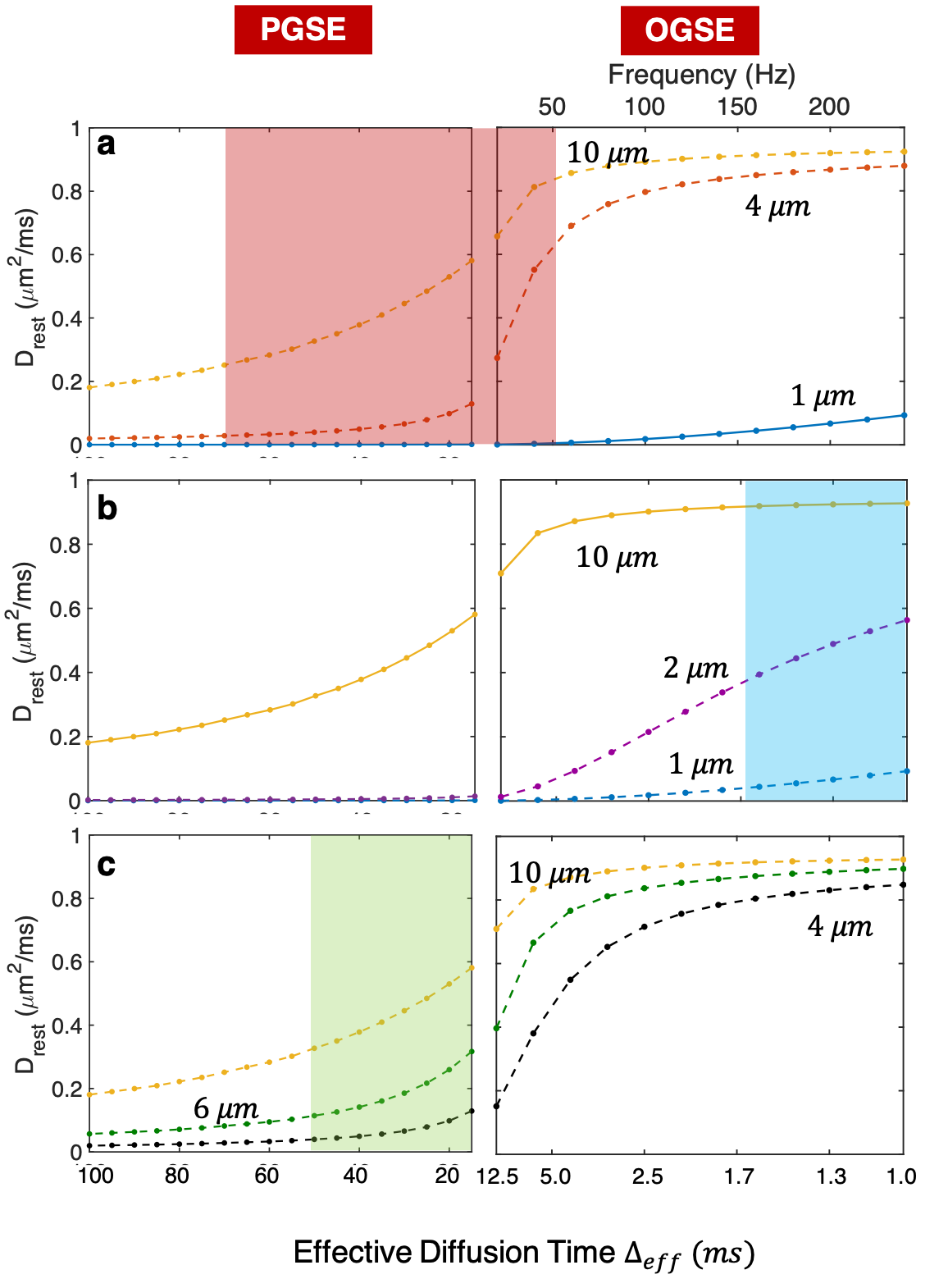
The detection sensitivity of DW-MRI depends on the effective diffusion time Δeff1. With some prior knowledge on the anticipated cells sizes, we can sensitize the measurement to different length scales, allowing the separation of the 2 populations in the following 3 scenarios:
- Scenario 1: Separating large cells from small cells
The Δeff range can be selected to remove signal sensitivity to small cells (pink box, Fig. 1a), where the intracellular diffusion coefficient Din,s ≈ 0 (e.g. R=1µm). Eq. 2 can be simplified to:
$$ S = v_{in,s} + v_{in,l}\cdot S_{in,l} + (1- v_{in,l}-v_{in,s})\cdot S_{ex}$$
allowing the estimation of Rl and vin,l of the large cells and vin,s of the small cells, but without sensitivity to Rs. This is termed the constrained 2-P IMPULSED method.
- Scenario 2: Separating small cells from large cells
The Δeff can also be selected in the high frequency OGSE range to desensitize the signal to large cells (blue box, Fig. 1b), where Din,l ≈ constant (e.g. R = 10µm). Eq. 2 becomes:
$$ S = v_{in,l}\cdot exp(-b\cdot D_{in,l}) + v_{in,s}\cdot S_{in,s} + (1- v_{in,l}-v_{in,s})\cdot S_{ex} $$
allowing the estimation of Rs, vin,s and vin,l, but without sensitivity to Rl. This is termed the constrained 2-P OGSE method.
- Scenario 3: Separating cells of close radius
For cells of similar radius, the signal cannot be selectively desensitized. We choose Δeff (green box, Fig. 1c) in the PGSE range . This decreases the number of fitted parameters by eliminating the frequency dependence of Din and Dex. This method is termed the 2-P PGSE method.
Method
The matrix based method was used to simulate diffusion signal from PGSE and cosine-modulated OGSE 5. The tissue was modeled as spherical cells of 2 different radii and volume fractions, with Din = 1μm2/ms and Dex = 2μm2/ms. The simulated signal was fitted with 100 random starting points to estimate radii and volume fractions, which were compared to ground truth values specified in the simulation. This process was repeated for a range of simulation parameters to ascertain the correspondence between the fitted parameter estimates and the ground truth (Table 1). Noise (SNR=80, 1500 realization) was subsequently added and simulations repeated.Results and Discussions
The apparent R from IMPULSED was a value between Rl and Rs (Fig. 2). The vin reflected the total volume fraction of both cell populations. The constrained 2-P IMPULSED method provided estimates of Rl, vin,l and vin,s with a median/maximum error of 0.4/2.4µm , 0.9/9%, and 0.6/4% for SNR=$$$\infty$$$, respectively. Similarly, the constrained 2-P OGSE method provided estimates Rs, vin,s and vin,l and (Fig.3) with a median/maximum error of 0.05/0.9µm, 0.5/22%, and 2/24 %, respectively. For cells of similar radius, we investigated 4 methods – IMPULSED, 2-P IMPULSED, 2-P PGSE and constrained 2-P PGSE. In the constrained 2-P PGSE, Din was fixed to 1µm2/ms to further reduce the number of free parameters6. The fitted R1, R2 and vin,1, vin,2 from constrained 2-P PGSE method agreed with the ground truth for R1 > 5µm (Figure 4). These observations appear to hold in preliminary simulation with noise (Figure 2 & 3).Conclusion
We introduced three scenarios where R1, R2 and vin,1, vin,2 can be accurately estimated when 2 cell populations co-exist in the same voxel. This method could be useful in studying biphasic tumours like round cell/myxoid liposarcoma, possibly influencing treatment decisions.Acknowledgements
The authors acknowledge Dr. Junzhong Xu, Dr. Xiaoyu Jiang, Zaki Ahmed, and Véronique Fortier for valuable discussions. This work is supported by Medical Physics Research Training Network Grant of the Natural sciences and Engineering Research Council (Grant number: 432290), and the Fonds de Recherche en Santé du Quebec.References
1.Jiang, X. et al.In vivo imaging of cancer cell size and cellularity using temporal diffusion spectroscopy. Magn. Reson. Med.00,1–9 (2016).
2. Panagiotaki, E. et al.Noninvasive Quantification of Solid Tumour Microstructure Using VERDICT MRI. Cancer Res.74,1902–1912 (2014).
3. Kilpatrick, S. E., Doyon, J., Choong, P. F. M., Sim, F. H. & Nascimento, A. G. The clinicopathologic spectrum of myxoid and round cell liposarcoma: A study of 95 cases. Cancer77,1450–1458 (1996).
4.Orvieto, E., Furlanetto, A., Laurino, L. & Dei Tos, A. P. Myxoid and round cell liposarcoma: a spectrum of myxoid adipocytic neoplasia. Semin. Diagn. Pathol.18,267–73 (2001).
5. Drobnjak, I., Zhang, H., Hall, M. G. & Alexander, D. C. The matrix formalism for generalised gradients with time-varying orientation in diffusion NMR. J. Magn. Reson.210,151–157 (2011).
6.Li, H., Jiang, X., Xie, J., Gore, J. C. & Xu, J. Impact of transcytolemmal water exchange on estimates of tissue microstructural properties derived from diffusion MRI. Magn. Reson. Med.(2016). doi:10.1002/mrm.26309
Figures