3539
A microstructure fingerprinting approach with heterogeneous axon diameters: validation on feline spinal cord1ICTEAM, Université catholique de Louvain, Louvain-la-Neuve, Belgium, 2Signal Processing Lab (LTS5), Ecole polytechnique fédérale de Lausanne, Lausanne, Switzerland, 3Computational Radiology Laboratory, Boston Children's Hospital, Harvard Medical School, Boston, MA, United States, 4Centre hospitalier universitaire vaudois (CHUV), University of Lausanne (UNIL), Lausanne, Switzerland
Synopsis
A microstructure fingerprinting approach is proposed in which an optimal single-fascicle configuration is selected from a pre-computed collection of Monte Carlo diffusion signals, or fingerprints, uniquely relating the diffusion MRI signals to the underlying axon properties. The simulated 3D geometries feature randomly-placed cylinders with diameter heterogeneity. The approach is validated on a public dataset of ex vivo cat spinal cord and exhibits indices of axon density and of the mean and standard deviation of the axon diameter distribution in agreement with histological measurements. The method is shown to outperform AMICO, which relies on approximate analytical expressions for the signal, and a simpler model using Monte Carlo simulations in a homogeneous packing of identical cylinders.
Purpose
Characterizing the axon density and diameter distribution (ADD) in the white matter could provide high clinical value but remains an open challenge. We propose a framework for quantitative microstructure estimation wherein the diffusion-weighted MRI (DW-MRI) signal is modeled by Monte Carlo (MC) simulations of the random walk of protons, known for their physical accuracy, in 3D geometries incorporating axon diameter heterogeneity. Optimal microstructural configurations are selected from a large dictionary of pre-computed diffusion signals or fingerprints based on agreement with measured data. On ex vivo cat spinal cord data, indices of the ADD and of axon density positively correlated with histological measurements. Our method compared favorably with a similar approach assuming homogeneous axon diameters and with the AMICO framework.
Methods
Microstructure fingerprinting with diameter heterogeneity
Signal model: In voxels containing one population of axons with orientation$$$\;\hat{u}_{fasc},\;$$$the DW-MRI signal1 $$$\;S\;$$$becomes$$S=M_0\left[\nu_{fasc}E_{fasc}(\hat{u}_{fasc})+\nu_{CSF}E_{CSF}\right]=w_{fasc}E_{fasc}(\hat{u}_{fasc})+w_{CSF}E_{CSF},$$where$$$\;M_0\;$$$is the thermal-equilibrium magnetization,$$$\;\nu_{fasc}\;$$$and$$$\;\nu_{CSF}\;$$$the physical volume fractions of respectively the fascicle and a partial volume of isotropic cerebrospinal fluid (CSF) and$$$\;w_{fasc}\;$$$and$$$\;w_{CSF}\;$$$their MR-visible signal weights. The normalized diffusion attenuation$$$\;E_{fasc}\;$$$is not modeled analytically but instead represented by a MC simulation of water diffusing in a geometry with parameters$$$\;{\Omega}.\;$$$
Estimation: Signals are precomputed for$$$\;N\;$$$geometries$$$\;\Omega_1,{\dots},\Omega_N\;$$$to form a single-fascicle dictionary$$$\;\left[E_{fasc}(\Omega_1;\hat{u}_{fasc}),{\dots},E_{fasc}(\Omega_N;\hat{u}_{fasc})\right]\;$$$of fingerprints uniquely relating the diffusion signal to microstructural properties. Given DW-MRI measurements$$$\;y,\;$$$the estimation consists in solving$$\begin{equation*}\begin{array}\\\mathbf{w}^*=&\underset{\begin{array}[l]\\\mathbf{w}_{fasc}\geq0\\w_{CSF}\geq0\end{array}}{\text{argmin}}&\left\|y-\begin{bmatrix} E_{fasc}(\Omega_1;\hat{u}_{fasc}),{\dots},E_{fasc}(\Omega_N;\hat{u}_{fasc}) |E_{CSF}\end{bmatrix}\cdot\begin{bmatrix}\mathbf{w}_{fasc}\\w_{CSF}\end{bmatrix}\right\|_2^2\\&&\\&\text{subject}\;\text{to}&\left|\mathbf{w}_{fasc}\right|_{0}=1,\\\end{array}\end{equation*}$$which becomes a simple dictionary look-up with non-negativity constraints:$$\hat{i}=\textrm{arg}\min\limits_{1\leq{i}\leq{N}}\,\min\limits_{w,w_{CSF}\geq{0}}\left\|y-wE(\Omega_i,\hat{u}_{fasc} )-w_{CSF}E_{CSF}\right\|_2^2,$$from which the optimal signal weight$$$\;\hat{w},\;$$$volume$$$\;$$$fraction$$$\;\hat{\nu}\;$$$and microstructural properties$$$\;\Omega_{\hat{i}}\;$$$are inferred.
In this study we performed MC simulations in$$$\;N=4120\;$$$geometries$$$\;$$$consisting of$$$\;N_{cyl}=1000\;$$$randomly-packed straight cylinders2-3(Figure A left) with diameters drawn from 109 different gamma distributions of pdf$$$\;\Gamma\left(\mu,\sigma\right)\;$$$with mean$$$\;\mu\in\left[0.4:0.4:8\right]\mu{m}\;$$$and standard deviation$$$\;\sigma\;$$$such that$$$\;\sigma/\mu\in\left[0.2:0.1:0.8\right],\;$$$verifying$$$\;\int_0^{20\mu{m}}\Gamma\left(\eta;\mu,\sigma\right)d\eta\geq{1-1/N_{cyl}}\;$$$to restrict axonal size and with intra-cylinder volume fractions$$$\;icvf\in\left[0.02:0.02:0.80\right],\;$$$leading to$$$\;\Omega=(\mu,\sigma,icvf).\;$$$
Validation
DW-MRI: The validation dataset is publicly-available and has been detailed previously4-5. Briefly, one slice of cervical spinal cord underwent PGSE imaging at 7T (Figure B), using high gradients and multiple short diffusion times for acute sensitivity to the ADD. Additional 4-shell imaging$$$\;$$$($$$\Delta/\delta=30/3ms,\;G=47.1,100.8,300,600mT/m,\;b=41,190,1680,6725s/mm^2$$$)$$$\;$$$with 195 gradient directions per$$$\;$$$shell was also performed. After Gibbs unringing6,15 images were discarded from each protocol based on visual inspection.
Histology: A second piece of contiguous tissue was imaged with an optical microscope and the axons were segmented automatically7, yielding maps of axon diameter mean$$$\;\mu\;$$$(volume-weighted), standard deviation$$$\;\sigma\;$$$and intra-axonal water fraction$$$\;icvf.\;$$$White matter was delineated by thresholding the histology-derived myelin volume fraction.
Data analysis: Our approach was compared to a simpler version1 , referred to as "homogeneous fingerprinting", in which the geometries consisted of hexagonally-packed cylinders with a unique diameter$$$\;\mu\;$$$(Figure A right) and where the single-fascicle dictionary comprised$$$\;N=986\;$$$combinations$$$\;\Omega_i=\left(\mu_i,icvf_i\right).\;$$$Comparisons were also made with AMICO8 which, unlike MC simulations, separates the intra-axonal contribution, assumed to arise from straight cylinders with diameters$$$\;d_1,\dots,d_{N_{in}}\;$$$from the extra-axonal signal modeled by a zeppelin-like diffusion tensor with perpendicular diffusivity$$$\;D_{\bot}.\;$$$AMICO solves$$\hat{\mathbf{w}}=\textrm{arg}\min\limits_{\mathbf{w}\geq{0}}\left\|y-\begin{bmatrix}E_{cyl}(d_1),\dots,E_{cyl}(d_{N_{in}})|E_{zep}(D_{\bot{1}}),\dots,E_{zep}(D_{{\bot}N_{ex}})\end{bmatrix}\cdot\begin{bmatrix}\mathbf{w}_{in}\\\mathbf{w}_{ex}\end{bmatrix}\right\|_2^2+\lambda\left\|\mathbf{w}\right\|_2^2,$$where$$$\;E_{cyl}\;$$$assumes the Gaussian Phase Distribution,$$$\;N_{in}=30,d_{30}=20\mu{m},N_{ex}=7\, D_{\bot{30}}\;$$$depends on the intrinsic (parallel) diffusivity$$$\;D\;$$$trough a tortuosity model and$$$\;\lambda\;$$$is a regularization parameter. From$$$\;\hat{\mathbf{w}}=\left[\hat{\mathbf{w}}_{in},\hat{\mathbf{w}}_{ex}\right]^T\;$$$one obtains$$$\;icvf=|\hat{\mathbf{w}}_{in}|_1/\left|\hat{\mathbf{w}}\right|_1,\;$$$the volume-weighted mean axon diameter$$$\;\mu=\sum_{j=1}^{N_{in}}\hat{w}_{in,j}d_j\;$$$and a standard deviation$$$\;\sigma\;$$$after converting to the number-weighted ADD$$$\;\hat{w}_{in,j}\leftarrow\frac{\hat{w}_{in,j}/d_j^2}{\sum_{k=1}^{N_{in}}\hat{w}_{in,k}/d_k^2}.\;$$$$$\;$$All three estimated models used an intrinsic diffusivity$$$\;D\;$$$and fascicle orientation$$$\;\hat{u}_{fasc}\;$$$from an initial Diffusion$$$\;$$$Tensor$$$\;$$$Imaging fit using the 3D acquisition protocol, and ignored$$$\;$$$CSF contributions$$$\;(w_{CSF}=0).\;$$$The voxel-wise correlation and mean absolute error (MAE) were performed between histology-derived$$$\;\mu,\sigma,icvf\;$$$and the corresponding model indices.
Results and discussion
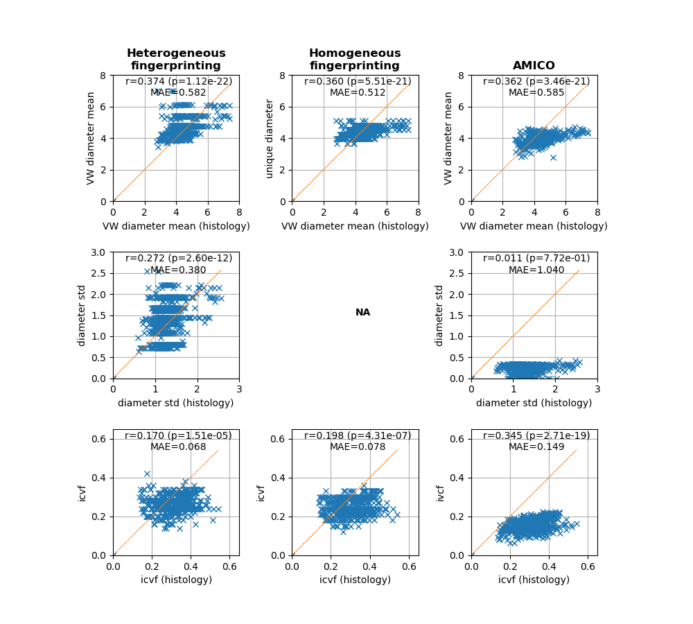
Our heterogeneous microstructure fingerprinting was the only model to produce statistically-significant correlations for the three axonal indices (Figs C-D).
The axon diameter indices of the three models exhibited similar correlations and MAEs; however the estimates of our approach were more evenly distributed around the histological values while AMICO underestimated larger diameters. Our approach yielded estimates of$$$\;\sigma\;$$$with higher correlation and lower MAE than those derived from AMICO, which all neared zero. AMICO obtained a stronger correlation for the icvf index but a larger MAE due to systematically underestimating the histological values.
These results suggest the potential of using a prior parameterization of the ADD: while AMICO can in theory reconstruct any distribution, its fitting may be less stable and predict degenerate distributions with$$$\;\sigma\approx{0}.\;$$$Geometric cylinder packings also enforce physical compatibility between the intra- and the extra-axonal contributions (e.g., tortuosity) as opposed to analytical approaches such as AMICO, where$$$\;\mathbf{w}_{in}\;$$$can be transferred to$$$\;\mathbf{w}_{ex},\;$$$thereby underestimating$$$\;icvf.\;$$$Using diffusion tensors for the extra-axonal signal fundamentally ignores diffusion hindrances and time-dependence effects9 especially visible when multiple diffusion times are used, all of which is naturally handled by MC simulations.
Conclusion
A microstructure fingerprinting approach was extended to account for axon diameter heterogeneity and yielded estimates of the axon density and the mean and standard deviation of axon diameters in line with histological measurements. Combined with more advanced acquisition sequences such as oscillating gradients, our framework could fully use the potential of MC simulations for the quantitative and reliable estimation of white matter microstructure in vivo.Acknowledgements
No acknowledgement found.References
[1] Rensonnet, G., Scherrer, B., Girard, G., Jankovski, A., Warfield, S.K., Macq, B., Thiran, J.P. and Taquet, M., 2019. Towards microstructure fingerprinting: Estimation of tissue properties from a dictionary of Monte Carlo diffusion MRI simulations. NeuroImage, 184, pp.964-980.
[2] Hall, M.G., Nedjati-Gilani, G. and Alexander, D.C., 2017. Realistic voxel sizes and reduced signal variation in Monte-Carlo simulation for diffusion MR data synthesis. arXiv preprint arXiv:1701.03634.
[3] Nedjati-Gilani, G.L., Schneider, T., Hall, M.G., Cawley, N., Hill, I., Ciccarelli, O., Drobnjak, I., Wheeler-Kingshott, C.A.G. and Alexander, D.C., 2017. Machine learning based compartment models with permeability for white matter microstructure imaging. NeuroImage, 150, pp.119-135.
[4] Duval, T., Perraud, B., Vuong, M.T., Lopez Rios, N., Stikov, N. and Cohen-Adad, J., 2016. Validation of quantitative MRI metrics using full slice histology with automatic axon segmentation. In Proceedings of the 24th Annual Meeting of ISMRM (Singapore). https://osf.io/gt4x2/
[5] Fick, R., Sepasian, N., Pizzolato, M., Ianus, A. and Deriche, R., 2017, April. Assessing the feasibility of estimating axon diameter using diffusion models and machine learning. In IEEE International Symposium on Biomedical Imaging (ISBI).
[6] Kellner, E., Dhital, B., Kiselev, V.G. and Reisert, M., 2016. Gibbs‐ringing artifact removal based on local subvoxel‐shifts. Magnetic resonance in medicine, 76(5), pp.1574-1581. (https://bitbucket.org/reisert/unring/src/master/ )
[7] Zaimi, A., Duval, T., Gasecka, A., Côté, D., Stikov, N. and Cohen-Adad, J., 2016. AxonSeg: open source software for axon and myelin segmentation and morphometric analysis. Frontiers in neuroinformatics, 10, p.37.
[8] Daducci, A., Canales-Rodríguez, E.J., Zhang, H., Dyrby, T.B., Alexander, D.C. and Thiran, J.P., 2015. Accelerated microstructure imaging via convex optimization (AMICO) from diffusion MRI data. NeuroImage, 105, pp.32-44.
[9] Burcaw, L.M., Fieremans, E. and Novikov, D.S., 2015. Mesoscopic structure of neuronal tracts from time-dependent diffusion. NeuroImage, 114, pp.18-37.
Figures