3529
VASE-RESOLVE: Accelerated Readout Segmented Echo Planar Imaging with Compressed Sensing and Variable-Width Readout Segments1University of Erlangen-Nuremberg, Erlangen, Germany, 2Siemens Healthcare GmbH, Erlangen, Germany, 3University of Glasgow, Glasgow, United Kingdom
Synopsis
Readout-segmented echo-planar imaging (EPI) with 2D navigator correction, also known as RESOLVE, is an established method for performing high-resolution diffusion weighted imaging in clinical studies. However, the method requires long acquisition times compared to single-shot EPI. A reduction in acquisition time could be achieved with Compressed Sensing (CS), but sub-sampling of EPI-based sequences is problematic because of the phase evolution during the echo train. This work introduces a new CS sampling scheme for readout-segmented EPI that varies the readout-segment width as a function of sampling density and preserves the phase relationship between data points.
Introduction
The REadout Segmentation Of Long Variable Echo trains (RESOLVE) technique for diffusion-weighted imaging uses readout-segmented EPI with 2D navigator correction to reduce echo spacing in the echo train1,2. This leads to reduced spatial distortion and blurring compared to single-shot EPI and allows an increased spatial resolution. However, this multi-shot approach results in an increased acquisition time compared to single-shot EPI. This can be reduced by using simultaneous multi-slice imaging3,4 or readout partial Fourier5. Still acquisition times can be prohibitively long, particularly for diffusion tractography studies. Compressed Sensing6 (CS) has been used for decreasing the acquisition time in a number of MRI sequences, especially in 3D imaging. This technique could also be used to reduce scan times with RESOLVE, but random sub-sampling during the EPI echo train would result in a disrupted phase evolution and image artefacts. To overcome this limitation, this study introduces a new sampling scheme that can be used to sub-sample 2D RESOLVE data sets without introducing phase discontinuities, called variable-segment (VASE) RESOLVE.Methods
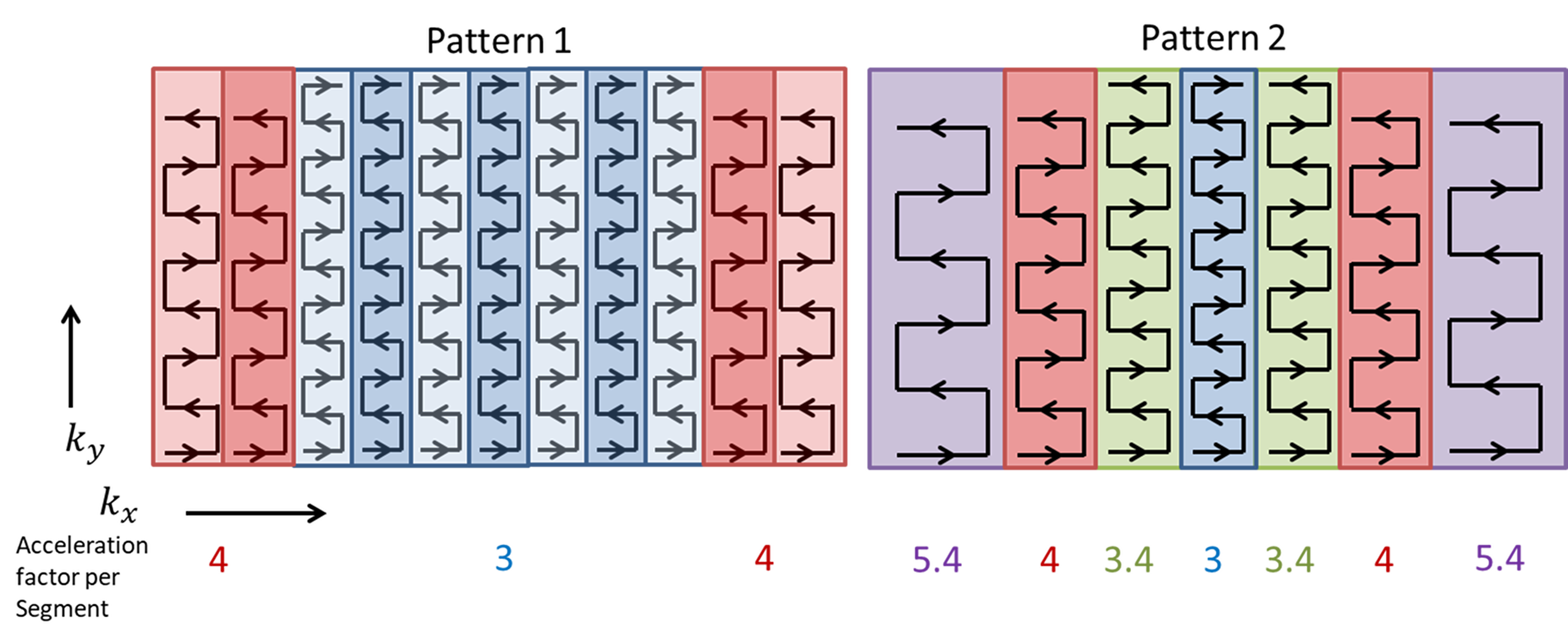
The principle idea of VASE-RESOLVE is to increase the width of the outer readout segments and to simultaneously increase the acceleration factor in these segments. The wider readout segment corresponds to an increased kx coverage, which is achieved by increasing the length of the readout gradient and consequently the EPI echo spacing. To offset the effects that the longer echo spacing has on phase evolution and signal decay due to T2*, the acceleration factor is adapted to maintain a constant effective echo spacing (true echo spacing divided by the acceleration factor). The increased kx coverage provided by the outer segments means that, for a given spatial resolution, fewer readout segments are required for image acquisition and the overall scan time is reduced. Fig. 1 shows two VASE-RESOLVE sampling schemes: one in which the segment widths are chosen to keep sample points on a Cartesian grid and a more generalised version, in which this condition is relaxed. A prototype sequence based on the Siemens RESOLVE product sequence was employed for the acquisition of pattern 1. Pattern 2 was retrospectively undersampled from a previously reconstructed and navigator corrected standard 3-fold accelerated acquisition with 15 segments. Images were acquired from healthy subjects on a 7-Tesla MAGNETOM Terra system (Siemens Healthcare GmbH, Erlangen, Germany) equipped with a 1Tx32Rx head coil (Nova Medical, Wilmington, MA, USA).Results
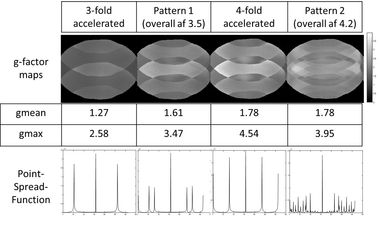
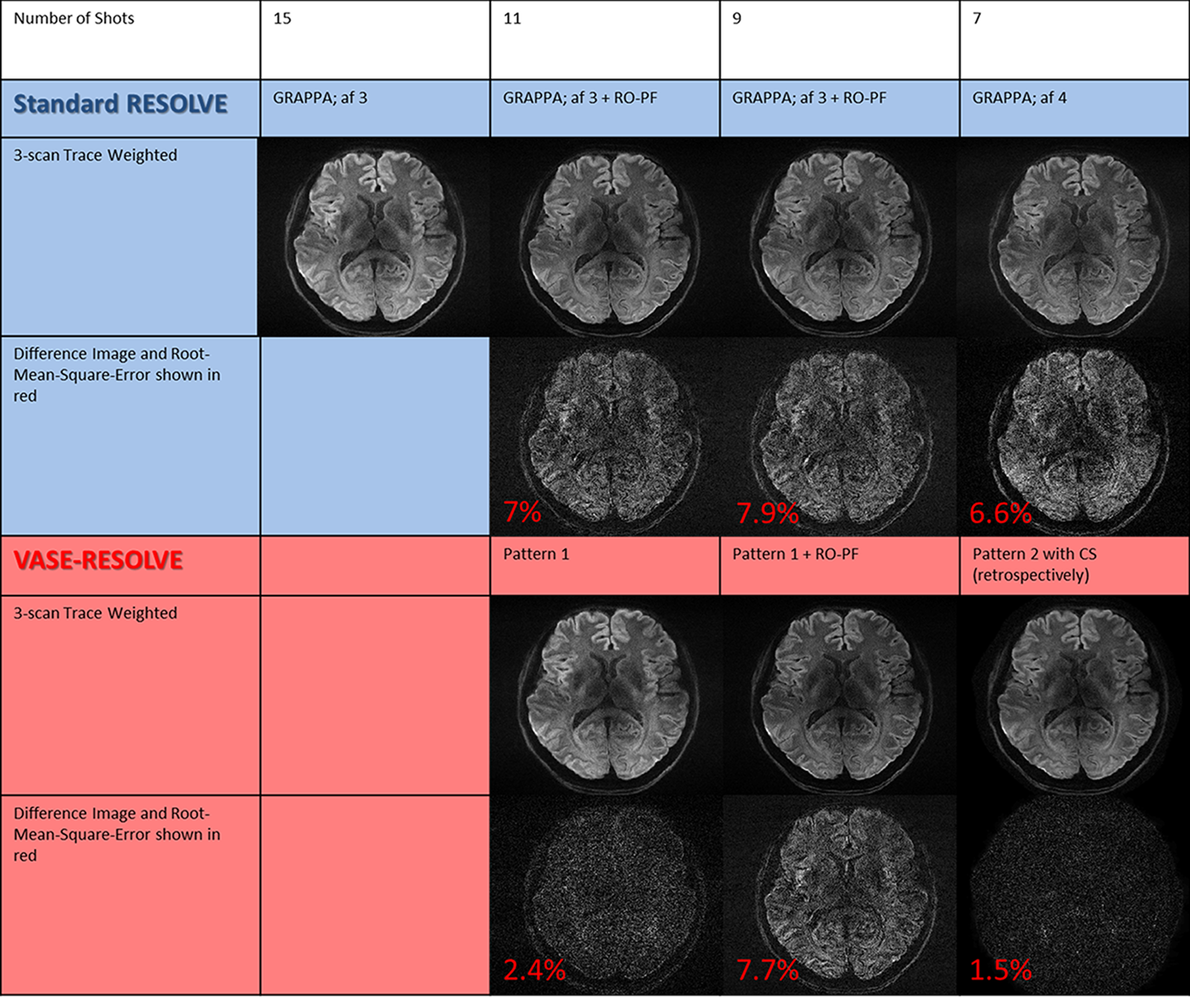
Figure 2 compares the respective g-factor maps and point spread functions (PSF) for a standard 3-fold and 4-fold accelerated acquisition and for the proposed sampling patterns 1 and 2 (from Fig. 1). The PSF for pattern 2 shows significantly reduced sidelobes, which makes it more suitable for a CS reconstruction. Also, pattern 1 shows reduced sidelobes, although not to the same extent as for pattern 2 due to the fact that undersampling is limited to a Cartesian grid. In Fig. 3 the respective reconstructed images are shown for VASE-RESOLVE and standard accelerated RESOLVE with readout partial Fourier. The RMSE is significantly reduced when using VASE-RESOLVE compared to a 4-fold accelerated acquisition (from 6.6 % to 1.5 %) and to readout partial Fourier (from 7% to 2.4 %).Discussion
VASE-RESOLVE allows a substantial reduction in scan time compared to the standard RESOLVE sequence. In the case of the pattern 2 sampling scheme, an acceleration of more than a factor of 2 can be achieved. When combined with readout partial Fourier and SMS, a whole-brain, trace-weighted protocol with 0.7 mm in-plane resolution can be acquired in less than 2 minutes. For pattern 2, the acquisition was performed retrospectively based on a fully reconstructed data set acquired using a standard 3-fold accelerated acquisition. This approach is acceptable, because the phase evolution is not changing between segments in Pattern 1. As the effective echo-spacing is kept the same, this is also true for the Non-Cartesian case in Pattern 2. Multi-shot diffusion-weighted imaging sequences, such as RESOLVE, are important techniques at 7 tesla, where field inhomogeneity and short T2* values limit the use of single-shot EPI. However, the VASE-RESOLVE technique will also be of value for acquisitions at 1.5 T and 3 T, where the standard RESOLVE method is well established in clinical application.Conclusion
The proposed sampling scheme with a variable-width readout segment allows readout-segmented EPI to be combined with CS without introducing image artefacts due to signal discontinuities in k-space. The technique can be combined with existing methods of acceleration, such as SMS and readout partial Fourier. The resulting VASE-RESOLVE method allows for fast, high-resolution diffusion-weighted imaging to be performed with a low level of distortion and image blurring.Acknowledgements
We would like to thank Tracy Hopkins and Rosie Woodward for helping us conduct the volunteer measurements.References
1. Porter, D. A. & Heidemann, R. M. High resolution diffusion-weighted imaging using readout-segmented echo-planar imaging, parallel imaging and a two-dimensional navigator-based reacquisition. Magn Reson Med 62, 468–475 (2009).
2. Naganawa, S. et al. Anatomical details of the brainstem and cranial nerves visualized by high resolution readout-segmented multi-shot echo-planar diffusion-weighted images using unidirectional MPG at 3T. Magn Reson Med Sci 10, 269–275 (2011).
3. Setsompop, K. et al. Blipped-controlled aliasing in parallel imaging for simultaneous multislice echo planar imaging with reduced g-factor penalty. Magnetic Resonance in Medicine 67, 1210–1224 (2011).
4. Frost, R. et al. Scan time reduction for readout-segmented EPI using simultaneous multislice acceleration: Diffusion-weighted imaging at 3 and 7 Tesla. Magn Reson Med 74, 136–149 (2015).
5. Frost, R., Porter, D. A., Miller, K. L. & Jezzard, P. Implementation and assessment of diffusion-weighted partial Fourier readout-segmented echo-planar imaging. Magn Reson Med 68, 441–451 (2012).
6. Lustig, M., Donoho, D. & Pauly, J. M. Sparse MRI: The application of compressed sensing for rapid MR imaging. Magn Reson Med 58, 1182–1195 (2007).
7. Uecker, M., Tamir, J., Ong, F., Holme, C. & Lustig, M. Bart: Version 0.4.01. (2017). doi:10.5281/zenodo.817472
Figures