3526
Sensitivity of 2D-Selective RF Excitations for Inner-Field-of-View Diffusion-Weighted Imaging to Field Inhomogeneities1Department of Systems Neuroscience, University Medical Center Hamburg-Eppendorf, Hamburg, Germany
Synopsis
Inner-field-of-view techniques based on 2D-selective RF (2DRF) excitations reduce geometric distortions of echo-planar imaging and improve diffusion-weighted imaging of the human spinal cord. A comparison of two common approaches revealed that a setup with the 2DRF excitation collinear to the image plane suffers from a reduced signal-to-noise ratio (SNR) for tall slice stacks. In this study, it is shown that the SNR loss is related to the larger sensitivity of the corresponding 2DRF excitation to frequency offsets caused by field inhomogeneities and is particularly pronounced for the large fields-of-excitation and 2DRF pulse durations needed for tall slice stacks.
Introduction
Geometric distortions of echo-planar imaging (EPI)1 can be reduced with inner-field-of-view techniques2-3 based on 2D-selective RF (2DRF) excitations4-5, which has been shown to improve diffusion-weighted imaging of the human spinal cord2-3. A comparison of two inner-field-of-view approaches2-3 revealed a lower signal-to-noise ratio (SNR) for 2DRF excitations being collinear to the image plane if a large volume is covered6. Here, it is shown that this SNR reduction is related to the larger sensitivity of the corresponding 2DRF excitation to frequency offsets caused by field inhomogeneities that are particularly pronounced for the large fields-of-excitation needed for tall slice stacks.Methods
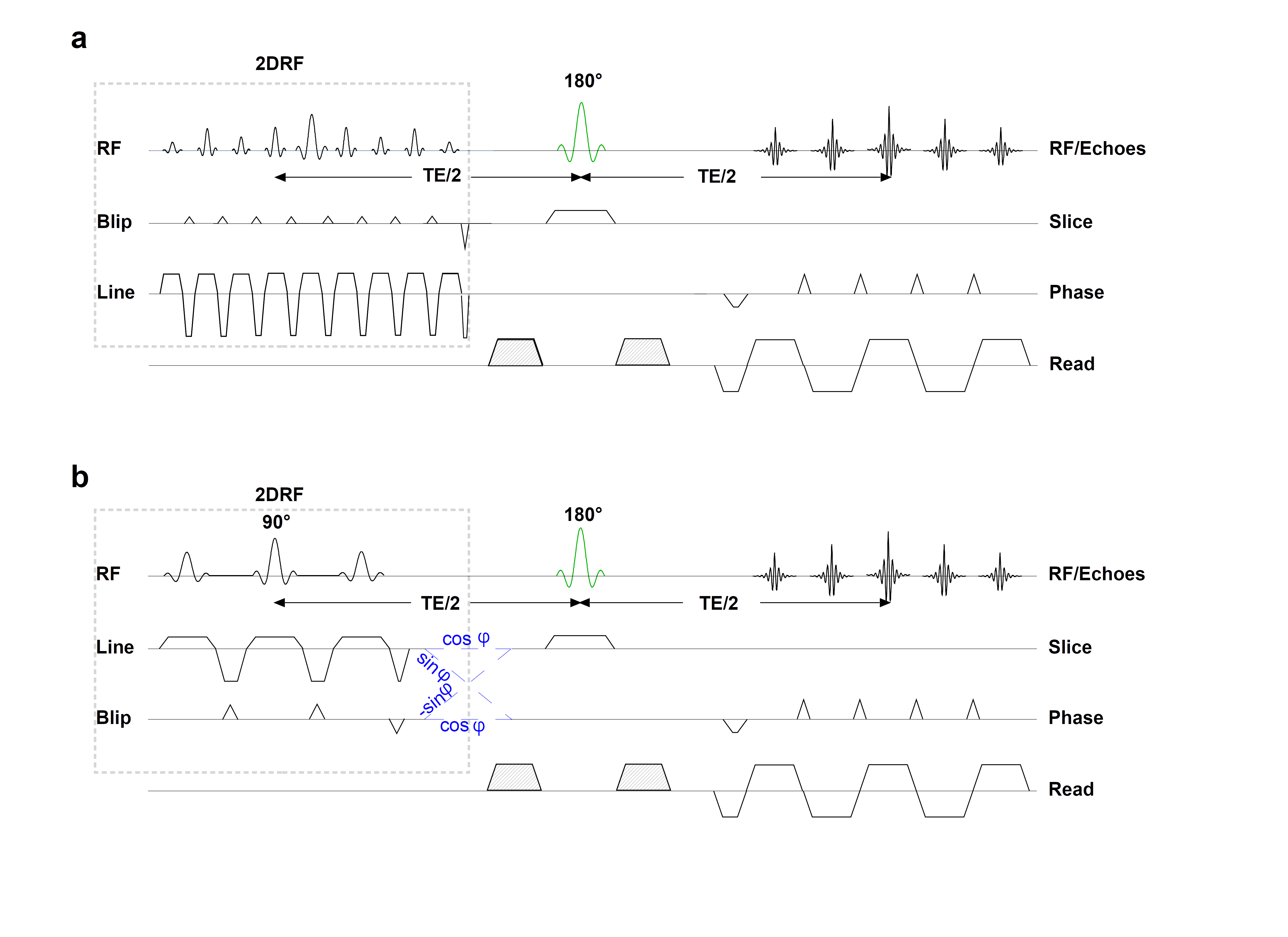
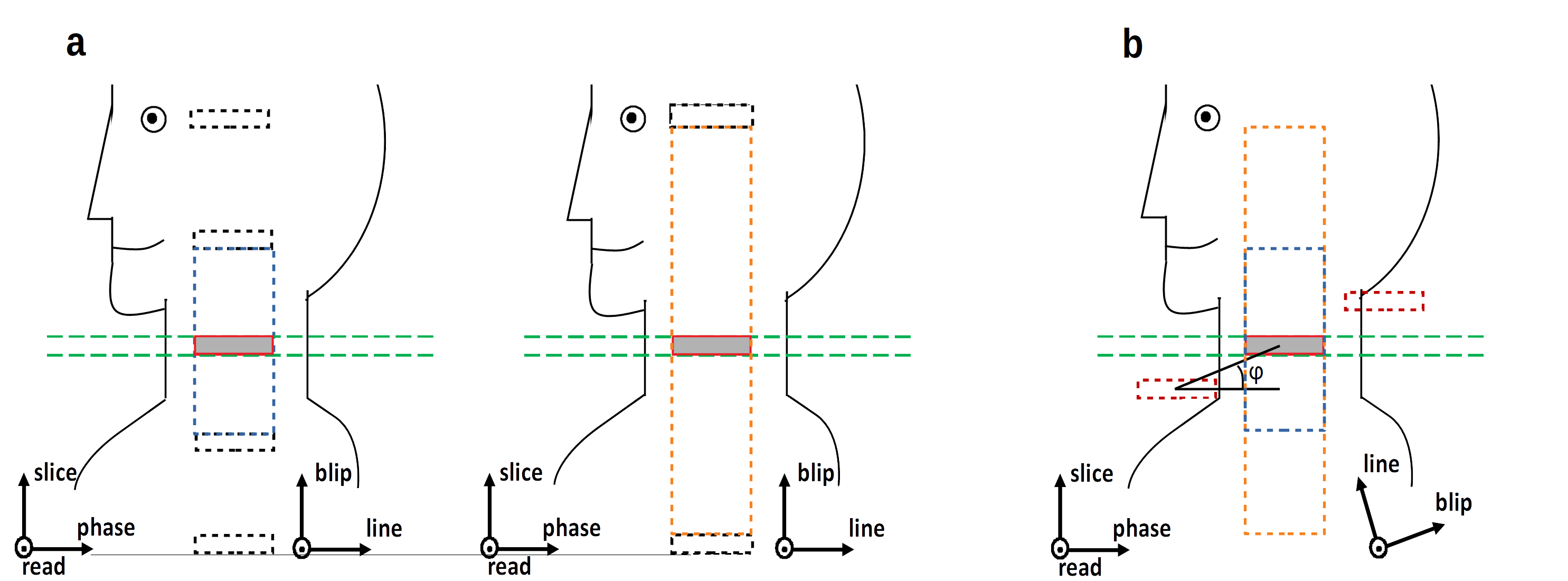
Figure 1 and 2 present the EPI pulse sequences and geometric setups used in the present study. Both sequences involve a 2DRF excitation based on a fly-back blipped-planar trajectory as the initial excitation but differ regarding its orientation relative to the image plane and the position of the side excitations occurring in the blip direction. In the “collinear” setup (Fig. 1a and 2a), the line and blip direction of the 2DRF trajectory coincide with the imaging phase and slice directions2, respectively. Thus, to avoid interferences, the side excitations must be positioned above and below the slice stack which requires a large field-of-excitation (FOE) and 2DRF pulse duration for tall slice stacks (cf. Fig. 2a). For the “tilted” setup (Fig. 1b and 2b), the 2DRF trajectory is rotated compared to slice and phase-encoding direction such that the side excitations neither are refocussed nor interfere with the slice stack to measure3. Thus, a small FOE is sufficient, independent of the number of slices, yielding short 2DRF pulses. Experiments were performed on a 3T whole-body MR system (PrismaFit, Siemens Healthineers, Erlangen, Germany) using a standard 64-channel head-neck coil in combination with a 32-channel spine array coil. Only coil elements with significant signal contributions were selected for the acquisitions. A water phantom and healthy volunteers that gave their informed consent prior to the examination, were investigated. 2DRF excitations were based on a trajectory resolution of 4.0×10.0mm² in the line×blip (tilted) and blip×line directions (collinear) and were designed to excite rectangular profile(s) with a size of 4×56mm² (slice×phase-encoding direction). 15 to 62 slices were targeted yielding FOEs of 60mm to 248mm for the collinear setup (2DRF pulse duration between (33.8 ms and 7.9 ms) and a fixed FOEs of 70mm for the tilted setup (6.4 ms). An inner FOV of 32×128 mm² with 16 mm oversampling in the phase encoding direction to account for profile imperfection was acquired with a resolution of 1.0×1.0×4.0mm3. Diffusion-tensor imaging was performed with six non-collinear directions of the diffusion weighting (b value 625 s mm-2) yielding echo times between 64 and 70ms and repetition times between 5600ms. Additionally simultaneous multi-slice (SMS) acquisitions7 with an acceleration factor of 2 (TR = 3400ms) and a correspondingly reduced FOE were performed for comparison.Results and Discussion
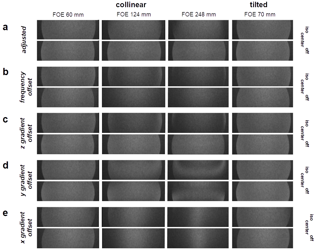
Figure 3 demonstrates the influence of frequency and gradient offsets
on the SNR in a water phantom. For the collinear setup with a small
FOE (15 slices) and the tilted setup (15-62 slices),
no significant impact on the SNR is observed. However, for the
collinear setup with larger FOEs (31 and 62 slices), the
frequency (100Hz) and z gradient offset (0.05mT m-1)
clearly show a reduced SNR due to the low bandwidth of the 2DRF
excitation in the blip, i.e. slice, direction: the offsets shift the
excited profile in the slice direction such that it is not fully
covered by the refocussing RF pulse. In contrast, the tilted setup
exhibits a shift mainly in the phase-direction making it much more
robust to frequency offsets and field inhomogeneities.
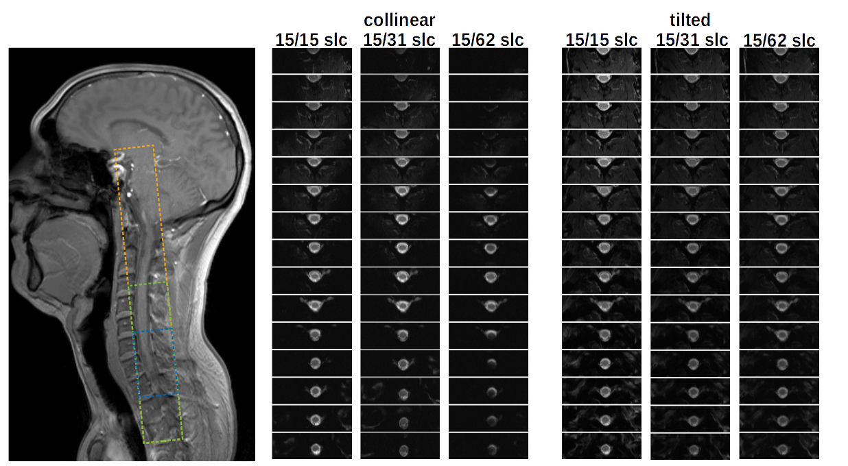
Example images of the human spinal cord are shown in Fig. 4. For
larger slice stacks and FOEs, the signal intensity is reduced in some
sections of the spinal cord for the collinear setup due to field
inhomogeneities. In contrast, the tilted setup shows a no SNR
reduction in the cord even for a tall slice stack.
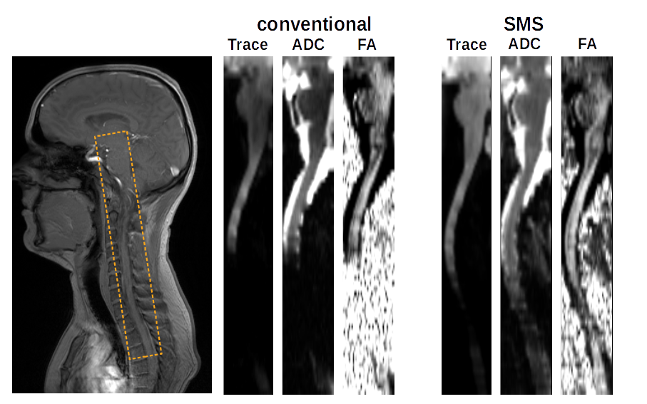
In Fig. 5, conventional and SMS DTI acquisitions obtained with
the collinear setup are presented. In the SMS acquisitions, the FOE
is halved because one of the side excitations is positioned to cover
the other slice to be acquired simultaneously which reduces the SNR
loss caused by field inhomogeneities.Conclusion
Compared to the tilted setup, the 2DRF excitation of the collinear setup suffer from an increased sensitivity to field inhomogeneities for tall slice stacks that can reduce the SNR in the spinal cord; however, simultaneous multi-slice acquisitions can help to ameliorate the problem.Acknowledgements
This work was supported by Wings for Life.References
1. Mansfield P, Multi-planar image formation using NMR spin echoes. J Phys C 1977; 10: 349-352
2. Saritas EU, Cunningham CH, Lee JH, Han ET, Nishimura DG. DWI of the spinal cord with reduced FOV single-shot EPI. Magn Reson Med. 2008; 60: 468-73.
3. Finsterbusch J. Improving the performance of diffusion-weighted inner field-of-view echo-planar imaging based on 2D-selective radiofrequency excitations by tilting the excitation plane. J Magn Reson. Imaging 2012; 35: 984-92.
4. Hardy CJ, Cline HE Spatial localization in 2 dimensions using NMR designer pulses. J Magn Reson. 1989; 82: 647-654
5. Pauly J, Nishimura DG, Macovski A. A k-space analysis of small-tip-angle excitation. J Magn Reson, 1989; 81: 43-56
6. Florin C, Finsterbusch J. Shorter Acquisition Times for Diffusion-Weighted Imaging of the Human Spinal Cord with Simultaneous Acquisition of Multiple Inner Fields-of-View. Proc Int Soc Magn Reson Med 2018, 1625
7. Setsompop K, Gagoski B, Polimeni J, Witzel T, Wedeen V, Wald LBlipped-controlled aliasing in parallel imaging for simultaneous multislice echo planar imaging with reduced g-factor penalty. Mag Reson Med. 2012; 67: 1210-1224
Figures