3521
Improvement of EPI-based DWI of the head/neck region using additional local shim coils at 3 Tesla1Department for Diagnostic and Interventional Radiology, University Hospital Tuebingen, Tuebingen, Germany, 2Department for Diagnostic and Interventional Radiology, Univeristy Hospital Tuebingen, Tuebingen, Germany
Synopsis
Implementing additional local shim coils into the head and neck surface coil may enhance the performance of EPI-based diffusion weighted imaging of the head/neck region at 3 Tesla. Thus, we evaluated single shot echo planar DWI and standard T1-weighted gradient echo acquisition without and with the use of additional local shim coils. In addition, apparent diffusion coefficients were quantified in specific anatomical regions. Local shim coils improved the homogeneity of the static magnetic field, resulting in marked improvement of image quality, signal loss, distortion and fat saturation. ADC values did not significantly differ between the measured anatomical compartments.
Introduction
In clinical practice, diffusion weighted imaging (DWI) is a widely used MR imagine technique which measures the movement of water molecules through the different kinds of tissue1-3. Usually it is employed for staging oncological malignancies and non-oncological diseases such as inflammatory lesions3,4,5.
Since global shimming gradient coils are not able to completely homogenize complex static field inhomogeneities, a potential alternative is thus the use of additional local shim coils integrated into the lower part of the head/neck surface coil for further improvement of the inhomogeneities of the local static magnetic field, which may result in an improved image quality for EPI based DWI.
The purpose was to assess the performance of local shim coils within the head and neck surface coil for diffusion weighted MRI of the head/neck region at 3 Tesla.
Methods
DWI of the head/neck of 10 healthy volunteers (7 male, mean-age 30.2±4.4 years) were obtained. MR acquisitions were performed on a clinical 3T scanner (MAGNETOM Vida, Siemens Healthcare, Germany).
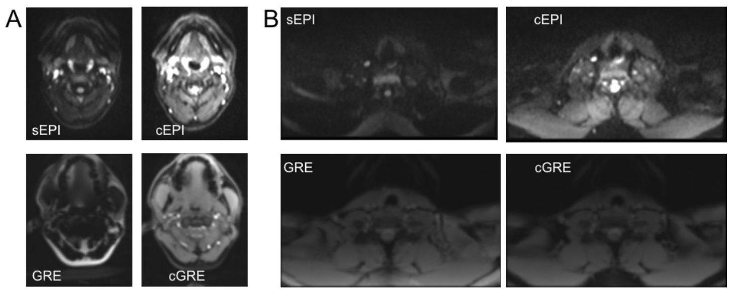
In all volunteers, a single shot EPI DWI acquisition of the head/neck region (skull base to shoulders) was performed without (sEPI) and with (cEPI) the use of local shim coils. In addition, a standard T1-weighted gradient echo with spectral fat saturation was performed without (GRE) and with (cGRE) the use of local shim coils. In order to quantify static field inhomogeneities, B0 field maps with and without active local shim coils were acquired using a dual-echo T1w gradient echo sequence. Sequence parameters are described in (Figure 1).
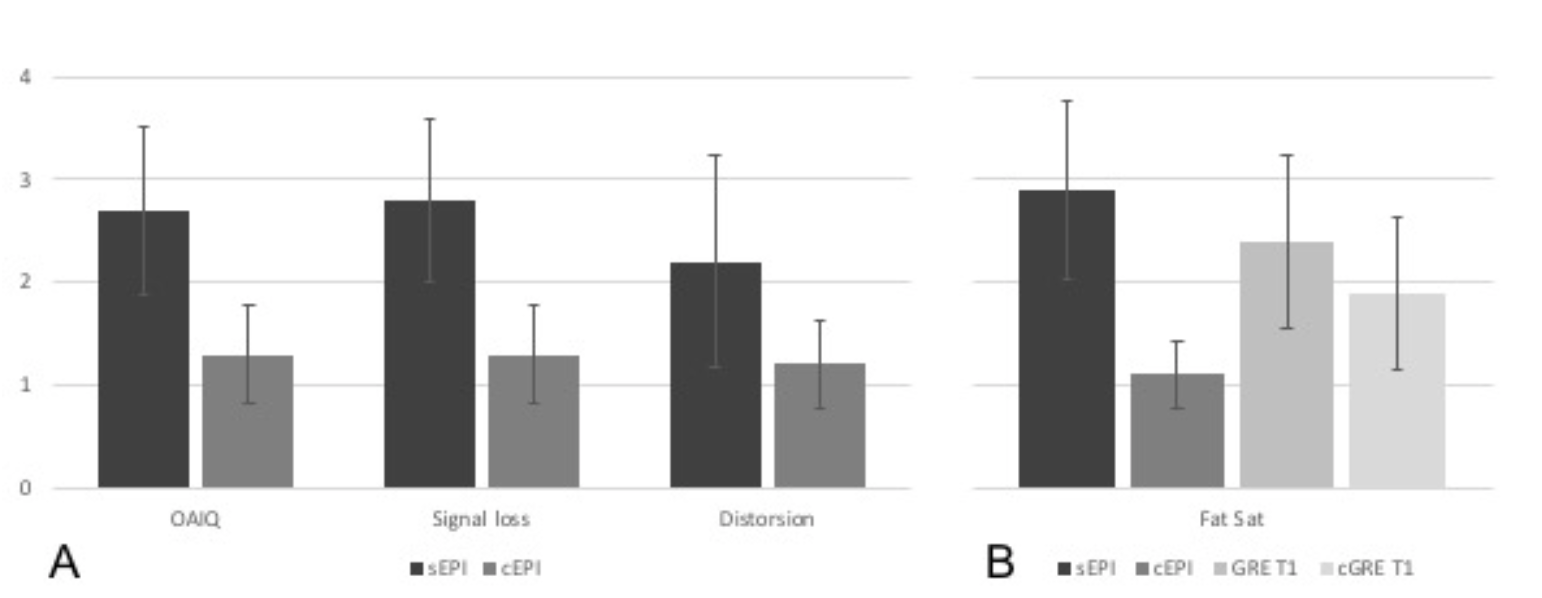
Evaluation was performed by two radiologists in consensus with 7 and 3 years of experience in magnetic resonance imaging. The EPI-based DWI sequences with and without additional local shim coils were assessed for overall image quality, fat saturation, signal loss as well as spatial distortions. GRE T1 without and with local shim coils were only assessed for quality of saturation. Visual evaluation for all categories were performed on a 4-point Likert scale (1 being the best possible outcome). In addition, we compared the effect of local shim coils on the static field distribution. To this end, a field map was computed from the dual-echo GRE sequence.
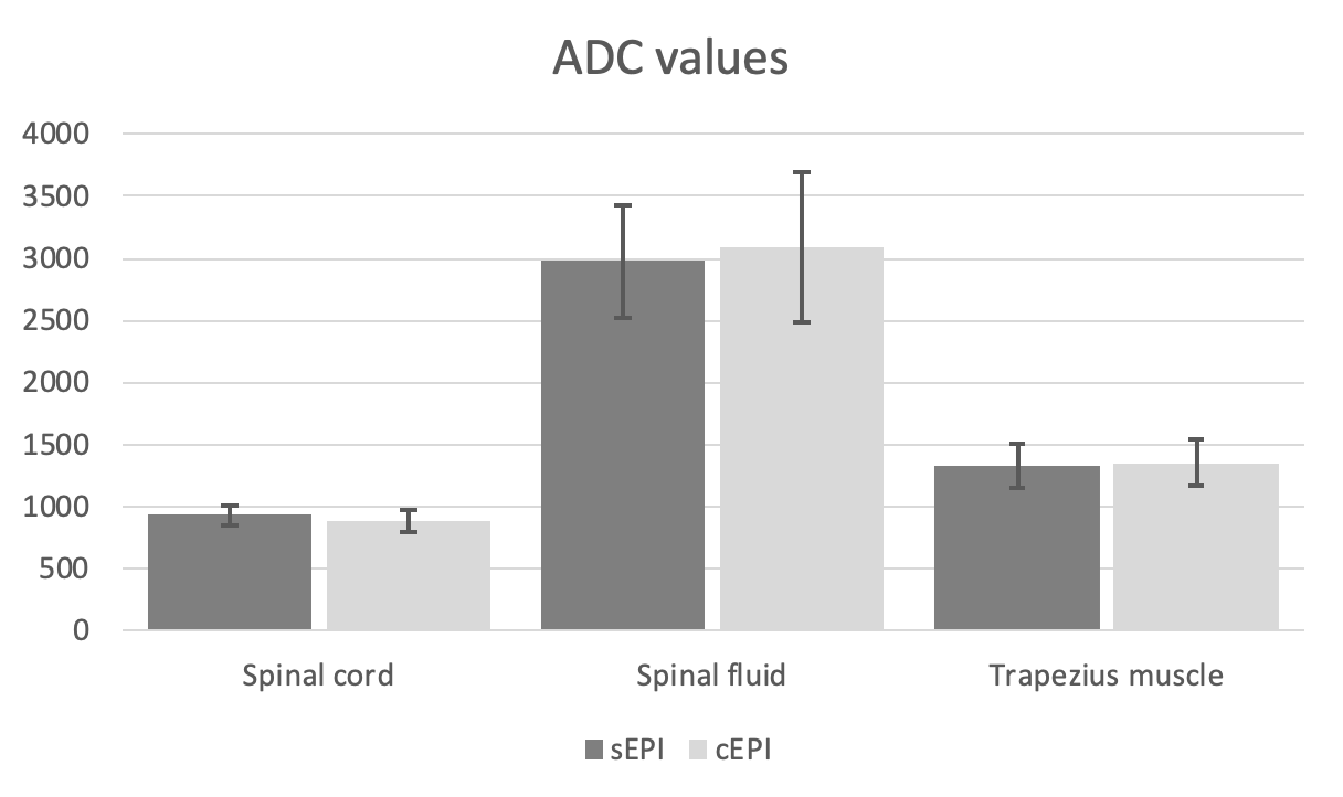
To assess quantitative effects of local shim coils on quantification of ADC in EPI and cEPI, the mean voxel value ± standard deviations of apparent diffusion coefficients (ADC) were compared in three separate anatomical compartments (spinal cord, spinal fluid and trapezius muscle) using circular regions of interest (ROI).
Results
The use of additional local shim coils for single shot echo planar DWI resulted in a marked improvement of overall image quality compared to EPI without the use of additional coils (p<0.001). Furthermore, signal loss was almost non-existing in cEPI, while sEPI showed significantly increased, intermediate levels (p<0.001). The same was seen for distortions between cEPI and sEPI. Although, the margin was lower than for overall image quality or signal loss, spatial distortions were significantly reduced in cEPI (p=0.008) (Figure 2, 3).Regarding the saturation of fat between single shot EPI without and with the use of local shim coils as well as T1-weighted GRE without and with additional local shim coils resulted in a clear improvement of cEPI (p<0.001). Comparing GRE and cGRE showed improved fat saturation in cGRE, however not significantly (p=0.177) (Figure 2, 3).
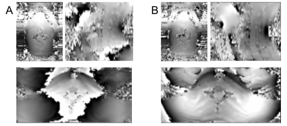
Comparing the B0 field maps without and with the use of local shim coils resulted in a marked homogenization of the static field, especially in the lower and posterior neck region (Figure 4).
ADC values were quantified in three different anatomical compartments (spinal cord, spinal fluid, trapezius muscle). The measured values did not show a significant difference between the compartments (p≥0.08) (Figure 5).
The use of additional local shim coils did not increase scan time (4:09 minutes) (Figure 1).
Discussion
In recent years, previous studies already showed significant improvement in shimming techniques5,6. Our results indicate that cEPI significantly improves overall image quality compared to sEPI, as well as markedly improves fat saturation compared to sEPI and GRE. However, cGRE showed no significant improvement for the saturation of fat compared to sEPI and GRE. Additionally, quantification of ADC values showed no significant difference between sEPI and cEPI.
Conclusion
In comparison to the use of standard single shot EPI-DWI, using head and neck surface coils with additionally integrated local shim coils resulted in an improvement of the homogenization of the static magnetic field, therefore, improving DWI image quality. For T1-weighted GRE the use of these additional coils has no effect on the saturation of fat.
Acknowledgements
No acknowledgement found.References
1. Bae YJ, Choi BS, Jeong HK, Sunwoo L, Jung C, Kim JH. Diffusion-Weighted Imaging of the Head and Neck: Influence of Fat-Suppression Technique and Multishot 2D Navigated Interleaved Acquisitions. AJNR Am J Neuroradiol. 2018;39(1):145-50.
2. Hirata K, Nakaura T, Okuaki T, Kidoh M, Oda S, Utsunomiya D, et al. Comparison of the image quality of turbo spin echo- and echo-planar diffusion-weighted images of the oral cavity. Medicine (Baltimore). 2018;97(19):e0447.
3. Mikayama R, Yabuuchi H, Sonoda S, Kobayashi K, Nagatomo K, Kimura M, et al. Comparison of intravoxel incoherent motion diffusion-weighted imaging between turbo spin-echo and echo-planar imaging of the head and neck. Eur Radiol. 2018;28(1):316-24.
4. Ma G, Xu XQ, Hu H, Su GY, Shen J, Shi HB, et al. Utility of Readout-Segmented Echo-Planar Imaging-Based Diffusion Kurtosis Imaging for Differentiating Malignant from Benign Masses in Head and Neck Region. Korean J Radiol. 2018;19(3):443-51.
5. Walter SS, Liu W, Stemmer A, Martirosian P, Nikolaou K, Notohamiprodjo M, et al. Combination of integrated dynamic shimming and readout-segmented echo planar imaging for diffusion weighted MRI of the head and neck region at 3Tesla. Magn Reson Imaging. 2017;42:32-6.
6. Andre JB, Bammer R. Advanced diffusion-weighted magnetic resonance imaging techniques of the human spinal cord. Top Magn Reson Imaging. 2010;21(6):367-78.
Figures