3520
Diffusion-Weighted ZOOM-EPI with Simultaneous Multi-Slice Imaging1Department of Systems Neuroscience, University Medical Center Hamburg-Eppendorf, Hamburg, Germany
Synopsis
Zonal oblique multi-slice (ZOOM) EPI uses cross-sectional RF excitations to focus the measurement volume to a small, inner volume which allows to reduce the FOV without aliasing in the phase-encoding direction. Thus, geometric distortions are reduced and the spatial resolution of diffusion-weighted acquisitions can be increased as has been demonstrated for the optic nerve and spinal cord. In this study, ZOOM-EPI is combined with simultaneous multi-slice (SMS), the boundary condition to avoid unwanted signal contributions is determined, and the feasibility to shorten acquisitions times is demonstrated for diffusion-tensor imaging (DTI) of the human spinal cord.
Introduction
Spin-echo echo-planar imaging (EPI)1 is the standard technique for diffusion-weighted acquisitions but suffers from geometric distortions in the presence of field inhomogeneities. The distortions increase with the echo train length and, thus, are more pronounced for larger fields-of-view (FOVs) and higher spatial resolutions which limits the applicability of EPI to small, inner structures like the optic nerve or the spinal cord. To shorten the echo train and reduced geometric distortions accordingly, zonal oblique multi-slice (ZOOM) EPI2,3 has been proposed. It involves cross-sectional RF excitations to focus the refocussed magnetization to an inner volume which allows to reduce the FOV without aliasing in the phase-encoding direction. In this study, ZOOM-EPI is combined with simultaneous multi-slice (SMS)4, the boundary condition to avoid unwanted signal contributions is determined, and the feasibility to shorten acquisitions times is demonstrated for diffusion-tensor imaging (DTI) of the human spinal cord.Methods
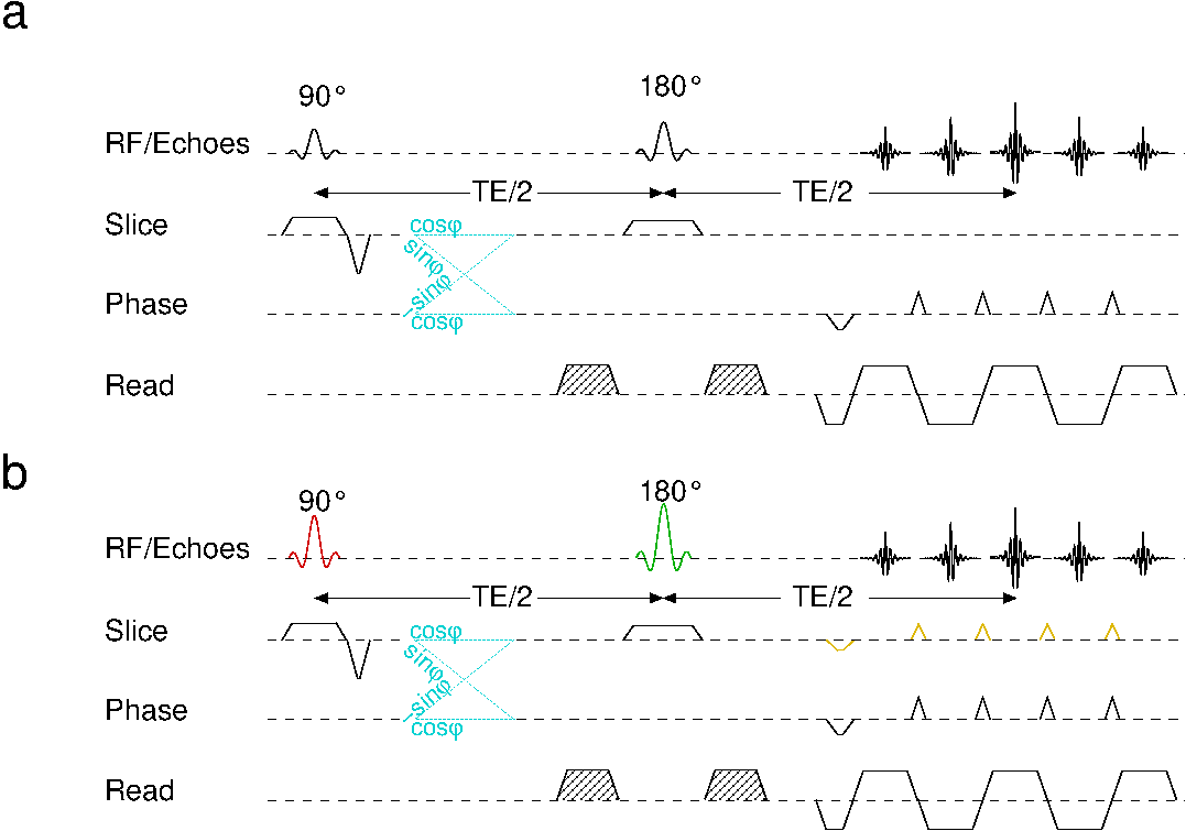
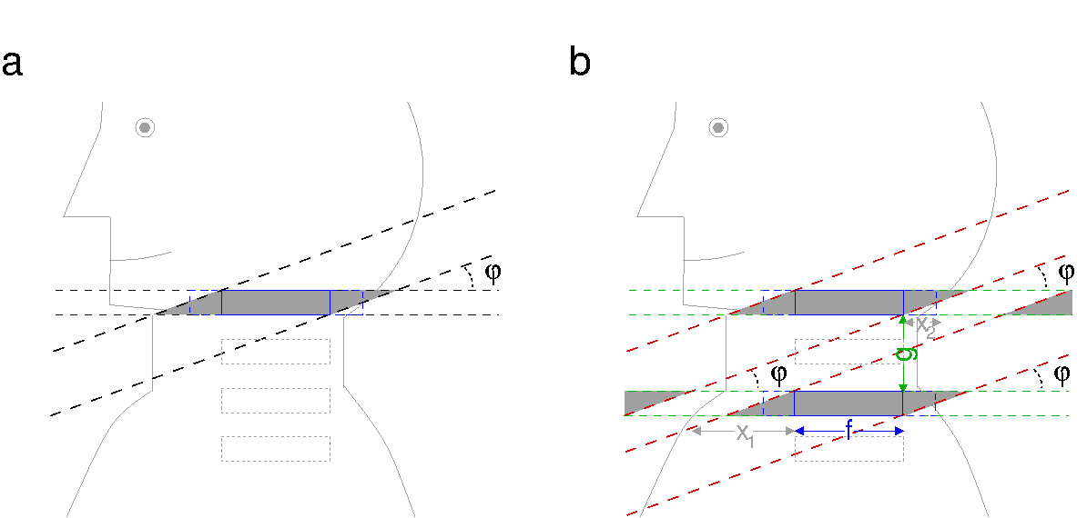
The pulse sequences and geometric setups used in the present study are sketched in Fig. 1 and 2. The plane of the initial RF excitation is tilted by an angle φ compared to the image plane defined by the refocussing RF pulse. On both sides of the desired inner FOV, transition zones are excited and refocused which requires phase-encoding oversampling to avoid aliasing. Furthermore, neighboured sections are partially covered and saturated by the tilted RF excitation. Thus, the angle φ must be chosen as a compromise between minimum saturation effects and small transition zones. To extend the conventional approach (Fig. 1a and 2a) to simultaneous multi-slice imaging, both RF pulses must cover multiple frequency bands (Fig. 1b and 2b). As an unwanted side effect, magnetization outside of the target region can be excited and refocused by the bands of different slices. However, they can be positioned outside of the object to avoid their unwanted signal contributions if the tilt angle φ obeys tan φ < g/(f+max(x1, x2)) (g: minimum gap between the simultaneously acquired slices, f: desired inner FOV; xi: distances from inner FOV to object boundary on different sides in the different slices; cf. Fig. 2b). Furthermore, additional gradient pulses to use the blipped-CAIPI approach4 were applied in the slice direction. Experiments were performed on a 3T whole-body MR system (PrismaFit, Siemens Healthineers, Erlangen, Germany) using a 64-channel head-neck coil together with a 32-channel spine array coil with only those coil elements selected that provide significant signal contributions for the chosen measurement volume. A water phantom was used for test experiments, in vivo acquisitions (62 slices, no gap) were performed in healthy volunteers after their informed consent was obtained. Images were acquired with a voxel size of 1.0×1.0×4.0mm3, a tilt angle φ of 5°, an inner FOV of 40mm plus 20mm phase-encoding oversampling to account for the transition zones, and involved an interleaved excitation order due to saturation in directly neighboured sections. Diffusion-tensor imaging was performed with six different directions of the diffusion weighting, a b value of 625s mm-2 yielding an echo time of 75ms, and 12 averages. SMS acquisitions were accelerated by a factor of 2 which allowed to shorten the repetition time from 7300ms to 3700ms and the total acquisition time from 10.5min to 5.7min, slightly less than a factor of 2 due to the extra reference acquisitions required for SMS imaging. Isotropic diffusion-weighted images and maps of the apparent diffusion coefficient (ADC) and fractional anisotropy (FA) were obtained with the analysis framework provided by the manufacturer.Results and Discussion
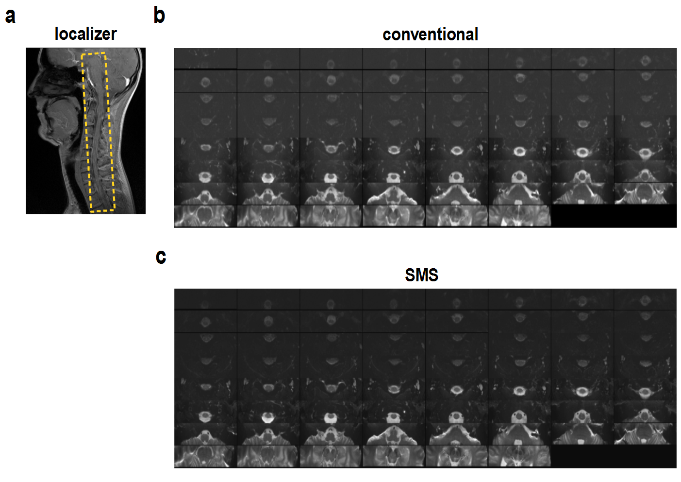
Non-diffusion-weighted images of the human brain stem and spinal cord are presented in Fig. 3. For lower cord sections, the signal intensity is considerably reduced due to the lower performance of the receive coils. No adverse effect like residual aliasing or noise amplification is observed for the SMS acceleration.
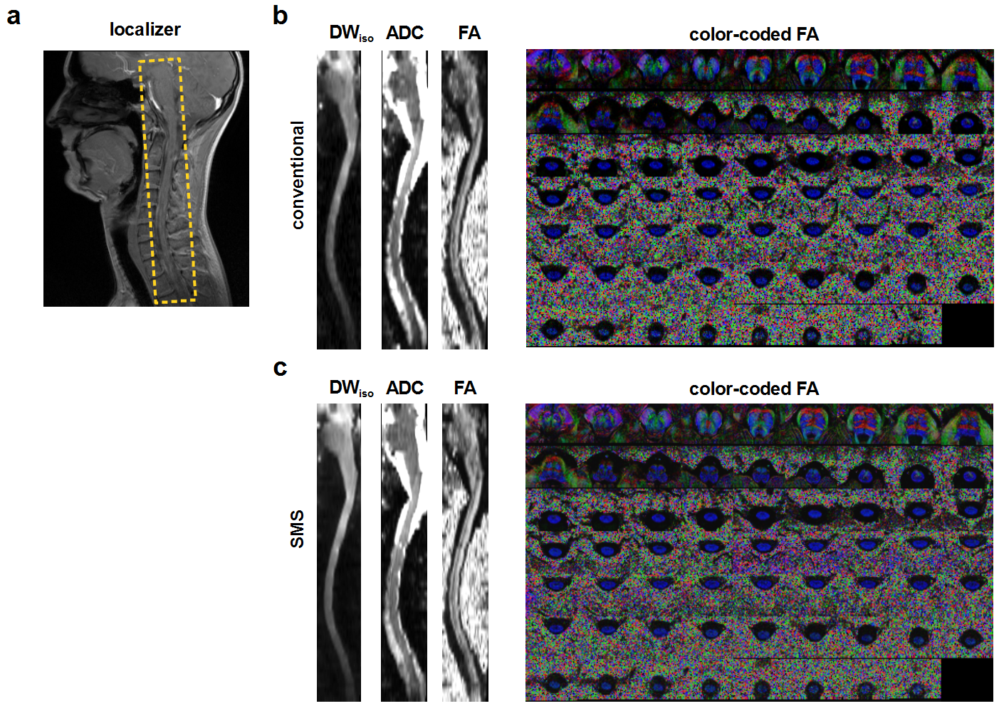
Results of a DTI experiment are summarized in Fig. 4. The conventional and SMS acquisitions show a very similar performance despite the much shorter repetition and acquisition time of the SMS acquisition. Due to the small FOV, only minor geometric distortions are visible in the spinal cord. Color-coded maps of the FA show a reduced anisotropy in spinal cord grey matter, reflect the nerve fibre orientation in white matter, and show some of the fibre crossing of transverse pontine fibres with the pyramidal tracts.
Conclusion
For an appropriately chosen tilt angle, ZOOM-EPI can be accelerated with SMS acquisitions without side effects. Thus, DTI acquisitions of small target regions like the optic nerve or spinal cord can be shortened which could improve the clinical applicability in patients.Acknowledgements
This work was supported by a grant from Wings for Life.References
1. Mansfield P. Multi-planar image formation using NMR spin echos. J Phys C. 1977; 10: 55-58.
2. Wheeler-Kingshott CA, Parker GJ, Symms MR, Hickman SJ, Tofts PS, Miller DH, Barker GJ. ADC mapping of the human optic nerve: increased resolution, coverage, and reliability with CSF-suppressed ZOOM-EPI. Magn Reson Med 2002; 47: 24-31.
3. Wheeler-Kingshott CA, Hickman SJ, Parker GJ, Ciccarelli O, Symms MR, Miller DH, Barker GJ. Investigating cervical spinal cord structure using axial diffusion tensor imaging. Neuroimage 2002; 16: 93-102.
4. Setsompop K, Gagoski BA, Polimeni JR, Witzel T, Wedeen VJ, Wald LL. Blipped-controlled aliasing in parallel imaging for simultaneous multislice echo planar imaging with reduced g-factor penalty. Mag Reson Med 2012; 67: 1210-1224
Figures