3513
Evaluation of Diffusion-Weighted Readout-Segmented EPI in Macaque Brain Imaging at 7T1Interdisciplinary Institute of Neuroscience and Technology, Qiushi Academy for Advanced Studies, College of Biomedical Engineering & Instrument Science, Zhejiang University, HangZhou, China, 2School of Medicine, Zhejiang University, Hangzhou, China, 3MR Collaboration Northeast Asia, Siemens Healthcare, Shanghai, China, 4Siemens Healthcare GmbH, Erlangen, Germany, 5Center for Magnetic Resonance Research, University of Minnesota, Minneapolis, MN, United States
Synopsis
Diffusion-weighted
imaging (DWI) has been widely used in brain structure and connectivity
investigations and other neuroscience applications. To implement an optimized
protocol for high spatial resolution DW imaging at ultra-high field, we have
conducted macaque brain imaging by using readout segmented echo planar imaging
at 7T. Our results indicate that, the rsEPI has potential to achieve DWI with
improved signal-to-noise ratio and with the intent of minimizing geometric
distortion.
Introduction
DWI has been widely used in brain structure and connectivity investigation in neuroscience research1. Single-shot EPI (ssEPI) as an well-established approach, however, suffers from well-known artifacts caused by susceptibility changes at air-tissue interfaces as well as the subtle T2* blurring due to significant T2* relaxation, which restrains the potential higher spatial resolution it can possibly reach at ultra-high field (UHF). The readout-segmented EPI (rsEPI) in conjunction with GeneRalized Autocalibrating Partially Parallel Acquisitions (GRAPPA)2 and two-dimensional navigator correction can provide lower susceptibility distortion, reduced T2* blurring, and a robust correction for susceptibility-induced artifact3. In the current study, we evaluated geometric distortion and signal-to-noise ratio (SNR) efficiency of DW images by utilizing rsEPI for in vivo macaque brain imaging on a 7T human MR scanner. Recommended protocols are provided through a series of image quality comparison.Methods
All MRI scans were performed on a 7T research system (Siemens Healthcare, Erlangen, Germany) equipped with a 1Tx/28Rx knee coil (QED, Mayfield Village, OH, USA). Three healthy female macaques (2-3 years old with weights of 3.6, 3.2 and 3.7 kg) were included in the study, and were maintained and anesthetized by 1.5~2% isoflurane – all procedures were in accordance with NIH standards and approved by local Institutional Animal Care Committee. All monkeys were fixed in the sphinx position with its head centered within the knee coil.
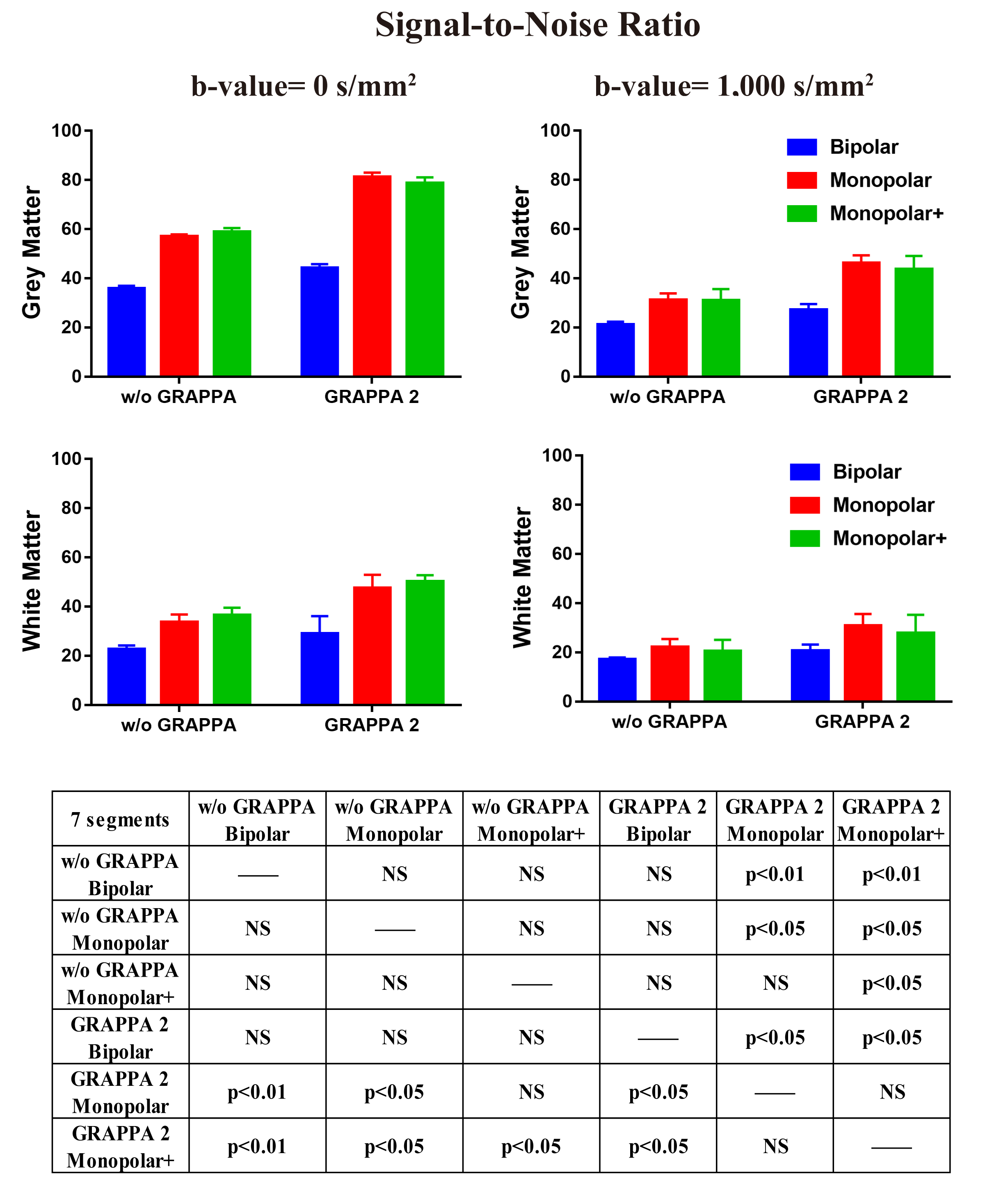
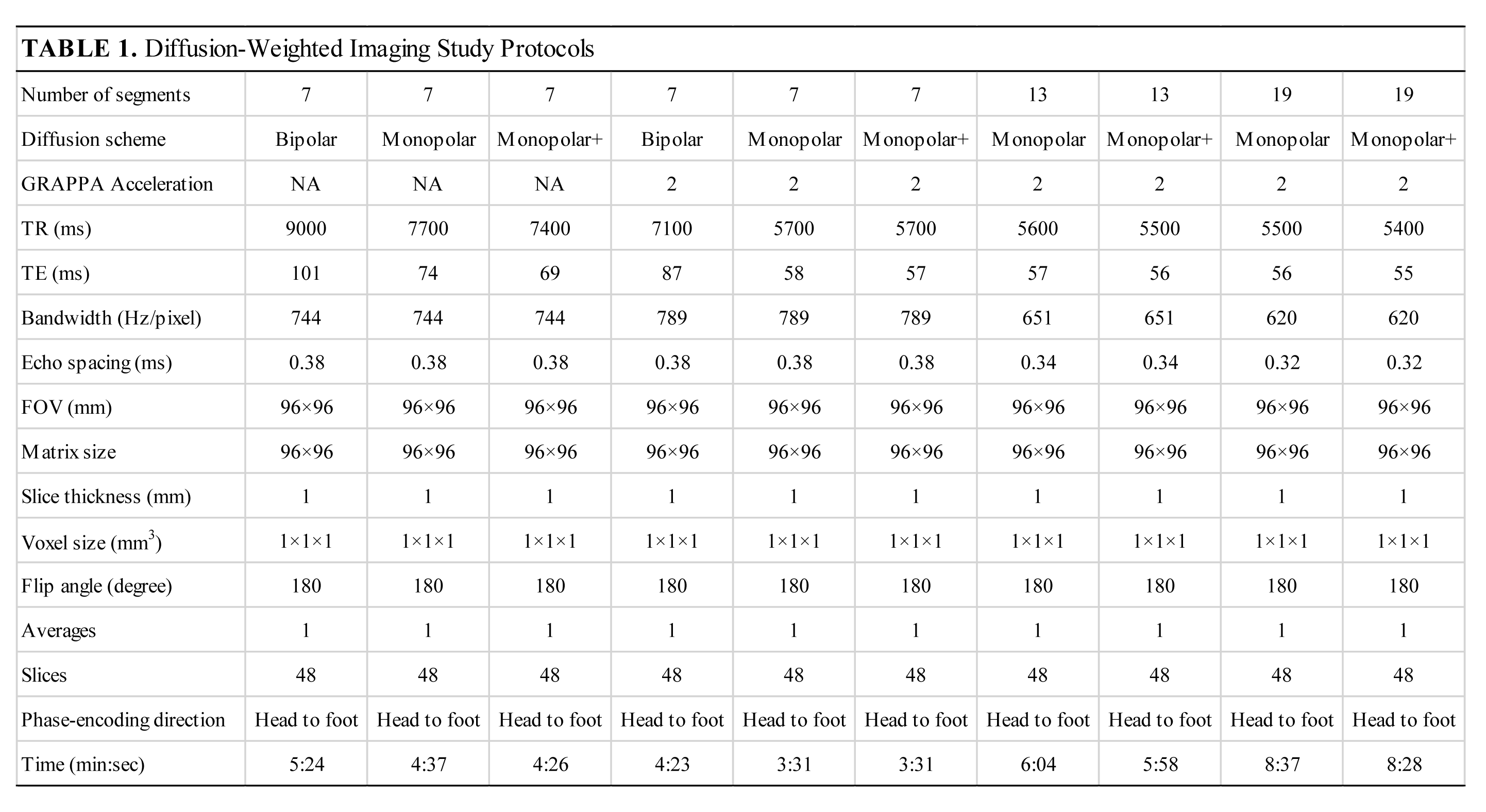
A prototype sequence4,5 was used to acquire 1 mm isotropic rsEPI DW images with different diffusion encoding schemes, acceleration modes, and number of readout segments. Evaluation of the image SNR (illustrated in Table 1) with optimized parameters such as TR, TE, EPI-factor, receiver bandwidth and echo spacing was performed. The diffusion encoding scheme include the conventional bipolar gradient scheme (twice refocused bipolar gradient scheme), the traditional Stejskal-Tanner sequence (single refocused, monopolar gradient) and a modified Stejskal-Tanner sequence (optimized monopolar scheme, monopolar+). The monopolar+ is an optimized Stejskal-Tanner scheme with a single bipolar gradient6. The SNR values were calculated by using the difference method and a two-factor repeated measures analysis of variance test7.
In addition, 3D MPRAGE T1-weighted images were obtained as structure reference, with isotropic spatial resolution of 0.5 mm, TR/TE 3,500/3.74 ms, TI 1,500 ms, flip angle 7 deg, bandwidth 170 Hz/pixel, FOV 96 mm and matrix size 192x192.
Results and Discussion
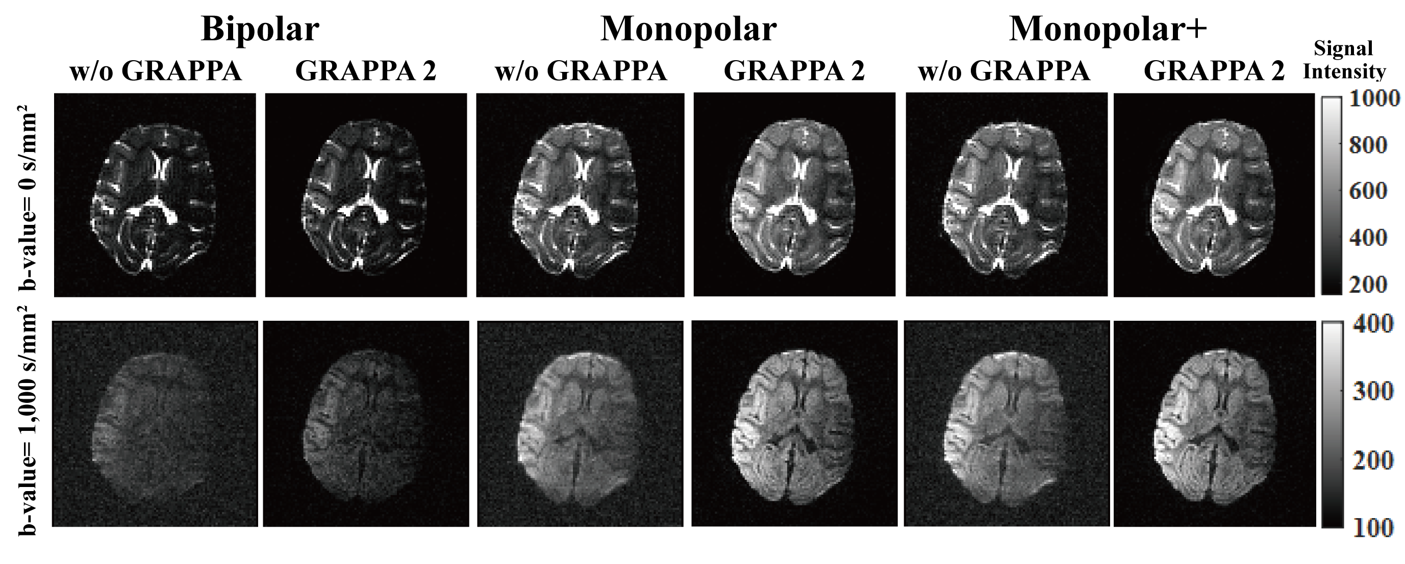
Figure 1 shows the SNR comparison results of DW images at b-value = 0 and 1,000 s/mm2 with 7 readout segments using different diffusion encoding schemes and accelerate modes. With GRAPPA acceleration, the mean SNRs of grey matter and white matter were consistently higher than those without GRAPPA. Similarly, the mean SNRs were higher under GRAPPA acceleration mode with monopolar and monopolar+ when compared with bipolar scheme, but the difference between monopolar and monopolar+ scheme did not reach statistical significance. The SNR benefit of using GRAPPA comes from the reduction of TE, despite of g-factor noise penalty. The GRAPPA technique reduces also the effective echo train length and thus improves the image quality3. Corresponding images are shown in Fig. 2.
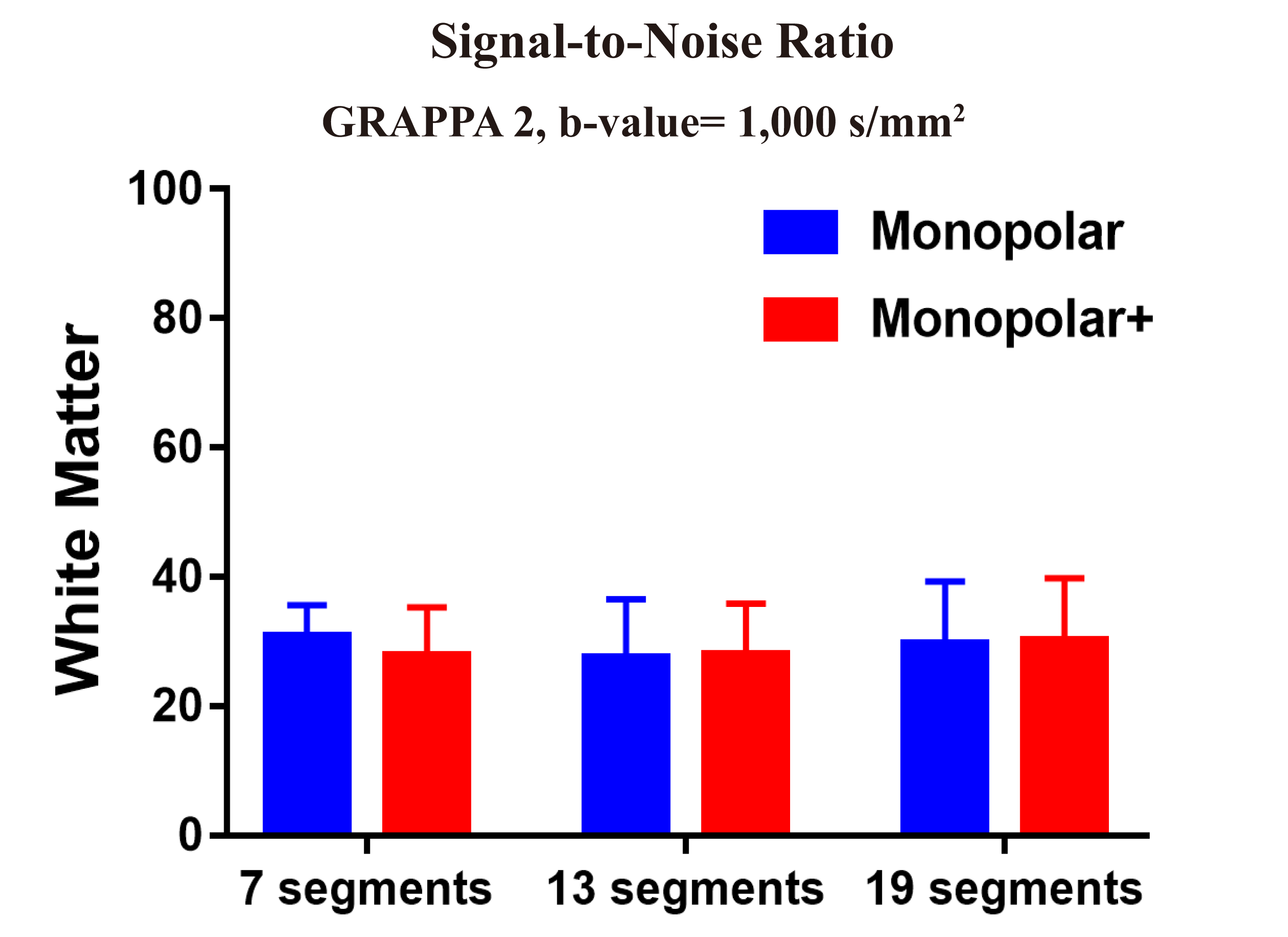
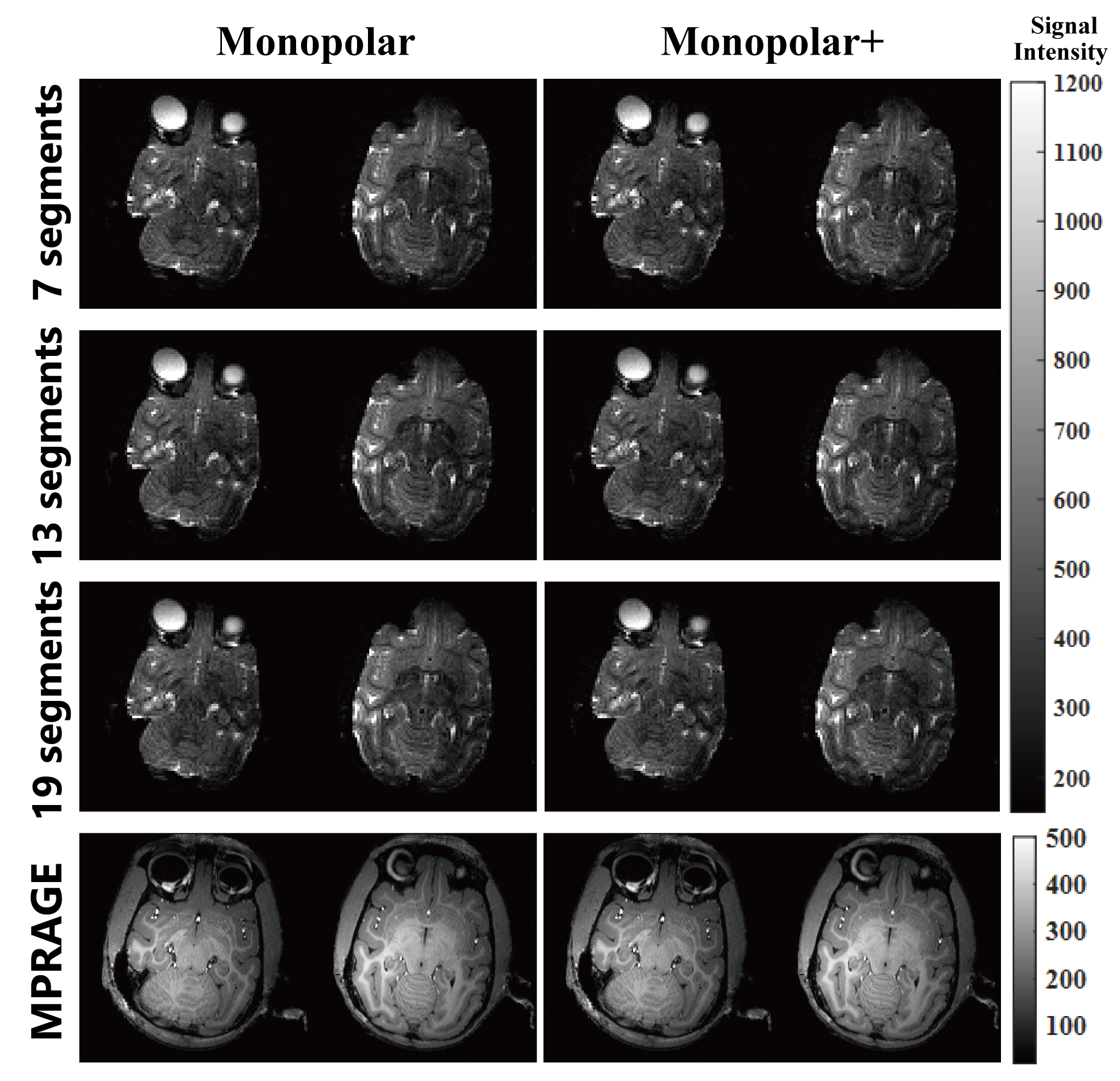
Figure 3 and 4 show the comparison of 1 mm isotropic DW images with monopolar and monopolar+ scheme between 7, 13, 19 readout segments with an GRAPPA acceleration factor of two. These DW images showed slight geometric distortion at air-tissue interfaces such as paranasal sinus, the nasal cavity and the ear cavity. Increasing readout segments of rsEPI leads to longer scan time but shorter readout echo spacing (see Table 1), the latter of which is supposed to alleviate image distortions due to the susceptibility effect8. However, the geometric distortion observed in Figure 4 were not apparently reduced with increased readout segments. In addition, there is still no significant SNR difference between monopolar and monopolar+ scheme.
The standard bipolar diffusion encoding is commonly used to reduce eddy currents, but limits the minimum achievable TE, because of an extra refocusing pulse and additional corresponding crusher gradients. The low eddy current environment provided by modern gradient coil designs allow the use of monopolar and monopolar+ diffusion encoding. Particularly, the monopolar+ diffusion encoding is designed to improve image SNR, shorten TE and minimize eddy current distortion, which may effectively merit diffusion-related investigations with higher sensitivity to eddy current effects (e.g., tractography)6.
Conclusion
In conclusion, rsEPI with GRAPPA, shorter readout segments in conjunction with monopolar+ diffusion encoding is optimal for DWI of macaque brain at UHF.Acknowledgements
National Natural Science Foundation 81701774 and 61771423.References
1. Mansfield P, Pykett IL. Biological and medical imaging by NMR. Journal of Magnetic Resonance, 1969 1978:29:355–373.
2. Griswold M A, Jakob P M, Heidemann R M, et al. Generalized autocalibrating partially parallel acquisitions (GRAPPA). Magn Reson Med, 2002, 47(6):1202-1210.
3. Porter DA, Heidemann RM. High resolution diffusion-weighted imaging using readout-segmented echo planar imaging, parallel imaging and a two-dimensional navigator-based reacquisition. Magn Reson Med. 2009:62:468–475.
4. Pipe JG, Farthing VG, Forbes KP. Multishot diffusion-weighted FSE using PROPELLER MRI. Magn Reson Med. 2002; 47:42–52.
5. Robert Frost, et al. High-Resolution Diffusion-Weighted Neuroimaging at 3T and 7T with Simultaneous Multi-Slice RESOLVE. MAGNETOM Flash | (63) 3/2015
6. Morelli J N, Runge V M, Feiweier T, et al. Evaluation of a modified Stejskal-Tanner diffusion encoding scheme, permitting a marked reduction in TE, in diffusion-weighted imaging of stroke patients at 3 T. Investigative Radiology, 2010, 45(1):29-35.
7. Fritz J, Fritz B, Zhang J, et al. Simultaneous Multislice Accelerated Turbo Spin Echo Magnetic Resonance Imaging: Comparison and Combination With In-Plane Parallel Imaging Acceleration for High-Resolution Magnetic Resonance Imaging of the Knee. Investigative Radiology, 2017, 52(9):529.
8. Wang Y, Ma X, Zhang Z, et al. A comparison of readout segmented EPI and interleaved EPI in high-resolution diffusion weighted imaging. [J]. Mag Reson Imag, 2017, 47:39.
Figures