3512
Effects of imperfect slice separation in simultaneous multi-slice for diffusion kurtosis imaging1Department of Neurosurgery, Medical College of Wisconsins, Milwaukee, WI, United States, 2Center for Imaging Research, Medical College of Wisconsin, Milwaukee, WI, United States, 3Department of Biophysics, Medical College of Wisconsin, Milwaukee, WI, United States, 4Department of Radiology, Medical College of Wisconsin, Milwaukee, WI, United States
Synopsis
Simultaneous multislice (SMS) technique accelerates MRI data acquisition. However, slice separation in reconstruction is imperfect. This results in residual leakage between the simultaneously excited slices. For diffusion MRI, repeated pairing of the same slices in the image series leads to repeated leakage of a consistent diffusion signal from the other slice. Studies of this effect on diffusion MRI is sparse. Here, we tested two different SMS acquisition and image reconstruction techniques and quantified resulting errors on all diffusion kurtosis (DKI) metrics in different ROIs. Large errors were observed with SMS compared to singleband acquisitions for all DTI and DKI metrics
Introduction:
Simultaneous multislice (SMS) technique accelerates MRI data acquisition. However, slice separation in reconstruction is imperfect. This results in residual leakage between the simultaneously excited slices. For diffusion MRI, repeated pairing of the same slices in the image series leads to repeated leakage of a consistent diffusion signal from the other slice. Studies of this effect on diffusion MRI is sparse1,2,3. An earlier study reported an average effect on the MD and mean kurtosis (MK) in gray and white matter of two separated slices2. Another group studied MK only in the pyramidal tracts3. We have previously demonstrated this effect using simulations4. Here, we expand those studies by testing different SMS acquisition and image reconstruction techniques and quantifying resulting errors on all diffusion kurtosis (DKI) metrics in different ROIs.Methods:
In addition to conventional SMS, a novel acquisition strategy was also tested. We randomized which slices were simultaneously excited across different diffusion weighted images (DWI). We call this randomized-slice (rs)SMS4. With rsSMS, leaking signal will be from a different anatomy for each DWI. Hence, leakage from the other slice should appear as random noise. Slice pair randomization was done with constraints to reduce T1 effects due to varying TR for each slice and also keep enough coil sensitivity variations for slice pairs.
The study was conducted on a GE 7T scanner. It was approved by the IRB and written consent was obtained from the participant. Data was acquired from a volunteer using conventional singleband, SMS and rsSMS. Each acquisition type was repeated twice to show reproducibility and the order was randomized to minimize systematic bias. All scan parameters were matched, except for the multiband factor of 2 for SMS and rsSMS. Single-shot SE-EPI was used with 3mm isotropic resolution and b = {1000, 2000} s/mm2 and 30 directions per shell. Each acquisition had two singleband, non-diffusion weighted reference images. In SMS and rsSMS, those reference images were used to train GRAPPA kernels.
After preprocessing to correct for motion and eddy currents, the multiband images were reconstructed with both Slice GRAPPA (SG) and Split-Slice GRAPPA (SPSG). SPSG has been introduced recently to reduce slice crosstalk5,6. Interslice and intraslice artifacts were calculated with the LSLA approach6.
For analysis, means and standard deviations from ROIs from different fiber tracks were calculated. Measurements from the first singleband acquisition were used as the reference and the corresponding metrics from second singleband, SMS and rsSMS with SG and SPSG were subtracted to quantify errors.
Results:
SPSG reduced slice leakage compared to SG for both SMS and rsSMS. The errors were slightly higher in rsSMS, compared to SMS. This is possibly an indication of suboptimal GRAPPA kernels in rsSMS due to varying slice separation distances. The separation between rsSMS slices was higher, on average, but the minimum slice separation was about half that of conventional SMS.
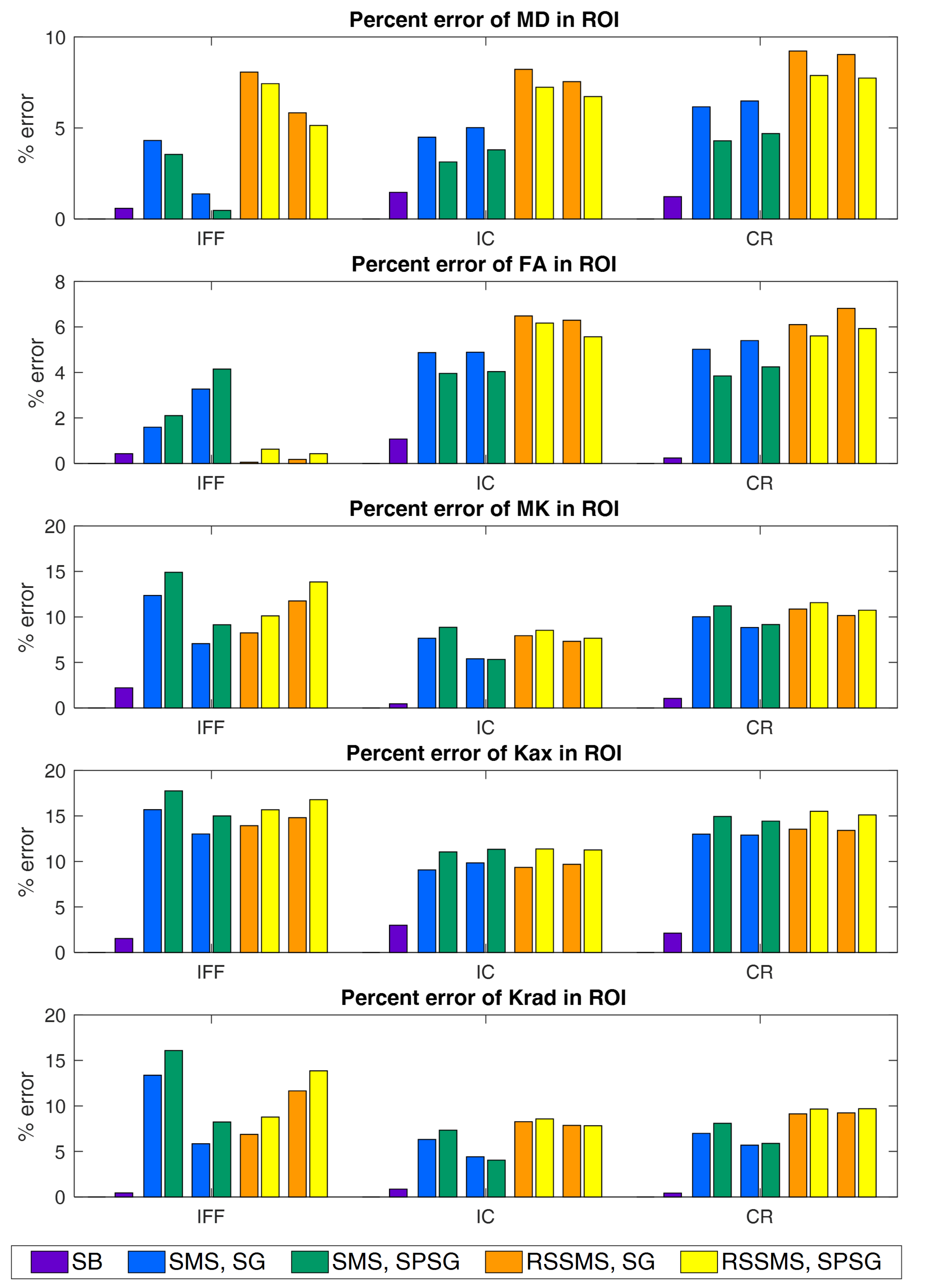
Fig.1 shows the errors in the ROIs for the second singleband image and all multiband images with respect to the first singleband. Note the small difference between the two singleband acquisitions compared to errors in SMS and rsSMS. In other words, multiband errors were much larger than typical scan-to-scan variability in conventional experiments.
MD and FA were less sensitive to slice separation errors than the kurtosis metrics. But, no acquisition or reconstruction technique consistently performed best. An interesting trend was observed for the reconstruction methods. For MD and FA, SPSG had lower error than SG, but the trend was reversed for the DKI metrics.
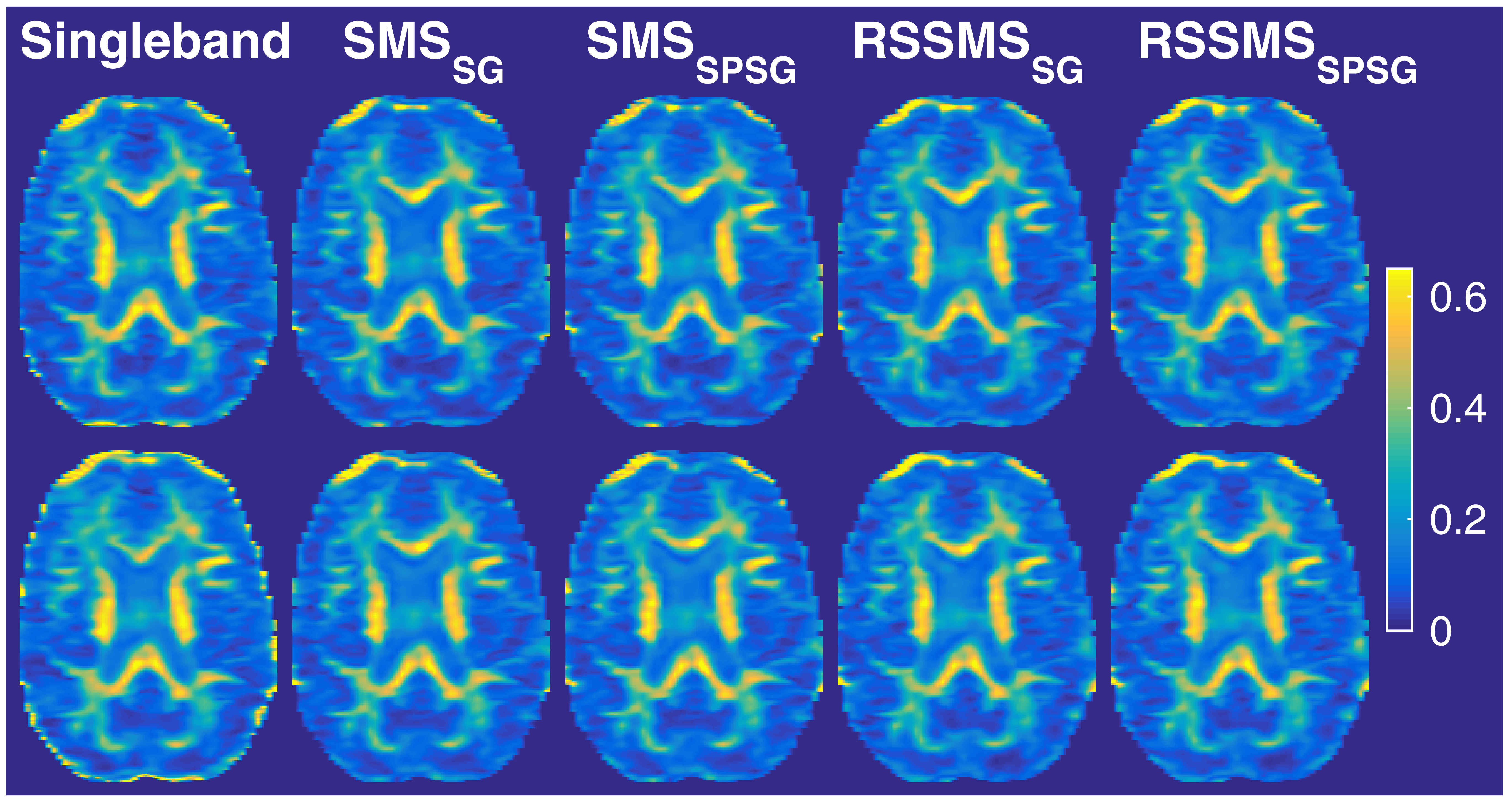
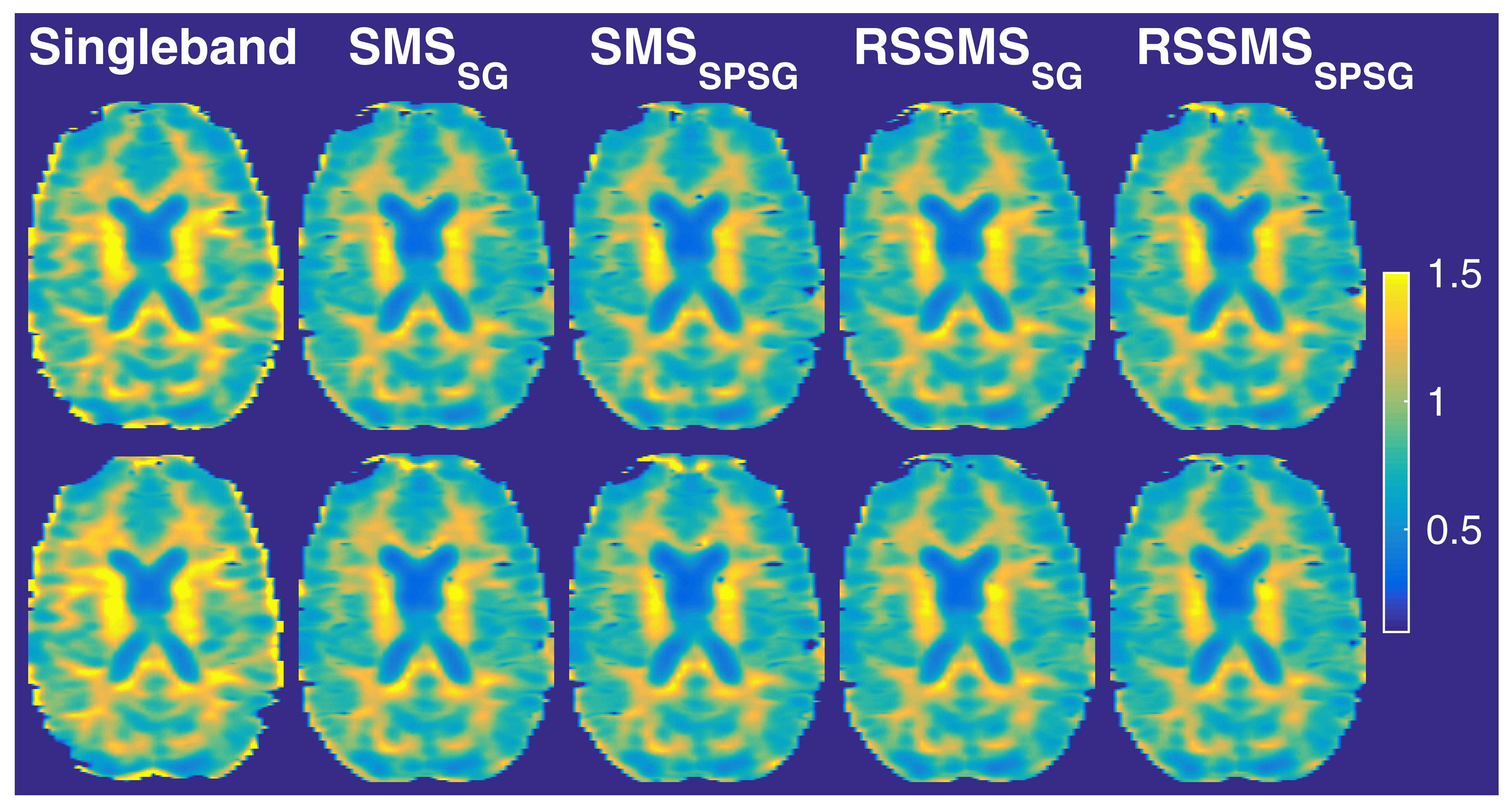
For a qualitative comparison, an axial slice is presented for FA (Fig.2) and MK (Fig.3). Subtle decreases in the multiband FA maps and more visible drop in MK values are observed for all multiband acquisitions relative to the singleband acquisition. This is particularly evident, for instance, in the internal capsule, which had 7% error. Errors are visible in the gray matter, as well, although we did not analyze gray matter.
Discussion:
Large errors were observed with SMS and rsSMS with respect to singleband acquisitions for all DTI and DKI metrics, regardless of the reconstruction method. In addition, these errors might also have strong spatial dependence. For instance, if a voxel with high diffusion (hence low intensity) mixes with a voxel with low diffusion (hence high intensity), for a given b-vector, the voxel will erroneously show low diffusion. Therefore, some fiber tracts might be compromised more than others, and this effect will vary from subject to subject. Furthermore, increasing the multiband factor could result in even larger errors. Studies using multiband diffusion MRI need to test such errors before planning experiments.Acknowledgements
This work is supported by funding provided by the Daniel M. Soref Charitable Trust.References
1. Setsompop K, Cohen-Adad J, Gagoski BA, et al. Improving diffusion MRI using simultaneous multi-slice echo planar imaging. NeuroImage 2012;63:569–80.
2. Xu J, Glenn GR, Bhat H, et al. Accelerated Diffusional Kurtosis Imaging using Simultaneous Multi-slice Echo Planar Imaging. Proc. Intl. Soc. Mag. Reson. Med. 21:3183 (2013).
3. Kasahara A, Suzuki Y, Mitsuda M, et al. Usefulness and Consideration of Multi-Band EPI using Simultaneous Multi-Slice for Diffusion Kurtosis Imaging at 1.5 Tesla Magnetic Resonance Imaging. Austin J Radiol. Volume 3, Issue 4 2016 (open access).
4.Olson DV, Arpinar VE, Nencka AS, and Muftuler LT. Randomized-Slice Simultaneous Multi-Slice for Diffusion Kurtosis Imaging. Proc. Intl. Soc. Mag. Reson. Med. 5331 (2018).
5. Setsompop K, Cauley SF, Bhat H, Polimeni J, Wald LL. Characterization and Mitigation of Signal Leakage in Simultaneous Multi-Slice (SMS) Acquisition. Proc. Intl. Soc. Mag. Reson. Med. 21, p. 3315 (2013).
6. Cauley SF, Polimeni JR, Bhat H, Wald LL, Setsompop K. Interslice leakage artifact reduction technique for simultaneous multislice acquisitions. Magn Reson Med. 72: 93 - 102 (2014).
Figures