3510
Multi-Echo Segmented Diffusion-Weighted MRI for Ex-Vivo Whole-Brain Measurements with 300mT/m Gradients1Max Planck Institute for Human Cognitive and Brain Sciences, Leipzig, Germany, 2Max Planck Institute for Evolutionary Anthropology, Leipzig, Germany
Synopsis
We provide a novel method to increase SNR of segmented diffusion-weighted EPI acquisitions. Multiple gradient echoes were acquired after each diffusion-preparation and combined in an SNR-optimized way using weightings from quantitative T2* maps. The combination of diffusion-weighted echoes yielded an SNR-gain of 58% compared to single-echo dMRI data with an increase in the segmented readout duration by only 23.1 ms. The multi-echo diffusion MRI acquisition and combination were employed to acquire high-quality ex-vivo diffusion-weighted MRI data from a wild chimpanzee brain.
Introduction
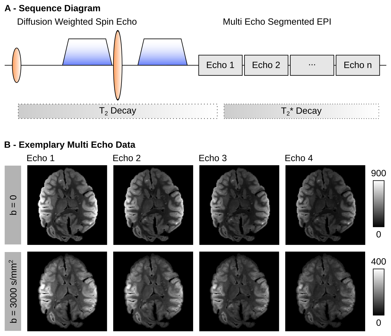
Typical challenges of ex-vivo diffusion MRI (dMRI) acquisitions include low diffusivity in fixed tissue requiring strong diffusion-weightings, signal drop from trapped air, image distortions and increased echo-times due to strong diffusion-weighting requirements. Highly segmented EPI (sEPI) acquisitions can be employed to counteract these challenges by shortening echo-times and reducing image distortions in ex-vivo dMRI1,2. The short sEPI readout trains can easily be repeated to acquire multiple gradient-echoes (GRE) with a negligible increase in acquisition time. However, such Multi-Echo (ME) acquisitions are not employed in dMRI, due to T2* signal decay between echoes. In functional MRI, T2* relaxation maps are employed for SNR optimal echo-combination3. In this work, an ME-sEPI Stejskal-Tanner dMRI sequence4 was developed to achieve high-quality dMRI acquisition for ex-vivo imaging of a wild chimpanzee brain on a human-scale scanner (Figure 1A). Individual echoes were combined using T2*-dependent weighting.
Methods
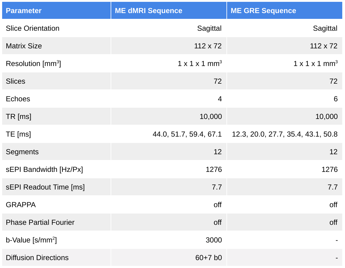
MRI data were acquired from the brain of a 6-year-old juvenile wild female chimpanzee from Taï National Forest (Ivory Coast). The animal died from natural cause without human interference. The brain was extracted on site by a veterinarian four hours after death and immersion-fixed with 4% paraformaldehyde. Further preparations included the removal of superficial vessels, washing out paraformaldehyde in phosphate-buffered saline and placement in Fomblin. ME-dMRI data were acquired using a 3T Connectom System (Siemens Healthineers, Erlangen, Germany) with maximum gradient strength of 300 mT/m and a flexible 23-channel surface coil5. With a total left-right brain extent of 85 mm (100 mm including container), measurement on a small-bore scanner was not possible. ME-dMRI-sEPI and ME-GRE-sEPI datasets were acquired with matched resolution and acceleration (parameters in Table 1). The ME acquisition increased the readout by only 23.1 ms, which is neglectable compared to the diffusion-preparation plus readout of a single-echo only.
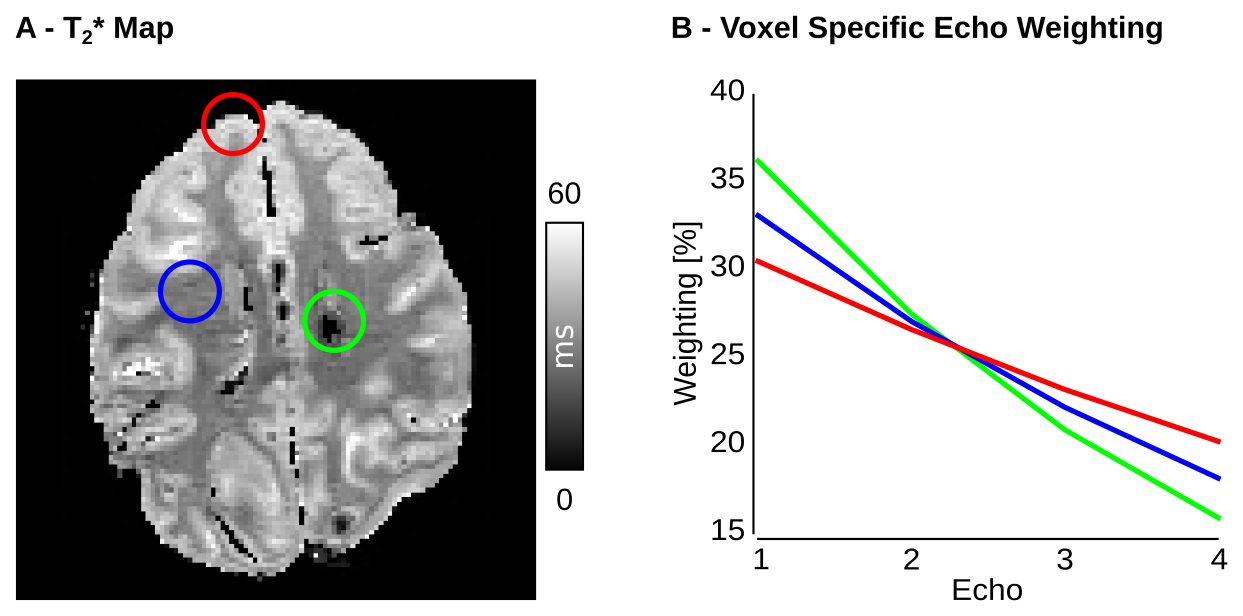
MP-PCA denoising6 was employed prior to echo-combination. A T2* map was calculated by fitting an exponential model to the ME-GRE data. A voxel-wise weighting-factor $$$w_i$$$ for each echo $$$S_i$$$ was calculated based on echo-time and T2*. For optimal SNR, the individual ME-dMRI echoes were combined using weighted averaging: $$S_{comb}=\sum_{i=1}^{n} w_i S_i=\sum_{i=1}^{n}\frac{\exp{-\frac{TE(i)}{T_2^*}}}{\sum_{j=1}^{n}\exp{-\frac{TE(j)}{T_2^*}}}S_i $$
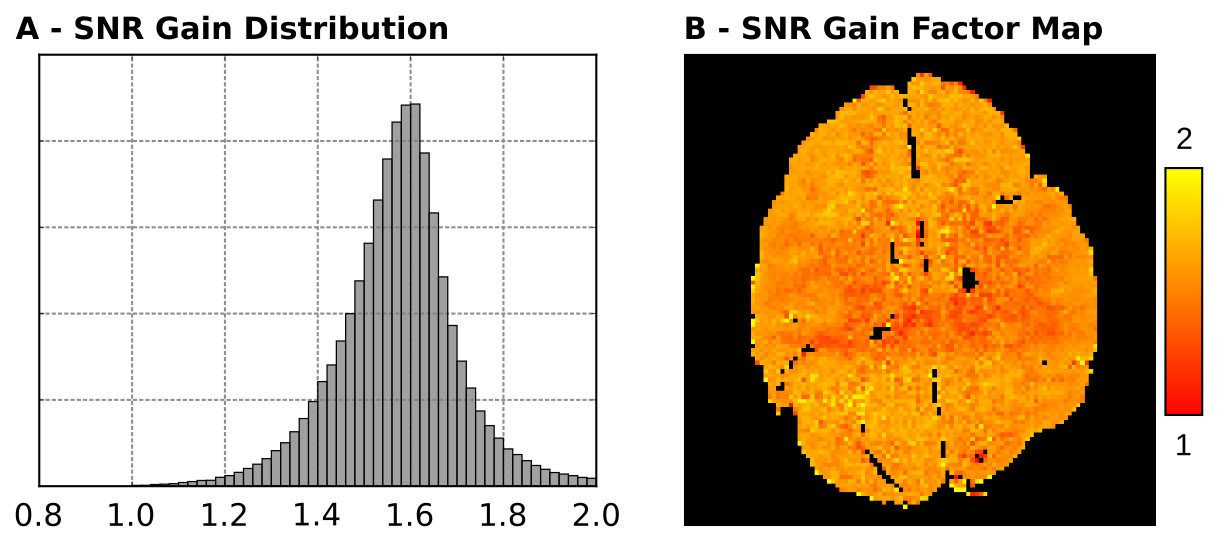
The T2*-dependence of $$$w_i$$$ yielded a voxel-specific SNR-gain compared to single-echo dMRI. To estimate the SNR-gain, the noise was approximated as Gaussian with similar variance, for all echoes. The SNR-gain was then computed as a weighted sum of Gaussian random variables.
$$\text{SNR}_{comb}=\frac{S_{comb}}{\sigma_{comb}}=\frac{S_{comb}}{\sqrt{\sum_i^nw_i^2}\sigma}$$
Deterministic diffusion tensor tracking and visualization of the processed dataset was performed using the software package brainGL [https://github.com/braingl].
Results
The ME-dMRI results from b=0 and one selected diffusion direction with b=3000s/mm2 are displayed in Figure 1B. The dMRI acquisition did not suffer from visible image distortions. A representative slice of the T2* reconstruction is displayed in Figure 2A. Weighting-factors $$$w_i$$$ for different regions of interest are displayed in Figure 2B. During echo-combination, voxels with reduced T2* received stronger weighting from earlier echoes compared to voxels with higher T2*. Figure 3 displays the spatial dependence of SNR-gain of ME combination compared to single-echo dMRI. The distribution shows an average SNR-gain of 58%, which is equivalent to 2.5 independent averages (Figure 3A). In the limiting case of neglectable T2* decay, the combination would have converged to a non-weighted average with a theoretical maximum SNR-gain factor of 2 (i.e $$$\sqrt{n_{\text{avg}}}$$$). The fiber tracking results of the dMRI data are displayed in Figure 4.Discussion
In this work, we employed an ME-dMRI sequence to acquire high-quality dMRI data of a fixed ex-vivo wild chimpanzee brain. We leveraged short readout trains to reduce image distortion and acquire multiple echoes for each diffusion-weighting. The ME-data were combined using a weighted average based on the underlying T2* map, yielding an overall SNR-gain of 58%. With an additional readout duration of only 23.1 ms per segment, this SNR-gain was achieved at a very small cost. We expect this method to further boost image quality and SNR of both segmented and single shot dMRI. Furthermore, ME-dMRI can potentially also provide information about microstructural T2*, by filtering restricted tissue compartments using diffusion-weighting. Future work on multiple echo-combination is required to investigate the extent to which individual diffusion-weighted echoes are affected by the compartmental T2* decay of the underlying microstructure.Acknowledgements
CE is supported by the SPP2041 program "Computational Connectomics" of the German Research Foundation (DFG)References
- Miller K. et. al, Diffusion imaging of whole, post-mortem human brains on a clinical MRI scanner. NeuroImage, 2011
- McNab JA. et. al, The Human Connectome Project and beyond: initial applications of 300 mT/m gradients. NeuroImage, 2013
- Kundu P. et. al, Differentiating BOLD and non-BOLD signals in fMRI time series using multi-echo EPI. NeuroImage, 2012
- Stejskal EO. and Tanner JE., Spin Diffusion Measurements: Spin Echoes in the Presence of a Time‐Dependent Field Gradient. J. Chem. Phys, 1965
- Frass-Kriegl R. et al. Flexible 23-channel coil array for high-resolution magnetic resonance imaging at 3 Tesla. Plos One, 2015
- Veraart J. et. al, Diffusion MRI noise mapping using random matrix theory. Magn Reson Med, 2016
Figures