3509
Sub-Millimeter Isotropic DTI and fiber tractography of the human Spinal Cord in-vivo1Duke University Medical Center, Durham, NC, United States
Synopsis
The ability to characterize complex microstructures in the small cross-sectional area of the spinal cord through diffusion tensor imaging has traditionally been limited by the achievable spatial resolution in-vivo. Through ultra-high spatial resolution diffusion imaging, this study presents a technique for accurately delineating complex fiber pathways such as the corticospinal tracts. When imaged with sub-millimeter isotropic spatial resolutions, it is possible to characterize intricate details in the spinal cord such as the bifurcations and decussations of the corticospinal tracts. The improved delineation of neural pathways in the spinal cord could facilitate the placement of stimulation electrodes for movement disorder treatments.
Introduction
An ability to characterize neural pathways that interconnect the brain with the peripheral nervous system through the spinal cord (SC) is of great importance in investigating potential neuronal mechanisms associated with many neurological diseases (e.g. movement disorder). While diffusion tensor imaging (DTI) has been the dominant method for delineating neural pathways in the brain, its in-vivo application in the SC has been hampered by technical limitations (e.g. sufficient spatial resolution to resolve the small cross sections) and physiological confounds (e.g. cerebrospinal fluid (CSF) pulsations). This study aims to achieve ultra-high (sub-millimeter) spatial resolution DTI in the SC to accurately delineate complex fiber pathways such as the corticospinal tracts (CST). The improved delineation of neural pathways in the SC will find broad applications, for example, in facilitating the placement of SC stimulation electrodes during pre-surgical planning for movement disorder treatments.Methods
In-vivo DTI data was acquired in a GE (Waukesha, WI) Premier Performance 3 T MRI scanner equipped with a high-power 60 cm torque-balanced gradient coil (with a peak strength at 115 mT/m) specifically designed for high-resolution DTI. Written informed consent was obtained from human subjects in accordance with our institutional IRB. A multiplexed sensitivity-encoded (MUSE)1 DTI pulse sequence was used with 4 interleaved excitations to achieve a 0.8 mm isotropic spatial resolution in 40 axial slices by using a 20.5cm FOV, a 256×256 sampling matrix and a 0.8 mm slice thickness. Such a high spatial resolution is expected to better resolve fiber tracts in the SC, and potentially the decussation of the CST. To mitigate CSF pulsation artifacts, cardiac gating was employed with a TR of 8 cardiac cycles. Diffusion weighting with a b-factor of 600 s/mm2 was applied across 15 directions in a total of seven scans (~9 min/scan). After averaging the seven scans to gain a sufficient signal-to-noise ratio, MRtrix32 was used to process the DTI images (specifically, de-noising, eddy-current correction and bias field correction) and generate the diffusion tensors. Subsequently, streamline fiber tracts were estimated through deterministic fiber tracking3.Results and Discussion
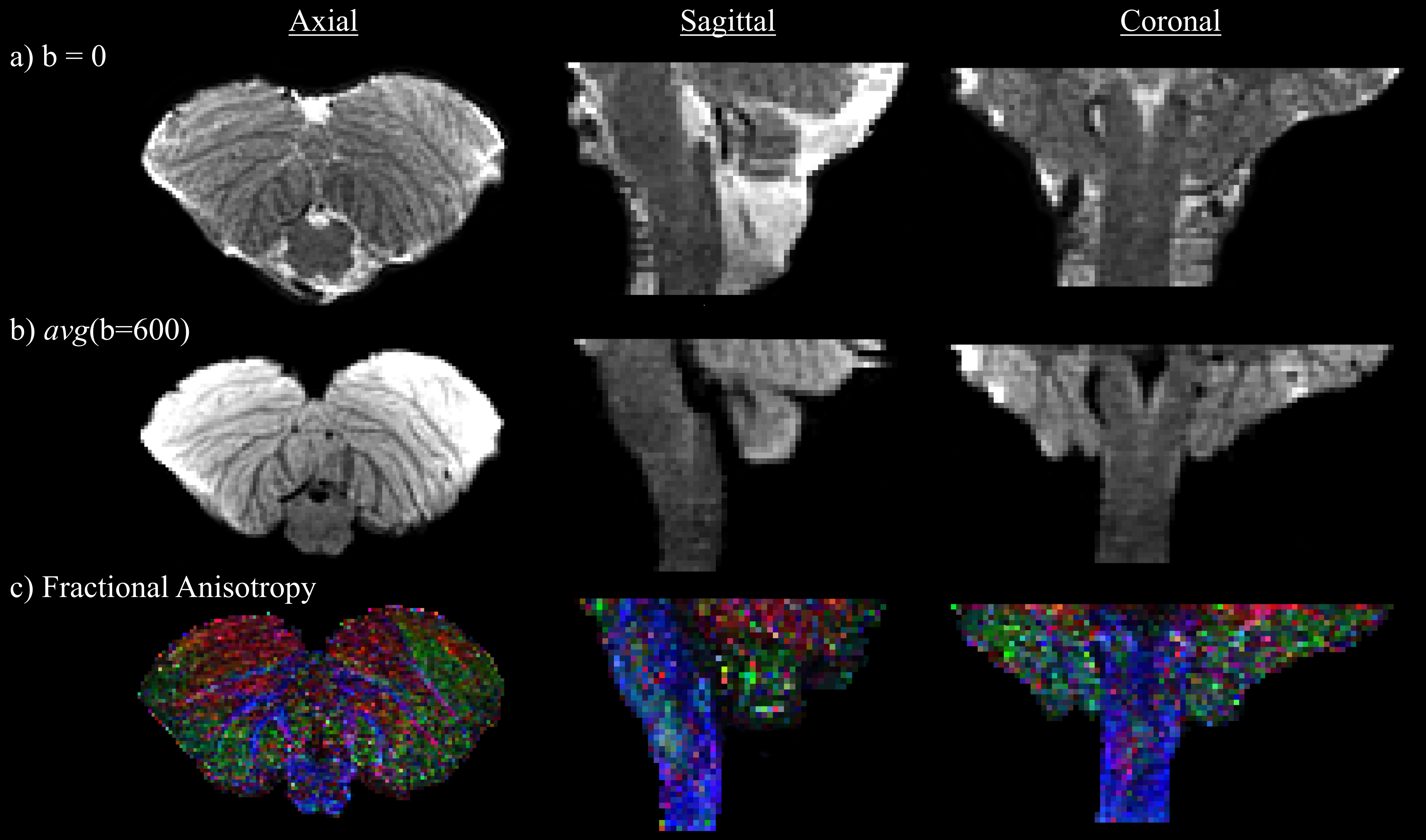
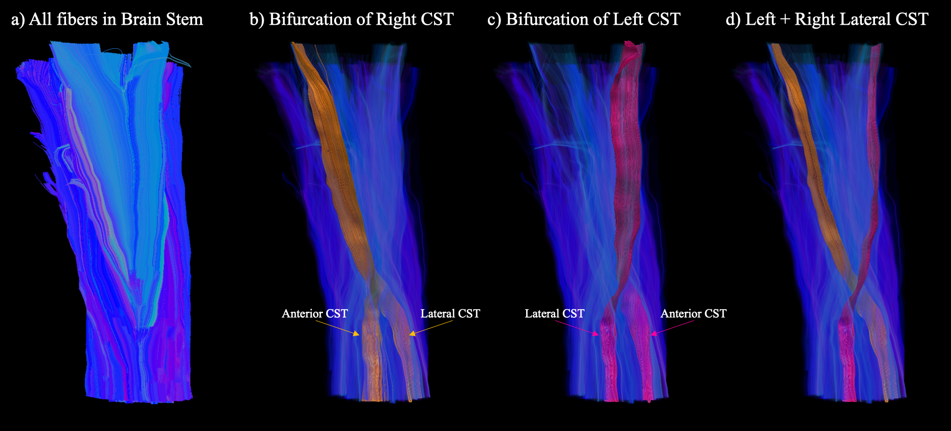
In Fig. 1, slice images with b=0, diffusion weighting (averaged over directions) and fractional anisotropy (FA) are presented with axial, sagittal and coronal views. Despite the small cross-sectional area of the SC shown in the axial slice images in Fig. 1, the ultra-high spatial resolution afforded in this study enabled a clear delineation of fiber tracts from the brain stem to the SC, as illustrated in Fig. 2a. From the fiber tracts in Fig. 2a, the bifurcation of the CST from the brain into the spinal cord can be readily characterized. Specifically, the right CST (orange) in Fig. 2b is shown to bifurcate in the inferior region of the medulla into the anterior and lateral CST, with the same characterization achieved for the left CST (pink) in Fig. 2c. Moreover, this delineation of the left and right lateral CSTs reveals the well-known pyramidal tract decussation (Fig. 2d), which to our knowledge is the first such demonstration using DTI at such a high spatial resolution in-vivo.Conclusion
The spinal cord has traditionally been a very challenging area to achieve accurate DTI, due to its small cross-sectional area as well as pronounced physiological motions due to CSF pulsations. As such, it has been difficult to characterize the complex microstructures and fiber pathways that comprise the SC. Despite the relatively straight nature of fiber tracts within the SC, it has not been possible to accurately delineate various well-known structures, such as the bifurcations and decussations of the CSTs from the brain to spinal cord. We demonstrate here that DTI data acquired at ultrahigh spatial resolution, with CSF pulsation minimized, can be used to characterize these intricate details in the human SC in-vivo. Such a fine-grained delineation of fiber tracts from the brain to the SC can find broad applications where a detailed knowledge of these structures is required. In light of the recent promise of SC stimulation to effectively treat many neurological diseases (e.g. movement disorders such as Parkinson’s Disease)4,5, such a delineation would facilitate in the accurate placement of stimulation electrodes to achieve the most efficient treatment possible.Acknowledgements
This work was funded in part by NIH grants R01 NS 075017, R24-106048, and S10-OD-021480.References
[1] Chen, N. K., Guidon, A., Chang, H. C., Song, A., W. A robust multi-shot scan strategy for high-resolution diffusion weighted MRI enabled by multiplexed sensitivity-encoding (MUSE). Neuroimage 72: 41-47, 2013.
[2] Tournier, J. D., Calamante, F., Connelly, A. MRtrix: Diffusion tractography in crossing fiber regions. Int. J. Imaging Syst. Technol., 22: 53-66, 2012.
[3] Basser, P. J., Pajevic, S., Pierpaoli, C., Duda, J., Albroubi, A. In vivo fiber tractography using DT-MRI data. Magn. Reson. Med. 44: 625-632, 2000.
[4] Yadav, A. P., Nicolelis, M. A. L. Electrical stimulation of the dorsal columns of the spinal cord for Parkinson’s disease. Mov. Disord. 32(6): 820-832, 2017.
[5] Fuentes, R., Petersson, P., Siesser, W. B., Caron, M. G., Nicolelis, M. A. Spinal cord stimulation restores locomotion in animal models of Parkinson’s disease. Science 323(5821):1578-82, 2009.
Figures