3508
The optimal b value study of endogenous water molecule diffusion imaging in the diagnosis of brain tumors1RenMin hospital of Wuhan University, Wuhan, China, 2Zhongnan Hospital of Wuhan University, Wuhan, China
Synopsis
In this study, thirty-six cases of meningioma were divided into benign and malignant meningioma. ADC values fitted by 11 b values and any pairs of two b values were used to distinguish between benign and malignant meningioma. We found that, the ADC values of benign, malignant, and all meningiomas showed a decreasing trend with the increase of b value, and the smaller the b value, the greater the decrease. when the b value was ≥ 200 s/mm2, any combinations of b values could discriminate between benign and malignant meningioma. The ADC values obtained by fitting b = 700 and b = 1000 s/mm2 had the highest AUC value.
Purpose
To evaluate the differential diagnostic value of ADC values fitted with different b-value combinations in benign and malignant meningiomas, and to investigate the optimal b value of DWI imaging in differential diagnosis of meningiomas.Methods
Thirty-six cases of meningioma were eventually included in the experiment. According to the pathological results, they were divided into two groups: benign and malignant meningioma. ADC values fitted by 11 b values (0, 50, 100, 150, 200, 300, 400, 500, 700, 900, 1000 s/mm2) and any pairs of two b values (56 pairs in total) were used to distinguish between benign and malignant meningioma. Independent sample t-test and Mann-Whitney U test were used to evaluate the differential diagnostic value of ADC values, and receiver operating characteristic (ROC) curves were used to determine the diagnostic performance, thresholds, sensitivity, and specificity of ADC values.Results
The ADC values of benign, malignant, and all meningiomas showed a decreasing trend with the increase of b value, and the smaller the b value, the greater the decrease (as shown in Table 1 and Fig.1). We exclued the sensitivity and specificity of combinations less than 60%, and found that, when the b value was ≥ 200 s/mm2, any combinations of b values could discriminate between benign and malignant meningioma (all P values < 0.05, as shown in Table 2 and Table 3). The ADC values obtained by fitting b = 700 and b = 1000 s/mm2 had the highest AUC value (0.785), and the threshold, sensitivity and specificity for distinguishing between benign and malignant meningiomas were 0.755×10⁻³ mm2/s, 72.0%, and 72.7%, respectively (as shown in Table 3 and Fig.2). The difference between b = 900 s/mm2 and any other b-values (including 200, 300, 400, 500, 700, and 1000 s/mm2) in benign and malignant meningiomas was statistically significant (P = 0.022, 0.010, 0.015, 0.004, 0.018, and 0.026, respectively, as shown in Table 3).Conclusions
ADC values of DWI obtained by fitting different combinations of b-values can effectively distinguish between benign and malignant meningiomas. The ADC values of benign, malignant, and all meningioma showed a decreasing trend with the increase of b value, and the smaller the b value, the greater the decrease. b = 700 and 1000 s/mm2 is considered to be the most significant b value.Acknowledgements
No acknowledgement found.References
No reference found.Figures


Table 1 The ADC value fitted with two different b and 11 b values between Benign and Malignant Meningioma
a was used with Mann–Whitney U test, P < 0.05 has a significant difference
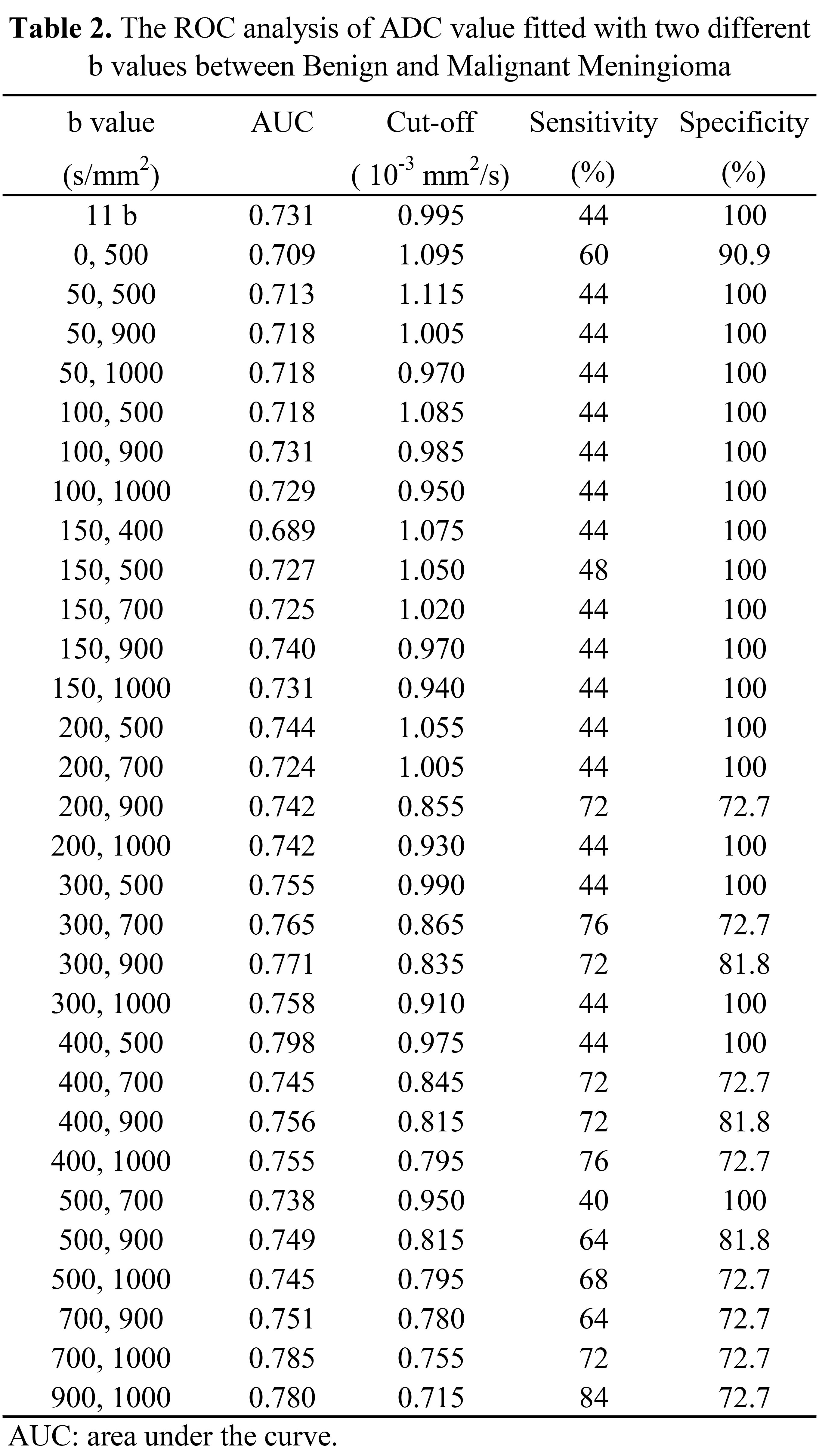
Table 2 The ROC analysis of ADC value fitted with two different b values between Benign and Malignant Meningioma
AUC: area under the curve

Table 3 The ROC analysis of ADC (Sensitivity > 60%) fitted with two different b values between benign and malignant meningioma
AUC: area under the curve