3506
In-vivo Diffusion Exchange Spectroscopy (DEXSY), to measure water exchange in tumours.1Centre for Advanced Biomedical Imaging, Division of Medicine, UCL, London, United Kingdom, 2Microstructure Imaging Group, Centre for Medical Image Computing, Department of Computer Science, UCL, London, United Kingdom, 3The Francis CRICK institute, London, United Kingdom
Synopsis
We describe the first use of Diffusion Exchange Spectroscopy (DEXSY) in-vivo, to measure water exchange in tumours. We present preliminary data suggesting that we can capture information about the micro-structure of tumors, with what appear to be intracellular, extracellular and perfusion peaks present in diffusion-diffusion exchange plots, acquired from subcutaneous tumor xenograft models, in-vivo using DEXSY.
Introduction
Experimental data were acquired in order to evaluate the DEXSY double diffusion encoding technique [1,2] in a subcutaneous tumour xenograft model in-vivo. Cell membrane permeability varies in both healthy and diseased tissues, in particular in tumors and the brain [3]. There has been recent interest in using diffusion MRI and Filter Exchange Imaging (FEXI) to image gene expression in cases where water exchange in tissues is altered due to modified aquaporin expression and UT-B reporter gene expression [4,5]. DEXSY could be more effective for studying these processes as it does not rely on a physiological model. DEXSY MRI has previously been used to measure diffusion exchange in-vitro in yeast [6,7,8,9,10]. Our previous simulations demonstrated that we can measure the movement of water molecules across the cell membrane using DEXSY in nervous tissue, over a wide range of permeabilities, and that measured diffusion exchange across the cell membrane increases with permeability [11,12]. We also established the Diffusion Exchange Index (DEI) as a quantitative indicator of cell membrane permeability. Here, for the first time, we extend this work to subcutaneous tumor xenograft models and in-vivo experiments.Methods
All in-vivo experiments were performed in accordance with the UK Home Office Animals Scientific Procedures Act, 1986 and United Kingdom Coordinating Committee on Cancer Research (UKCCCR) guidelines. 5 CD-1 mice were inoculated with 3 million SW1222 cells in their left flank, in order to create a subcutaneous xenograft tumour model. Data were acquired using a 20 cm horizontal bore 9.4 T Varian scanner with a 39 mm Rapid RF coil, with a slice selective DEXSY sequence. Slices were positioned coronally through the tumour. The DEXSY scan parameters used were δ = 15 ms, ∆ = 17 ms, tm = 200 ms, G1 and G2 = 0 − 640 mT /m in 16x16 steps. The mice were scanned under anaesthetic with a mixture consisting of 1-2.5 % Isoflurane in 1 L/min of oxygen. The four scans presented here are acquired from slices that included the whole tumour and little in the way of surrounding tissue. 2D Inverse Laplace transform software [13], was used to reconstruct DEXSY data in order to produce diffusion-diffusion exchange plots.Results
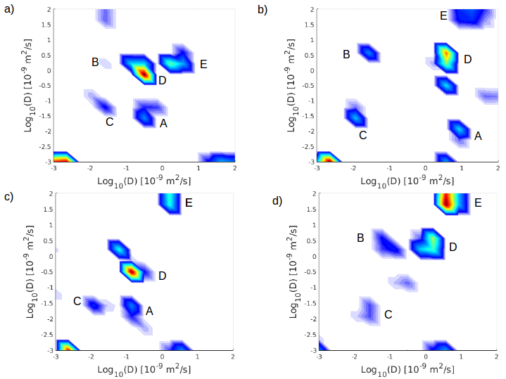
Figure 1: a) and b) show diffusion-diffusion exchange plots from the same subcutaneous tumor, with b) scanned 10 days after a). The tumor volume in b) was 1.6 times the volume in a), which could in part explain differences between exchange plots. Figure 1:c) and d) show diffusion-diffusion exchange plots from tumors in two different mice. In diffusion-diffusion exchange plots, diagonal peaks represent diffusion within discrete compartments (here, intracellular and extracellular) and cross peaks represent diffusion exchange between compartments. Peaks with very high diffusivity potentially correspond to vascular perfusion. In Figure 1: a), b), c) and d) we can see potential diffusion exchange peaks are labeled A and B, whilst potential intracellular, extracellular, and perfusion peaks are labeled C, D and E, respectively (there are no signs of vascular exchange). There is a great deal of variation in scans a), b), c) and d), which could reflect the variation in the size and shape of the tumors.Discussion and conclusion
Further work is required to replicate these findings and establish the cause of variability within the data. This first application of DEXSY in-vivo suggests, that DEXSY is highly sensitive to tissue microstructure and capable of detecting diffusion exchange, in subcutaneous xenograft tumour models.Acknowledgements
We are grateful to Petrik Galvosas (Victoria University of Wellington) for providing the inverse Laplace software.References
[1]P. T. Callaghan, I Furo, Journel of Chemical Physics, 120(8):4032-4038, (2004)
[2]Callaghan et al. Magnetic Resonance Imaging, 21(3-4):243–248,(2003)
[3]S. Lasic et al, Magnetic Resonance in Medicine, (2011).
[4]A. Mukherjee et al, Nature Communications 7, 13891 (2016)
[5]F. Schilling et al. Nature Biotechnology 35, 75–80 (2017)
[6]B. Siow et al., Proc. ICMRM 12, Cambridge, UK (2013), P43
[7]B. Siow at al., Proc. MRPM 12, Wellington, NZ, (2014), P06
[8]D. Benjamini et al., Phys. Rev. Lett. 118, 158003 (2017)
[9]J.O. Breen-Norris et al., Proc ISMRM 18, Paris, France, (2018), 1104
[10]J.O Breen-Norris et al. , Proc MRPM, Gainsville, Florida, USA, P26
[11]J.O. Breen-Norris et al., Proc ISMRM 17, Honolulu, USA, (2017), 1740
[12]J.O. Breen-Norris et al., Proc ICMRM 17, Halifax, CAN, (2017), P64
[13]Y.-Q.-Song et al, Journal of Magnetic Resonance, (2002)
Figures