3504
Diffusion MRI of the Brain with a Gradient Strength of 100 mT/m and a Slew Rate of 1200 T/m/s1Institute for Biomedical Engineering, University of Zurich and ETH Zurich, Zurich, Switzerland
Synopsis
The performance of diffusion-weighted single-shot EPI is limited by off-resonance artifacts and low signal-to-noise. To address both problems, we employ a recently developed gradient insert (strength: 100 mT/m, slew rate: 1200 T/m/s). Thereby the time for the diffusion encoding as well as the EPI readout train duration can be significantly shortened, resulting in higher signal yield and robustness against off-resonance artifacts. First in-vivo results are presented.
Introduction
Diffusion weighted (DW) MRI is most commonly performed with
single-shot Echo-Planar Imaging (EPI) due to is robustness against motion [1,2].
However, single-shot EPI is prone to
image distortions due to magnetic susceptibility differences, e.g. between
tissue and air, an effect increasing with resolution and magnetic field
strength. Moreover, the long echo time required for the long EPI readout reduces
the achievable signal-to-noise ratio (SNR) of diffusion images.
To address both problems, we employ a gradient insert to
shorten the duration of both diffusion-weighting and the EPI readout. An evaluation
of the imaging performance and first in-vivo results are presented.
Methods
MR scanning was performed on at 3T Achieva (Philips Healthcare, Best, The Netherlands) using an 8 channel transmit-receive coil array [3]. A custom-built head gradient insert was employed with 100 mT/m maximum gradient and 1200 T/m/s slew rate [4]. A diffusion-weighted spin-echo sequence with typical imaging parameters (FOV: 22cm, in-plane resolution: 1.9mm, slice thickness: 3mm, SENSE: R=1.5, DW: b=0, and b=1000s/mm2 in slice, read and phase direction) was performed on a healthy subject. The echo time using the gradient insert was 42 ms; the readout duration was 19 ms, corresponding to a bandwidth per pixel of 52.2 Hz in the phase encoding direction. In comparison, the same parameters using a regular gradient system (30 mT/m, 200 T/m/s) would yield an echo time of 68 ms with a readout duration of 49 ms and 20 Hz/pix bandwidth. From these values the expected relative SNR performance was calculated.
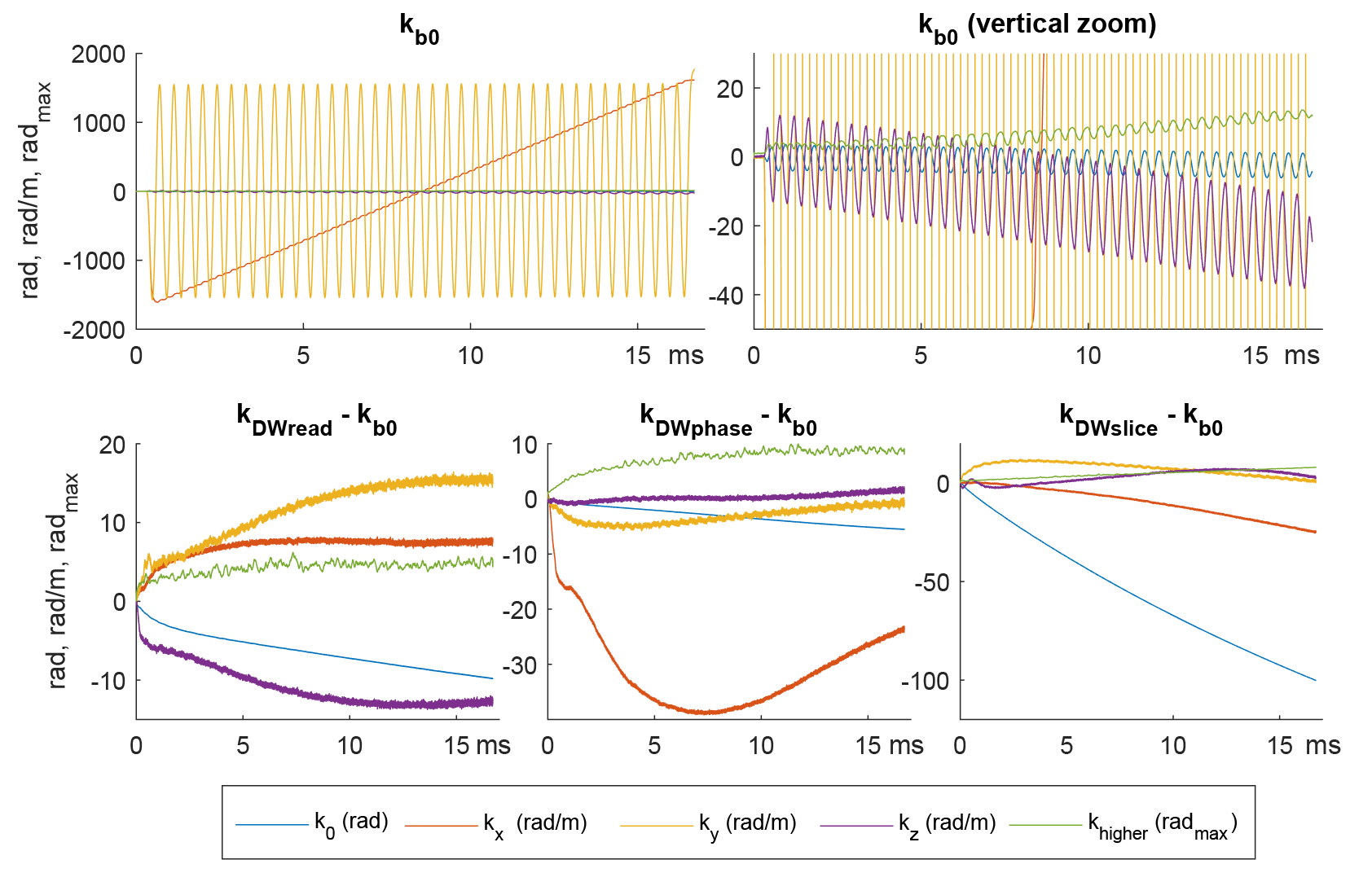
Operating gradient systems at such strength and slew rates imply increased eddy currents and concomitant fields, which have to be accounted for. To address this problem, the field evolution of all readouts was recorded using a Dynamic Field Camera (Skope MRT, Zurich, Switzerland). To capture eddy current effects of local spatial extent, the sequences were played out 11 times, each time repositioning the field camera within the imaging volume. From this data, a 10th order spherical harmonic model was calculated. Concomitant fields were calculated from the measured 1st order gradient fields and known relationship to the transverse fields. Images were reconstructed using all encoding information along with the object data in an iterative higher-order SENSE reconstruction [5]. A set of images also including map-based static-B0 correction was reconstructed for in addition.
Results
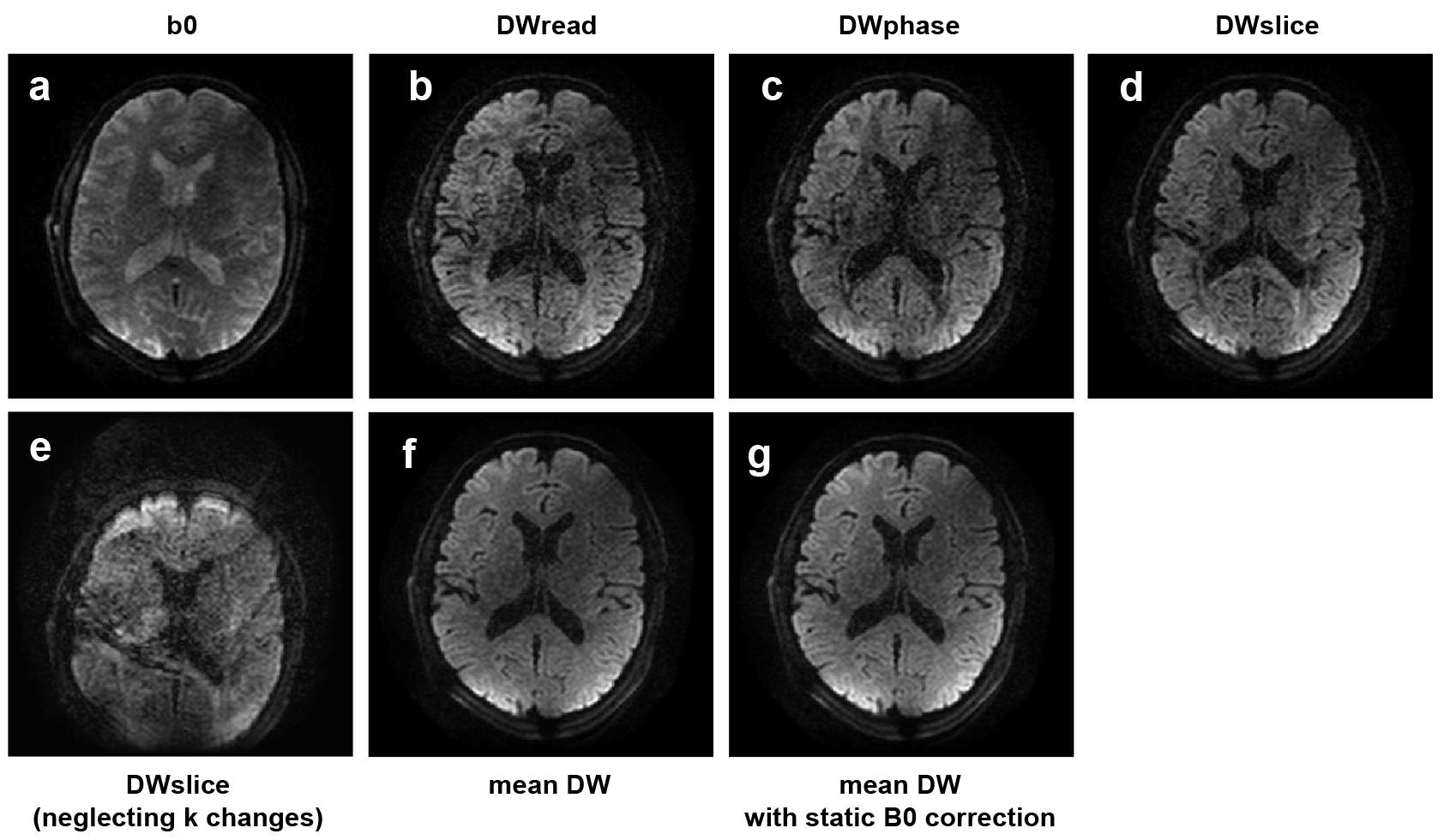
Assuming a T2 of 70 ms (white matter), shortening the echo time from 68 ms to 42 ms results in an expected signal gain of 45%. On the other hand, the shortened readout duration leads to a reduction in acquisition duty cycle from 46% to 30%. Hence, a net gain in SNR of 16% is expected. The recorded field evolutions showed strong undesired 0th order, 1st order as well as higher order terms (Fig.1). The encoding varied also when applying different diffusion weighting (Fig. 1, lower row). Using the recorded field evolutions allowed for a faithful image reconstruction (Fig.2a-d), whereas neglecting only the encoding changes due to the applied DW (Fig,2e) resulted in distorted and shifted images. The images (Fig. 2a-d) do not show apparent blurring artifacts in the phase encoding direction, nor distortions that are commonly visible in the region of the frontal lobe. The mean diffusion weighted image (Fig.2f) did not show any loss of resolution, indicated that geometrical congruence was achieved among the images despite excessive eddy currents present during the acquisition. Applying B0-correction (Fig.2g) in the reconstruction has virtually no effect on the image, probably owing to the high pixel-bandwidth.Discussion and Conclusion
This is the first report of in-vivo DWI using such extreme gradient specifications. A key element of the gradient coil design lies in the short gradient extent which allows to effectively limit peripheral nerve stimulation (PNS). Other gradient systems [6,7] that also achieve high gradient strength, cannot fully utilize it for fast readouts due to slew rate and PNS limitations, whereas a 2.5-fold increase in acquisition bandwidth-per-pixel was achieved with the current setup. The in-vivo DWI showed no visible EPI image distortions (even without static B0 correction) and were geometrically congruent, probably owing to the increased acquisition bandwidth as well as to the accurate characterization of the encoding fields. As compared to readout shortening by parallel imaging, no g-factor penalty has to be paid. The available gradient amplitude also significantly reduced the achievable echo time by 26 ms. For DWI of white matter an overall SNR gain of 16 % is expected despite the shortened acquisition duty cycle. Echo time shortening in this order of magnitude may be crucial to significantly improve DWI of structures with shorter T2, such as myelin water [8,9], where much higher SNR benefit can be expected.Acknowledgements
No acknowledgement found.References
- R. Turner et al. Magn. Reson. Med., 19 (1991), pp. 247-253
- Bammer et al., Magn Reson Med 2001; 46: 548–554.
- Roesler et al, 2019, submitted to ISMRM
- Weiger et al., Magn Reson Med. 2018 Jun;79(6):3256-3266.
- Wilm et al., Magn Reson Med. 2011 Jun;65(6):1690-701
- Lee et al., Magn Reson Med, 76 (2016) 1939-1950.
- Setsompop, NeuroImage, 80 (2013) 220-233.
- Ashish et al., PLOS One, 2014 Jun; https://doi.org/10.1371/journal.pone.0098391
- Avram et al., Neuroimage, Volume 53, Issue 1, 15 October 2010, Pages 132-138
Figures