3501
Motion-Robust and Distortion-Corrected Diffusion MR Imaging of the Liver with Optimized Motion-Compensated Waveforms and Multi-shot EPI acquisition1Medical Physics, University of Wisconsin Madison, madison, WI, United States, 2Radiology, University of Wisconsin Madison, madison, WI, United States, 3Applications and Workflow, GE Healthcare, Boston, MA, United States
Synopsis
Conventional liver diffusion MRI acquisitions suffer from several challenges including low spatial resolution, B0-induced distortions, and elastic motion-induced signal voids. In this work, motion-robust and distortion-corrected liver diffusion-weighted imaging (DWI) was enabled by combining optimized motion compensated diffusion waveforms with multi-shot EPI acquisitions. Diffusion-weighted images of healthy volunteers were acquired to evaluate the effect of motion compensation and distortion correction. Preliminary results demonstrate the feasibility of the proposed approach, including reduced ADC bias in the left lobe (due to the motion-robust waveforms) and reduced distortion (due to the multi-shot acquisition) compared to conventional liver DWI.
Introduction
Diffusion-weighted (DW)-MRI has wide-ranging applications, including the detection, staging, and treatment monitoring of cancer. However, conventional diffusion MRI acquisitions suffer from several challenges, including low spatial resolution, B0-induced distortions1, and elastic motion-induced signal voids2-3. In recent years, advanced techniques have been proposed to address these challenges. For example, multiplexed sensitivity encoding (MUSE)4 has been proposed for reduced-distortion, high resolution multi-shot EPI (msEPI) acquisition and reconstruction with applications in brain4 and prostate5 DW-MRI. Further, advanced diffusion gradient waveform design techniques6-7 enable optimized motion-robust DWI to avoid motion-induced signal voids, particularly in the left lobe of the liver. However, there is an unmet need for motion-robust, reduced-distortion DW-MRI of the abdomen. This work developed motion-robust and reduced-distortion DW-MRI of the abdomen by combining motion-robust DW gradient waveforms and MUSE-based msEPI.Methods
After IRB approval and informed written consent, four healthy volunteers were scanned on a 3T scanner (GE Signa Premier) with high channel density posterior and anterior receive array coils (AIR coil, GE Healthcare, Waukesha, WI).
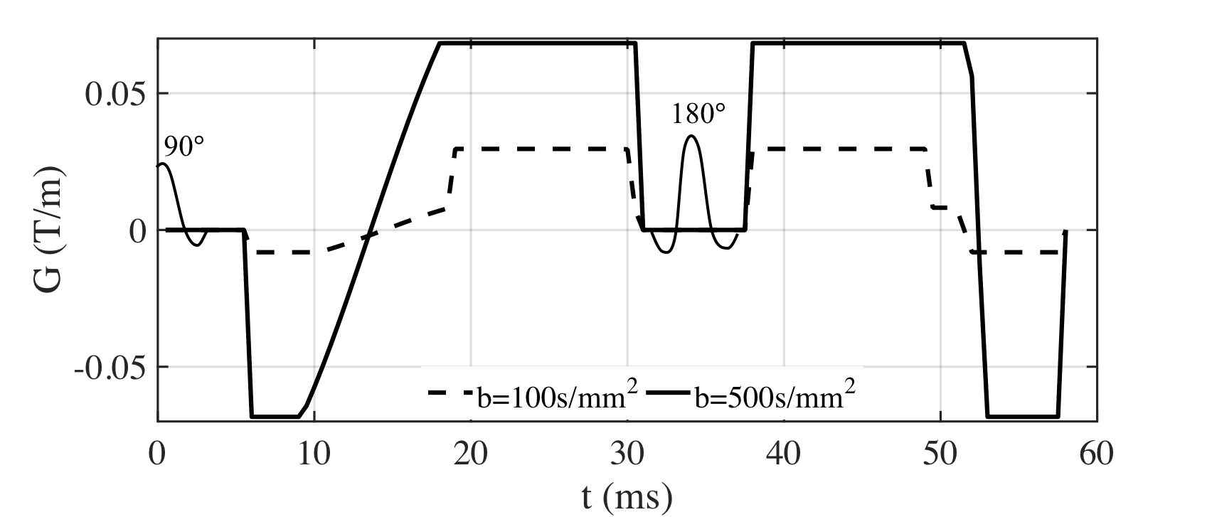
Diffusion waveform design: In this work, a M1-Optimized Diffusion Imaging (MODI) design was used to achieve motion-robust diffusion waveforms. We used an extension of an echo-time optimized motion-robust diffusion weighting gradient waveform design7, with a moderate non-zero first-moment (M1≠0) value to enable blood signal suppression. The waveforms designed in this study are shown in Figure 1.
Image acquisition: High-resolution T2-weighted (T2w) images were acquired as a reference of anatomic structure. DW images of conventional DWI and MODI were obtained with ssEPI and msEPI readout, respectively. DWI parameters included: FOV = 36 cm$$$\times$$$36 cm, in-plane resolution = 2.8 mm$$$\times$$$2.8 mm, slice thickness = 6 mm, acquisition bandwidth = $$$\pm$$$62.5 kHz, acceleration factor = 2 with partial Fourier acquisition and three orthogonal diffusion directions; b-values (#averages) = [100(4), 500(8)]s/mm2 for ssEPI and b-values (#averages) = [100(2), 500(4)]s/mm2 for msEPI to achieve similar acquisition time. Respiratory triggering was used with effective TR ranging from 5 to 8 seconds. TEs for conventional DWI with ssEPI and msEPI were 47 ms and 45.9 ms, respectively; while TEs for MODI with ssEPI and msEPI were 68.5 ms and 67.5 ms, respectively.
Image reconstruction and analysis: A phased-corrected multi-shot reconstruction technique4 was performed on msEPI data for both conventional DWI and MODI. ADC maps were calculated for all DWI series.
Results
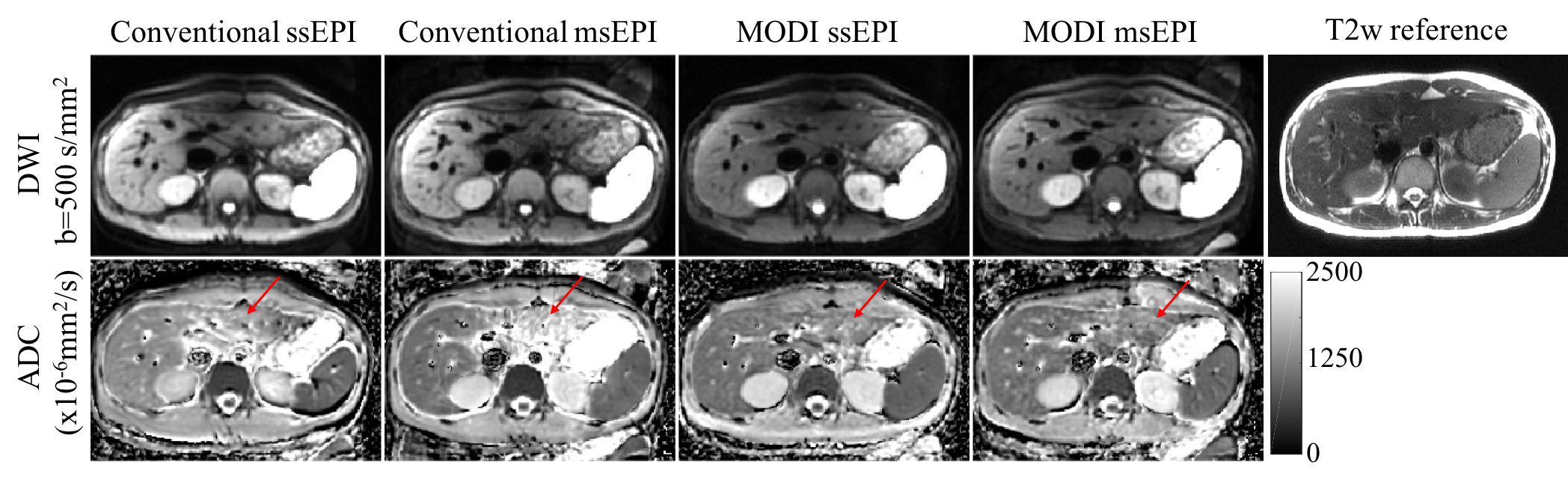
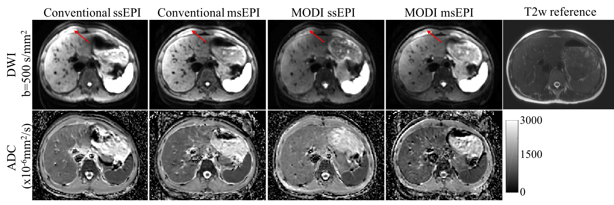
Figure 2 shows an example of motion compensation in a healthy volunteer using MODI ssEPI and MODI msEPI. Both MODI ssEPI and msEPI are able to compensate for the signal voids in DWI and reduce the quantitative ADC bias in the left lobe of liver, in comparison to conventional DWI with monopolar diffusion gradients. Figure 3 provides an example of a slice with severe geometric distortion artifacts due to susceptibility variations, where msEPI is able to provide DW images and ADC maps with substantially reduced distortion artifacts. Some residual errors were observed in msEPI reconstructions (using both conventional and MODI gradients) near the regions of of most intense motion near the heart.Discussion
In this study, we have investigated the feasibility of implementing motion-robust and distortion-corrected diffusion MRI by combining optimized motion compensated diffusion waveforms and msEPI. Preliminary results show promising image quality with reduced ADC bias in the left lobe and less overall distortion. Importantly, this work leveraged state-of-the-art hardware including high-performance gradient systems and high channel density of coil arrays, to enable advanced diffusion MRI features (M1-optimized diffusion gradients and multi-shot EPI) while maintaining high SNR.
Upon further validation, this technique may enable improved diffusion MRI of the liver, eg: for the assessment of metastases, where high spatial resolution and high image quality over the whole liver constitute a significant unmet need. This work has several limitations, including remaining challenges in msEPI reconstruction in the presence of intense motion of the liver, which leads to large phase variation between shots.
Conclusion
Motion-robust and distortion-corrected diffusion-weighted imaging can be enabled by combining optimized motion compensated diffusion waveforms with multi-shot EPI acquisitions.Acknowledgements
The authors would like to acknowledge research support from GE HealthcareReferences
[1] Naganawa, S., Kawai, H., Fukatsu, H., Sakurai, Y., AOKI, I., MIURA, S., MIMURA, T., KANAZAWA, H. and ISHIGAKI, T., 2005. Diffusion-weighted imaging of the liver: technical challenges and prospects for the future. Magnetic Resonance in Medical Sciences, 4(4), pp.175-186.
[2] Murphy, P., Wolfson, T., Gamst, A., Sirlin, C. and Bydder, M., 2013. Error model for reduction of cardiac and respiratory motion effects in quantitative liver DW‐MRI. Magnetic resonance in medicine, 70(5), pp.1460-1469.
[3] Kwee, T.C., Takahara, T., Niwa, T., Ivancevic, M.K., Herigault, G., Van Cauteren, M. and Luijten, P.R., 2009. Influence of cardiac motion on diffusion-weighted magnetic resonance imaging of the liver. Magnetic Resonance Materials in Physics, Biology and Medicine, 22(5), pp.319-325.
[4] Chen, N.K., Guidon, A., Chang, H.C. and Song, A.W., 2013. A robust multi-shot scan strategy for high-resolution diffusion weighted MRI enabled by multiplexed sensitivity-encoding (MUSE). Neuroimage, 72, pp.41-47.
[5] Zhang, Y., Holmes, J., Rabanillo, I., Guidon, A., Wells, S. and Hernando, D., 2018. Quantitative diffusion MRI using reduced field-of-view and multi-shot acquisition techniques: Validation in phantoms and prostate imaging. Magnetic resonance imaging, 51, pp.173-181.
[6] Aliotta, E., Wu, H.H. and Ennis, D.B., 2017. Convex optimized diffusion encoding (CODE) gradient waveforms for minimum echo time and bulk motion–compensated diffusion‐weighted MRI. Magnetic resonance in medicine, 77(2), pp.717-729.
[7] Peña-Nogales, Ó., Zhang, Y., Wang, X., Luis-Garcia, R., Aja-Fernández, S., H Holmes, J. and Hernando, D., 2018. Optimized Diffusion-Weighting Gradient Waveform Design (ODGD) formulation for motion compensation and concomitant gradient nulling. Magnetic resonance in medicine, early view.
Figures