3499
A novel approach to diffusion phase imaging using non-linear gradients in anisotropic media1School of Biomedical Engineering & Imaging Sciences, King's College London, London, United Kingdom, 2Department of Radiology, Athinoula A. Martinos Center for Biomedical Imaging, Boston, MA, United States, 3Philips Healthcare UK, Guildford, Surrey, United Kingdom
Synopsis
Classical methods for Diffusion-Weighted MRI use linear gradients to obtain information about tissue microstructure encoded in the loss of MR magnitude. These linear gradients cause diffusing spins to dephase which prevents the use of phase information. When, however, gradient fields are applied that vary quadratically in space, diffusion in anisotropic media results in a shift of net phase with only a minimal loss in signal magnitude enabling the exploration of MR phase. Both the theory of this new concept and Monte-Carlo simulations predict net phase for diffusion experiments using quadratic gradients in anisotropic media in excellent agreement.
Introduction
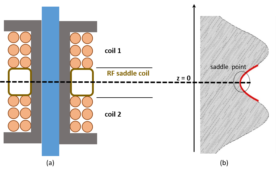
Diffusion-Weighted (DW) MRI exploits the thermally induced diffusion of water molecules to generate image contrast. In classical approaches, linear gradient pulses are applied that sensitize the MR signal to the motion of water molecules. Since diffusion is usually symmetric in space (i.e. an equal number of spins "moving to the left and to the right"), a spin ensemble moving randomly in this magnetic gradient field does not experience a net phase shift. The spins rather de-phase, resulting in a loss of signal magnitude. This means the phase of a classical diffusion experiments does not contain information about the probed tissue. In this novel approach, the linear diffusion gradient is replaced by a second order gradient field (e.g. Z2 with $$$B_z(x,y,z) = z^2 – \frac{1}{2}(x^2 +y^2)$$$). Such a gradient yields a net phase shift for diffusion in anisotropic media. For isotropic media, no net phase shift is expected due to $$$\Delta \vec{B} = 0$$$. In close proximity to the saddle point (x=y=z=0), the gradient field does not have linear components dephasing the signal. In contrast to classical DW MRI, in the presence of a quadratic gradient diffusion is encoded in the signal phase while preserving large portions of the signal magnitude at the center of the saddle point (see Figure 1(b)). This offers new possibilities to explore domains inaccessible to existing DW MRI methods, especially in complex tissue environments.1Material & Methods
A theoretical concept was developed that allows predictions about the effect of a Z2 gradient field on the phase of a spin ensemble diffusing in three dimensions. The analytic expression
$$\varphi = -2 \gamma \beta T (T + \Delta t)(D_z - \frac{1}{2}D_x - \frac{1}{2}D_y)$$
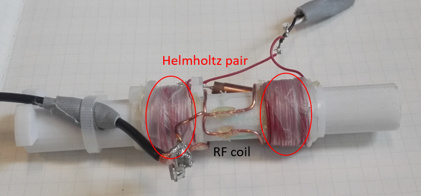
predicts the net phase φ for a spin-echo sequence. Two identical gradient pulses of duration T are applied, one before and one after the refocusing pulse. Δt is the time between the pulses, β the curvature of the quadratic gradient and γ the gyromagnetic ratio of water. Since diffusion is generally anisotropic, a unique diffusion coefficient is assigned to each direction (Dx, Dy and Dz). Thus, in case of isotropic diffusion there in no net phase shift due to Maxwell’s law stating $$$\Delta \vec{B} = 0$$$. Monte-Carlo simulations were used to examine the correctness of the analytic calculus. An experimental set-up was developed for initial imaging on a 1.5T MR scanner. Core of the set-up is a coil insert shown in Figure 1(a) (schematic) and Figure 2 (picture). A Helmholtz pair serves as gradient coil to generate the quadratic gradient. A small-scale RF saddle coil was positioned in the separation between the individual coils and was used for transmit/receive. Away from the saddle point, the gradient field shows increasingly linear behavior. In these regions, diffusion effects are expected to destroy signal magnitude. To verify this, initial experiments on isotropic water (with 1% salt and 0.1% gadolinium, $$$D = 2.3\times10^{-9} m^2/s$$$) and sunflower oil ($$$D = 1.1\times10^{-12} m^2/s$$$) phantoms were performed.2Results
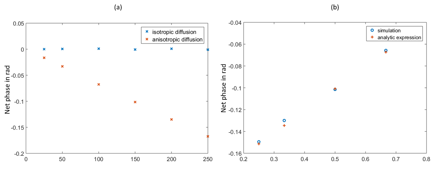
The equation predicts no net phase for isotropic diffusion ($$$D_x = D_y = D_z$$$). However, net phase is expected for anisotropic diffusion. The same findings were obtained in the simulations, see Figure 3(a). The plots for theoretical and simulated net phase are shown in Figure 3(b) for different degrees of anisotropy (varying ratio $$$D_x/D_z$$$) for symmetric diffusion in x- and y-direction ($$$D_x = D_y$$$). Comparison of theory and simulation results show excellent agreement.
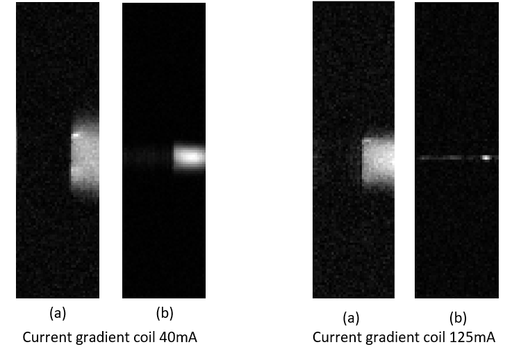
Figure 4 shows the magnitude images obtained in the initial experiments for water and oil phantoms. The images clearly show that in presence of the quadratic gradient, signal away from the saddle point is dephased. The gradient has a stronger effect on water (higher diffusivity) than on oil. These effects are more pronounced for stronger gradient fields. These observations confirm that diffusion effects due to increasingly strong linear
components away from the saddle point dephase the MR signal. They also confirm that the MR signal in around the saddle point is preserved.
Discussion & Conclusion
Classical diffusion MRI is a powerful imaging
modality that provides unique insight into tissue microstructure. More recently
developed methods employ more advanced models3 or double oscillating
gradient pulses4 to extract more complex information.
However, all these methods encode diffusion in the magnitude of the MR-signal
and, hence, suffer from inherently poor SNR which restricts sensitivity to
tissue microstructure. The presented novel approach overcomes these limitations
by employing a second order gradient field. The diffusion information
is encoded in the signal phase while preserving large portions of the signal
magnitude at the center of the field.
Both theory and simulation predict net phase for diffusion in anisotropic
material which can provide valuable insight into tissue architecture inaccessible
to other DW MRI methods.Acknowledgements
This work is funded by the King’s College London & Imperial College London EPSRC Centre for Doctoral Training in Medical Imaging (EP/L015226/1) and Philips Healthcare.References
1. Hall MG, Barrick TR. From diffusion‐weighted MRI to anomalous diffusion imaging. Magnetic Resonance in Medicine. 2008 Mar 1;59(3):447-55.
2. Perez EE, Carelli AA, Crapiste GH. Temperature-dependent diffusion coefficient of oil from different sunflower seeds during extraction with hexane. Journal of Food Engineering. 2011 Jul 1;105(1):180-5.
3. Panagiotaki E, Walker-Samuel S, Siow B, Johnson SP, Rajkumar V, Pedley RB, Lythgoe MF, Alexander DC. Noninvasive quantification of solid tumor microstructure using VERDICT MRI. Cancer research. 2014 Feb 3.
4. Ianuş A, Shemesh N, Alexander DC, Drobnjak I. Double oscillating diffusion encoding and sensitivity to microscopic anisotropy. Magnetic resonance in medicine. 2017 Aug;78(2):550-64.
Figures