3488
Reduced Eddy Current induced image distortions and Peripheral Nerve Stimulation based on the Optimal Diffusion-weighting Gradient Waveform Design (ODGD) formulation1Universidad de Valladolid, Valladolid, Spain, 2Medical Physics, University of Wisconsin-Madison, Madison, WI, United States, 3Radiology, University of Wisconsin-Madison, Madison, WI, United States, 4University of Wisconsin-Madison, Madison, WI, United States
Synopsis
Diffusion-Weighted MRI (DW-MRI) often suffers from signal attenuation due to long TE, motion-related artefacts, dephasing due to concomitant gradients (CGs), and image distortions due to eddy currents (ECs). Further, the application of rapidly switching gradients may cause peripheral nerve stimulation (PNS). These challenges hinder the progress, application and interpretability of DW-MRI. Therefore, based on the Optimized Diffusion-weighting Gradient waveforms Design (ODGD) formulation, in this work we design optimal (minimum TE) nth-order moment-nulling diffusion-weighting gradient waveforms with or without CG-nulling able to reduce EC induced distortions and PNS-effects. We assessed the feasibility of these waveforms in simulations and phantom experiments.
Introduction
Diffusion-Weighted MRI (DW-MRI) has a unique ability to probe tissue microstructure through the application of strong switching diffusion-weighting gradients. However, the application of these gradients results in significant challenges including 1) signal attenuation[1], 2) motion-related artifacts[2,3], 3) signal dephasing due to concomitant gradients (CGs)[4,5], 4) image distortions (shearing and scaling) due to eddy currents (ECs)[6,7,8], and 5) might cause peripheral nerve stimulation (PNS)[9,10].
Particularly, ECs and PNS appear when the gradient amplifiers are switched fast to drive large gradient intensities. Firstly, switching gradients result in current inductions that persist after the gradients are switched off[6,7]. Although, ECs are usually tackled through pre-emphasis and post-processing, there are also suboptimal (long TE) diffusion-weighting waveforms (e.g., BIPOLAR[11]) that achieve intravoxel rephasing and EC-nulling. Secondly, they cause an electric depolarization that may lead to PNS[9]. PNS is usually diminished by limiting the maximum slew rate of the waveform resulting in sub-optimal waveforms.
For these reasons, there is an unmet need for optimized diffusion-weighting waveforms able to overcome these challenges. Recently, a nonlinear constrained optimization formulation for the design of high-order moment-nulled and/or CG-nulled waveforms, termed Optimized Diffusion-weighting Gradient waveform Design (ODGD)[12], has shown great potential in-vivo DW-MRI. Therefore, the purpose of this work is to design optimized waveforms (minimum TE) based on the ODGD formulation that achieve EC- and PNS-nulling, while preserving previous properties.
Methods
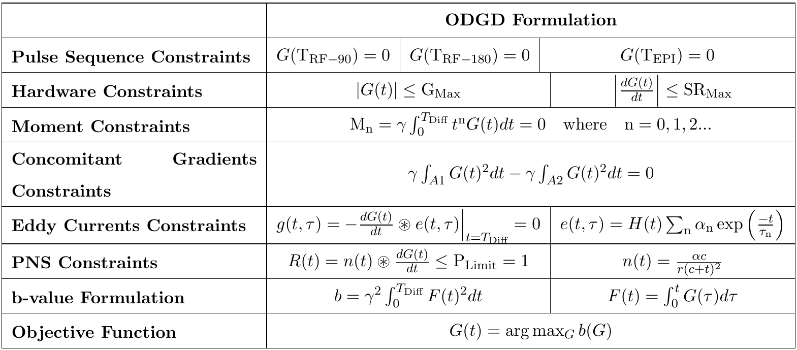
The ECs impulse response is given by $$$e(t,\tau)=H(t)\sum_\text{n}\alpha_\text{n}\exp{\left(-\frac{t}{\tau_\text{n}}\right)}$$$, where $$$\tau_\text{n}$$$,$$$\alpha_\text{n}$$$, n and H(t) are a characteristic time constant, amplitude, number of induced currents, and the step function, respectively. Thus, we can reduce EC induced distortions (EC-nulling) for a set of time constants ($$$\tau$$$) by adding the linear constraints $$$g(t,\tau)=-\frac{dG(t)}{dt}\circledast~e(t,\tau)\Bigr\rvert_{t=\text{T}_\text{Diff}}=0$$$ to the formulation[6,8].
A conservative nerve impulse response is given by[10,13] $$$n(t)=\frac{αc}{r(c+t)^2}$$$, where the gradient-coil specific constants $$$\alpha$$$, r, and c are the effective coil length, rheobase, and chronaxie time, respectively. Thus, for waveforms designs with the maximum slew rate supported by the MRI unit we can reduce the nerve stimulation (PNS-nulling) by adding the linear constraints $$$R(t)=\frac{dG(t)}{dt}\circledast~n(t)<Plimit=1$$$, where the PNS limit ($$$\text{P}_\text{Limit}$$$) is established by the International Electrotechnical Commission (IEC 60601-2-33). For the sake of clarity, Table 1 summarizes all constraints of the extended ODGD formulation.
In this work, MONO[12], BIPOLAR, MOCO[12] and nth-order moment-nulled ODGD waveforms without and with EC- and PNS-nulling were designed for b-values=100-2000s/mm², and TEPI (EPI readout time to the center of the k-space) of 16.4-46.4ms.
A 7.5L cylindrical water phantom doped with NaCl and NiCl2[14] was scanned at a 3T unit (GE Healthcare, Waukesha, WI) using a spin echo DW-EPI sequence to assess EC induced image distortions for several diffusion-weighting waveforms without and with EC-nulling, and to assess the implementation feasibility and SNR of PNS-nulling waveforms. Acquisition parameters were TR=4s, slice thickness=5mm, FOV=24x24cm, in-plane resolution=1.88x1.88mm, full k-space, six diffusion-weighting directions (±A/P,±R/L,±S/I), and b-values(averages)=[0(1),100(2),1000(4)]s/mm2. SNR maps were computed following Ref[15].
Results
Figure 1 shows examples of various waveforms designed for the experiments. Figure 2 shows the TE dependence on the b-value of MONO, BIPOLAR, MOCO and various ODGD waveforms, and the TE reduction achieved using ODGD-M1-CG compared to ODGD-M1-CG-EC, and ODGD-M1-EC without and with CG-nulling compared to BIPOLAR. ODGD-M1-CG results in TE reductions between 3.8-14.7% compared to ODGD-M1-CG-EC. Relative to BIPOLAR, ODGD-M1-EC and ODGD-M1-CG-EC result in TE reductions between 1.9-27.9%, and -7-11.22%, respectively.
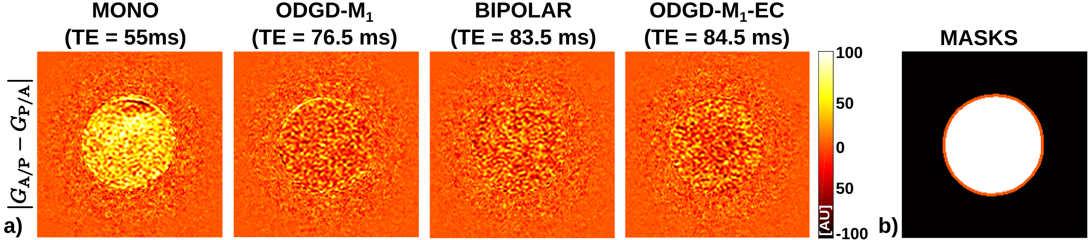
Figure 3 and 4 show the difference between DW images of opposite diffusion-weighting polarity, and their distribution, respectively. MONO is visually severely affected by EC induced distortions showing strong mismatch between opposite DW images. BIPOLAR, and ODGD-M1-EC are slightly more homogeneous than ODGD-M1. This is clearly visible at the phantom’s border where the shearing and scaling effects are apparent when showing the difference between opposite DW images. Further, BIPOLAR, and ODGD-M1-EC have tight confidence intervals with no bias on the phantom’s border.
Slew rate of ODGD-Mn-PNS are slightly different to ODGD-Mn (Figure 1.e)) resulting in TE reductions between 0-0.94%, 0-0.74%, and 0-0.75% for M0, M1, and M2 respectively. Further, there is no SNR difference between ODGD-M0 and ODGD-M0-PNS, neither between ODGD-M1 and ODGD-M1-PNS (results not shown).
Discussion
We implemented and assessed the feasibility of the extended ODGD formulation[12] in phantom experiments. ODGD-Mn waveforms, where n=1,2, without and with EC-nulling have promising performance to reduce EC-effects. Nevertheless, this formulation needs to be further validated in-vivo studies (note that PNS-effects only appear on in-vivo experiments). Extension of the formulation to optimize the imaging gradients in addition to the diffusion gradients is also desirable[13].Conclusion
ODGD provides optimized motion-compensated, CG-, EC- and/or PNS-nulled diffusion-weighting gradient waveforms, and have the potential to improve image quality compared to state-of-the-art DW-MRI methods.Acknowledgements
The authors acknowledge the Consejería de Educación of Junta de Castilla y León and the Fondo Social Europeo for the predoctoral grant of the first author. The authors also thank the National Institute of Health, NIDDK Wisconsin Multidisciplinary K12 Urologic Research Center Development Program K12DK100022. Finally, the authors would like to acknowledge research support from GE Healthcare.References
[1] Taouli B, et al. Diffusion-weighted MR Imaging of the Liver1. Radiology. 2009;254(1):47-66
[2] Norris DG. Implications of bulk motion for diffusion-weighted imaging experiments: Effects, mechanisms, and solutions. J Magn Reson Imaging. 2001;13:486-495.
[3] Murphy P, et al. Error Model for reduction of cardiac and respiratory motion effects in quantiative liver DW-MRI. Magn Reson Med. 2013;70(50):1460-1469.
[4] Baron C, et al. The effect of concomitant gradient fields on diffusion tensor imaging. Magn Reson Med. 2012;68:1190-1201.
[5] Bernstein MA, et al. Concomitant Gradient Terms in Phase Contrast MR: Analysis and Correction. Magn Reson Med. 1998;39(2):300-308.
[6] Bernstein, M. A, et al. Handbook of MRI pulse sequences. Elsevier, 2004.[7] Jezzard P, et al. Characterization of and correction for eddy current artifacts in echo planar diffusion imaging. Magn Reson Med. 1998;39(5), 801-812.
[7] Jezzard P, et al. Characterization of and correction for eddy current artifacts in echo planar diffusion imaging. Magn Reson Med. 1998;39(5), 801-812.
[8] Aliotta E, et al. Eddy Current-Nulled Convex Optimized Diffusion Encoding (EN-CODE) for Distortion-Free Diffusion Tensor Imaging With Short Echo Times. Magn Reson Med. 2018;79(2), 663-672.
[9] Reilly J. P, Peripheral nerve stimulation by induced electric currents: exposure to time-varying magnetic fields. Medical and Biological Engineering and Computing. 1989;27.2:101.
[10] Den Boer J. A, et al. Generalization to complex stimulus shape of the nerve stimulation threshold based on existing knowledge of its relation to stimulus duration for rectangular stimuli. In Proc Int Soc Magn Reson Med. 1989;7:108.
[11] Alexander A. L, et al. Elimination of eddy current artifacts in diffusion‐weighted echo‐planar images: the use of bipolar gradients. Magn Reson Med. 1997;38(6), 1016-1021.
[12] Peña-Nogales Ó, et al. Optimized Diffusion-Weighting Gradient Waveform Design (ODGD) formulation for motion compensated and concomitant gradient nulling. Magn Reson Med. 2018;00: 1-15.
[13] Schulte R. F, et al. Peripheral nerve stimulation‐optimal gradient waveform design. Magn Reson Med. 2015;74(2):518-522.
[14] Imaging B. 150028 USA Instruments INC. SNR Phantom for GE MRI. https://parts.blockimaging.com/150028-USA-Instruments--INC.-SNR-Phantom-for-GE-MRI-for-GE--Closed-MRI/. [Online; accessed 30-October-2018].
[15] Aja-Fernández, et al. Spatially Variant Noise Estimation in MRI: A Homomorphic Approach. Medical Image Analysis. 2015;20184-197.
Figures