3483
Framework to Identify Motion-Related Artifacts in Non-Diffusion Weighted Reference Images1Waisman Center, University of Wisconsin Madison, Madison, WI, United States, 2Medical Physics, University of Wisconsin Madison, Madison, WI, United States
Synopsis
Motion related artifacts can significantly degrade the quality of quantitative measures estimated from diffusion weighted images. Here, we present a method to identify images with significant motion artifact by inspecting the high and low frequency domain from a 1-D Fourier transform along the phase-encode direction of the data. Our results demonstrate the feasibility of such an approach for identifying images with significant motion artifacts, which can ultimately be used to improve the quality of diffusion parameter estimates. This framework may provide a more objective approach to identify images affected by motion-artifact compared to traditional visual inspection.
Introduction
Diffusion magnetic resonance imaging (dMRI) is a powerful imaging technique used to assess the brain’s white matter microstructure1. However, the need to acquire multiple DWIs for quantitative parameter estimation, such as with diffusion tensor imaging (DTI), can cause the total acquisition to be long, making individual DWIs susceptible to different types of image artifacts, particularly motion. While recent efforts to identify and correct for such artifacts has been an active area of research2,3, many of these methods fail to identify motion-related artifacts in the non-diffusion weighted reference (b=0) images. In particular, artifacts in these images will propagate and introduce significant errors in the quantitative diffusion measures4. Visual inspection of each reference image volume is therefore necessary to determine whether an individual non-DWI is usable or if it should be removed before performing model fitting. Such visual inspection can be time consuming and may introduce subjective bias into the dMRI dataset, particularly if more than one person reviews the data. As motion in DTI often causes image variations between odd and even slices, which have inherently high spatial frequency content in the slice direction, we propose a method to identify artifacts by examining the total energy of the high and low frequency domains computed from a 1-D FFT along the slice direction. We examine the method in three dMRI datasets collected from differing cohorts – a 1-month infant, an adolescent with autism spectrum disorder, and a typically developing adolescent and compare the method to that of visual inspection.Methods
Data were all acquired using a 32-channel head RF coil on a GE MR750 3T scanner. The 1-month infant dataset was acquired during non-sedated sleep, while the two adolescent datasets were acquired with the participants awake in the scanner. A multiple b-value diffusion imaging protocol was acquired from each participant, each with 6 non-diffusion weighted (b~0) images, while one adolescent dataset included 6 additional non-diffusion weighted images acquired in the reverse phase-encode direction. Each dataset underwent similar processing that included removal of Rician noise5, removal of Gibbs ringing artifact6, and correction for eddy-currents and motion using FSL’s EDDY tool7with outlier replacement8 enabled. The dataset that included additional non-diffusion weighted images acquired in the reverse phase-encode direction underwent distortion correction using FSL’s topup tool9. Non-diffusion weighted images were then analyzed for motion artifacts by taking the 1-D FFT, along the slice direction. The average energy of the 10th and 90th percentile (high frequency domain) of the 1-D FFT was measured and compared to the that of the average energy of the 50th percentile (low frequency domain). The ratio of the average energy from the high-to-low frequency domain was computed and used to identify non-diffusion weighted images containing significant image artifacts. Images were additionally visually inspected for motion artifact (i.e. signal dropout) and identified whether to be removed or included in the DTI parameter estimation.Results
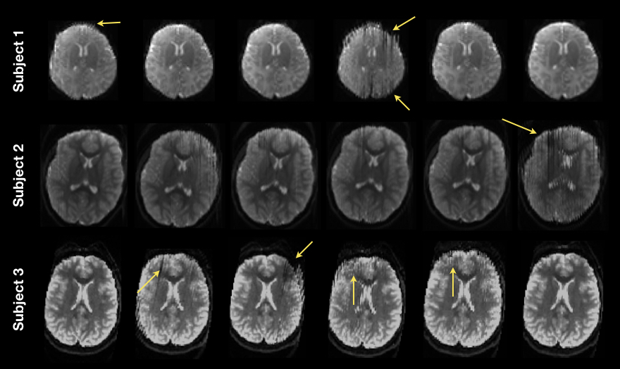
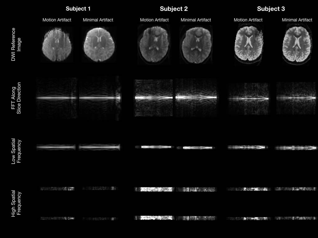
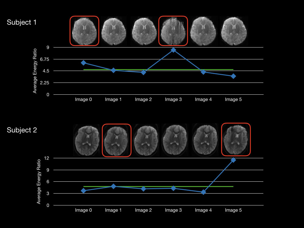
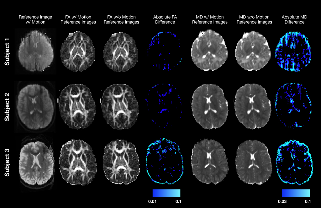
Representative non-diffusion weighted images with and without visible motion artifacts from each subject are displayed in Fig. 1. Taking the 1-D FFT along the slice direction, we observe that images containing significant motion artifacts have increased signal energy at higher spatial frequencies compared to the images without motion artifact (Fig. 2). This is further reflected in computing the ratios of average energy for the high-to-low frequency bands, with images with motion artifact having elevated average energy ratios (Fig. 3). In particular, across the different subjects, images with an average high frequency band energy greater than 4.75% of the average energy from the low-frequency band (Fig. 3) were identified to have significant motion artifact via visual inspection. Removing such non-diffusion weighted images improved the quality of the DTI parameter estimation (Fig. 4).Discussion
We provide a framework for identifying motion related artifacts from non-diffusion weighted images using the 1-D FFT along the slice direction by computing and examining the ratio of image energy from the high and low frequency bands. This proof-of-concept implementation demonstrates the feasibility to identify non-diffusion weighted images affected by motion artifact, and ultimately help improve the overall quality of the diffusion parameter estimates. Further work is needed to optimize the frequency band ratio threshold for sensitive and specific detection of artifacts. The presented framework should reduce subjective bias from visual inspection. Future work will examine the sensitivity of the approach to differing amounts of motion from larger cohorts of subjects as well as investigate whether this technique is applicable to the entire dMRI dataset. Finally, this framework may not be limited to dMRI datasets but may help identify motion-related artifacts in other interleaved multi-slice acquisitions.Acknowledgements
We’d like all of the participants and their families. This work was supported by the National Institutes of Mental Health (P50 MH100031; R01MH097464, and K99MH110596). Infrastructure support was also provided, in part, by a core grant to the Waisman Center from the National Institute of Child Health and Human Development (U54 HD090256).References
1. Alexander AL, Lee JE, Lazar M, Field AS. Diffusion tensor imaging of the brain. Neurotherapeutics. 2007;4(3):316-29.
2. Oguz I, Farzinfar M, Matsui J, Budin F, Liu Z, Gerig G, et al. DTIPrep: quality control of diffusion-weighted images. Front Neuroinform. 2014;8:4.
3. Bastiani M, Cottaar M, Fitzgibbon SP, Suri S, Alfaro-Almagro F, Sotiropoulos SN, et al. Automated quality control for within and between studies diffusion MRI data using a non-parametric framework for movement and distortion correction. Neuroimage. 2018;184:801-12.
4. Pierpaoli, C., 2011. Artifacts in diffusion MRI. In: Press, O.U. (Ed.), Diffusion MRI: Theory, Methods and Applications. Oxford University Press, Oxford, pp. 303–318.
5. Veraart J, Novikov DS, Christiaens D, Ades-Aron B, Sijbers J, Fieremans E. Denoising of diffusion MRI using random matrix theory. Neuroimage. 2016;142:394-406.
6. Kellner, E; Dhital, B; Kiselev, V.G & Reisert, M. Gibbs-ringing artifact removal based on local subvoxel-shifts. Magnetic Resonance in Medicine, 2016, 76, 1574–1581.
7. Andersson JLR, Sotiropoulos SN. An integrated approach to correction for off-resonance effects and subject movement in diffusion MR imaging. Neuroimage. 2016;125:1063-78.
8. Andersson JLR, Graham MS, Zsoldos E, Sotiropoulos SN. Incorporating outlier detection and replacement into a non-parametric framework for movement and distortion correction of diffusion MR images. Neuroimage. 2016;141:556-72.
9. Andersson JL, Skare S, Ashburner J. How to correct susceptibility distortions in spin-echo echo-planar images: application to diffusion tensor imaging. Neuroimage. 2003;20(2):870-88.
Figures