3477
Microstructural Variations in Intra-axonal and Extra-axonal Spaces Associated with Punctate White Matter Lesions in Preterm Infants Reveal by White Matter Tract Integrity Metrics1Department of Radiology, the First Affiliated Hospital, Xi’an Jiaotong University, Xi'an, China, 2MR Research China, GE Healthcare,Beijing,People's Republic of China, BengJing, China
Synopsis
Punctate white matter lesions (PWML) are common in preterm infants. Extensive microstructural changes were observed previously. However, specific changes related to axons and extra-axonal structures remain to be investigated. White matter tract integrity (WMTI) metrics derived from diffusion kurtosis imaging provide information of intra-axonal or extra-axonal spaces. This study aimed to use these metrics to quantify specific variations associated with PWML. Besides reduced fractional anisotropy and increased radial diffusivity, increased diffusivities in extra-axonal and intra-axonal spaces were found. Furthermore, tortuosity and fiber dispersion decreased obviously. These results suggested that PWML would influence axonal reorganization as well as extra-axonal structural integrity.
Introduction
Punctate white matter lesions (PWML) have been found in more than 20% of preterm infants 1, 2. Severe PWML can cause extensive changes in white matter microstructure, which has been found by using diffusion tensor imaging (DTI). However, conventional DTI parameters cannot distinguish the development of axons themselves from the growth of myelin3. White matter model based on diffusion kurtosis imaging (DKI) has been proposed to detect information of the intra-axonal and extra-axonal microstructure 4,5. White matter tract integrity (WMTI) metrics can be derived from this model. Metrics in the extra-axonal space include extra-axonal radial diffusivity (RDe), extra-axonal axial diffusivity (ADe), and tortuosity. Metrics in the intra-axonal space include intra-axonal diffusivity (Da) and fiber dispersion (FD). This study aimed to use these metrics to quantify specific variations associated with PWML.Methods
This study was approved by the local Internal Review Board and all parents of participants had signed the informed consents. Inclusion criterion was the evidence of punctate lesions in the cerebral white matter, which presented on T1WI and T2WI. Preterm infants without any MRI abnormality were selected as controls. Subjects with clinical diagnosis of congenital malformations of the central nervous system, hydrocephalus, gray matter lesions or major destructive white matter lesions were excluded.
Single-shot EPI diffusion kurtosis imaging was performed on a 3.0T scanner (General Electric Signa HDXT, WI, USA) with an eight-channel head coil. The other parameters were: b values = 500, 1000, 2000, 2500 s/mm2; 18 gradient directions; TR/TE = 8000~11000/91.7~126.1 ms; thickness = 4 mm; FOV = 180 × 180 mm2 ~ 240 × 240 mm2 (according to brain sizes); acquisition matrix = 128 × 128 ~ 172 × 172 (to keep the same resolution). Diffusion and kurtosis tensors were estimated by using constrained weighted linear least squares. Fractional anisotropy (FA), axial diffusivity (AD), radial diffusivity (RD), and WMTI metrics were calculated according to the white matter model for DKI.
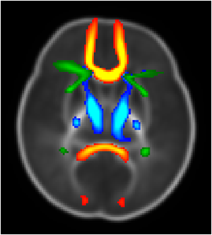
Tractography atlas for preterm infants were obtained by using the registration between local infants and the template from Johns Hopkins University (Figure 1). Ten regions of interests (ROI) were selected according to the labels: left anterior thalamic radiation (ATR_L), right anterior thalamic radiation( ATR_R), left corticospinal tract (CST_L), right corticospinal tract (CST_R), forceps major(F_major), forceps minor(E_minor), left inferior fronto-occipital fasciculus (IFOF_L), right inferior fronto-occipital fasciculus(IFOF_R), temporal part of left superior longitudinal fasciculus (SLF_temp_L), temporal part of right Superior longitudinal fasciculus (SLF_temp_R). Chi-Square test was performed to evaluate the gender ratios differences across groups. Mann-Whitney U test was used to evaluate differences in gestational age (GA), postnatal age, postmenstrual age (PMA), and regional values of metrics across groups. Tests were considered significant at P<0.05.
Result
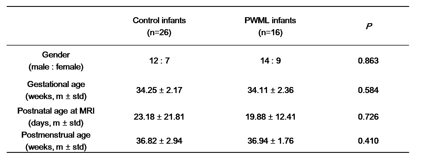
A total of 16 PWML and 26 control preterm infants were included. There were no significant differences in gender ratio, GA, postnatal age and PMA between PWML and control groups (Table 1).
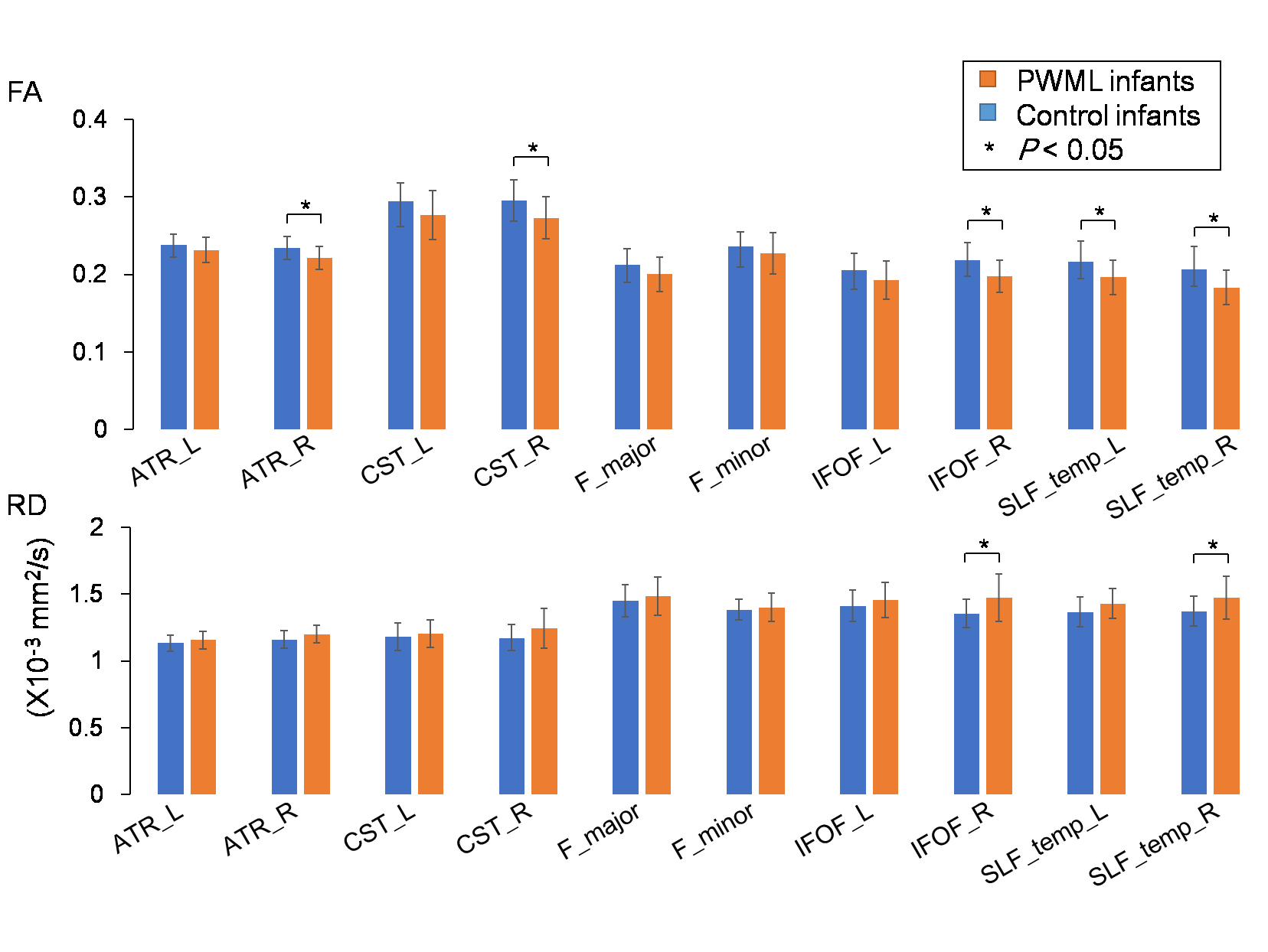
Compared to controls, decreased FA could be found in PWML infants on ATR_R, CST_R, IFOF_R, SLF_temp_L, and SLF_temp_R. Increased RD were found on IFOF_R and SLF_temp_R (Figure 2).
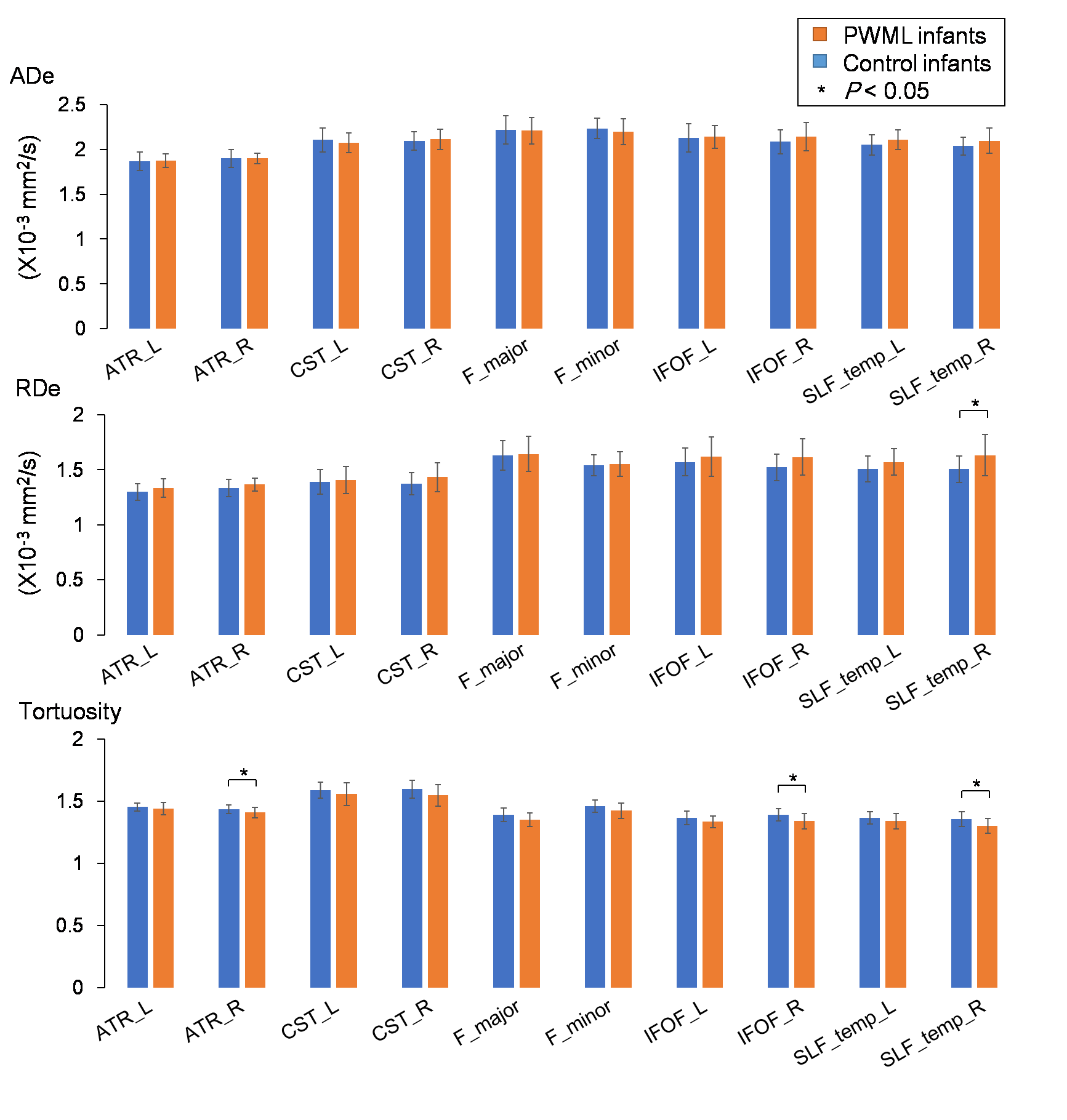
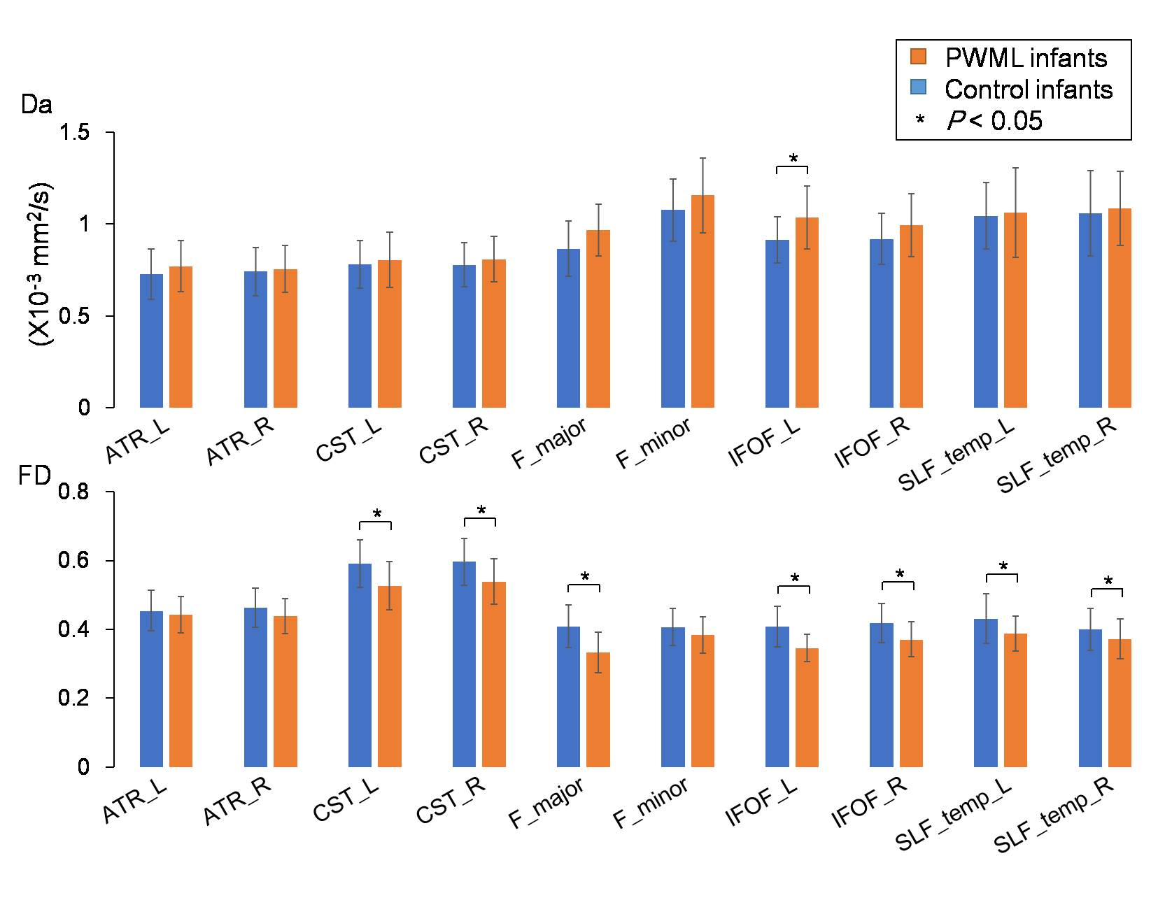
In the extra-axonal space (Figure 3), PWML infants held higher RDe on SLF_temp_R and lower tortuosity on ATR_R, IFOF_R, SLF_temp_R than controls. In the intra-axonal space (Figure 4), higher Da on IFOF_L, lower FD on CST_L, CST_R, F_major, IFOF_L, IFOF_R, SLF_temp_L, and SLF_temp_R were observed in PWML infants compared to controls.
Discussion
This study demonstrated extra-axonal and intra-axonal structural changes associated with PWML. Compared to previous DTI studies 6,7, our current work provided more specific information by using WMTI metrics derived from DKI. Conventional DTI parameters were also investigated in this work. And the change trends of FA and RD are in agreement with previous findings. Based on WMTI metrics, increased RDe and decreased tortuosity reflected structural changes in the extra-axonal spaces. Histological study has found that extra-axonal metrics are sensitive to myelin-related alterations 8. Results here indicate that PWML may cause dysmyelination in infants. Furthermore, changes in the intra-axonal space, especially the FD, suggest that axons themselves undergo variations. These may be related to the fiber organization during white matter development 9.Conclusion
PWML would influence axonal reorganization as well as extra-axonal structural integrity.Acknowledgements
This study was supported by the National Key Research and Development Program of China (2016YFC0100300), National Natural Science Foundation of China (81471631, 81771810 and 81171317), the 2011 New Century Excellent Talent Support Plan of the Ministry of Education, China (NCET-11-0438), the Fundamental Research Funds for the Central Universities (xjj2018265), the Fundamental Research Funds of the First Affiliated Hospital of Xi'an Jiaotong University (2017QN-09).References
1. Bassi L, Chew A, Merchant N, et al. Diffusion tensor imaging in preterm infants with punctate white matterlesions. Pediatr Res 2011;69:561-566.
2. de Bruïne FT, van den Berg-Huysmans AA, Leijser LM, et al. Clinical implications of MR imaging findings in thewhite matter in very preterm infants: a 2-year follow-up study. Radiology 2011;261:899-906.
3. Paus T.Growth of white matter in the adolescent brain :myelin or axon?.Brain and Cognition,2010,72(1):26-35.
4. Jelescu IO,Ve raart J,Adisetiyo V,et.al.One diffusion acquisition and different white matter models:how dose microstructure change in human early development based on WMTI and NODDI?Neuroimage,2015,107:242-256.
5. Fieremans E,Jensen JH,Helpern JA.White matter characterization with diffusional kurtosis imaging.Neuroimage,2011,58(1):177-188.
6. Bassi L, Chew A, Merchant N, et al. Diffusion tensor imaging in preterm infants with punctate white matter lesions. Pediatr Res 2011;69:561-566.
7. Li X, Gao J, Wang M, et al. Charaterization of extensive microstructural variations associated with punctate white matter lesions in preterm neonates. AJNR Am J Neuroradiol 2017;38:1228 -1234
8. Falangola, M.F., Guilfoyle, D.N., Tabesh, A., et al. Histological correlation of diffusional kurtosis and white matter modeling metrics in cuprizone‐induced corpus callosum demyelination. NMR Biomed. 2014; 27: 948-957.
9. Dubois J, Dehaenelambertz G, Kulikova S, et al. The early development of brain white matter: a review of imaging studies in fetuses, newborns and infants.[J]. Neuroscience, 2014, 276(6):48-71.
Figures