3470
Quantifying voxel-wise differences between diffusion propagators across multiple MAP-MRI datasets1National Institute of Biomedical Imaging and Bioengineering, National Institutes of Health, Bethesda, MD, United States, 2National Institute of Child Health and Human Development, National Institutes of Health, Bethesda, MD, United States, 3Biomedical Engineering, University of Arizona, Tucson, AZ, United States, 4Department of Surgery, University of Arizona, Tucson, AZ, United States, 5Center for Neuroscience and Regenerative Medicine, Henry Jackson Foundation, Bethesda, MD, United States, 6Clinical Center, National Institutes of Health, Bethesda, MD, United States, 7National Institute of Neurological Disorders and Stroke, National Institutes of Health, Bethesda, MD, United States
Synopsis
We describe a technique for voxel-wise analysis across multiple mean apparent propagator (MAP) MRI datasets warped using a diffeomorphic tensor-based registration algorithm. By measuring propagators from co-registered MAP datasets with the same MAP basis functions we can directly quantify voxel-wise differences using angular dissimilarity metrics. We show examples from a cohort of healthy volunteers, and from a longitudinal clinical dataset of a patient undergoing carotid endarterectomy. This approach could provide improved sensitivity in the detection and characterization of subtle microstructural tissue changes in cross-sectional group and longitudinal single-subject clinical studies.
Introduction
Mean apparent propagator (MAP) MRI1 is an powerful framework for measuring the probability density functions of net displacements of diffusing water molecules in tissues, i.e., the diffusion propagators, and quantifying important features, such as diffusion anisotropy or non-Gaussianity. MAP-MRI data can be acquired clinically2 and has the potential to quantify subtle features of tissue microstructure with a new family of microstructural parameters. Because MAP-MRI subsumes and extends diffusion tensor imaging (DTI)3, it is well-suited for tensor-based image registration4.
A recently proposed framework4 allows registration of multiple MAP-MRI datasets by first generating a DTI template, then transforming the diffusion-weighted images (DWIs) and their corresponding diffusion-encoding gradients from native space to template space using tensor-based deformation fields for each dataset (Fig.1). Each dataset comprising the transformed DWIs and corresponding voxel-wise diffusion-encoding gradients5 in template space can then be fit with MAP-MRI using the same MAP basis functions with orientations and scaling parameters defined by the template DTI (Fig.1).
We describe a technique for voxel-wise analysis of multiple MAP-MRI datasets registered using this approach. By measuring propagators in registered MAP datasets with the same MAP basis functions we can directly quantify voxel-wise differences between them using an angular dissimilarity metric. We show examples from a cohort of healthy volunteers, and from a longitudinal clinical dataset of a patient undergoing carotid endarterectomy. This approach could provide improved sensitivity in the detection and characterization of subtle microstructural tissue changes in cross-sectional group and longitudinal single-subject clinical studies.
Methods
MAP-MRI data was acquired in 5 healthy volunteers using spin-echo diffusion EPI on a clinical scanner and TE/TR=94/6000ms, 42 slices, 3-mm slice thickness, a 70x70 imaging matrix on a 210x210mm2 field-of-view, and parallel imaging acceleration of 2. Each dataset contained 498 DWIs with diffusion encoding on 6 b-shells from 0-6000s/mm2, with an increasing number of uniformly distributed diffusion-encoding orientations at larger b-values, and gradient pulse width and separation of δ=34.2ms and Δ=40.2ms, respectively. In addition, a longitudinal dataset was acquired from a subject who underwent carotid endarterectomy for asymptomatic carotid atherosclerosis and was imaged at three timepoints: prior to the surgery, one month after surgery, and six months after surgery.
All DWIs were preprocessed in native space to correct for subject motion and imaging distortions6. Subsequently, the transformed DWIs were co-registered using the pipeline from Fig.14, and propagators were estimated in template space using the MAP functional basis with orientation and scaling defined by the template DTI. A template of MAP propagators was obtained by averaging the registered MAP propagators across all subjects in the cohort of healthy volunteers. Voxel-wise differences between propagators from multiple datasets were quantified using the angular dissimilarity metric1,
$$\sin(\theta)=\sqrt{1-\frac{(\mathbf{a} \cdot \mathbf{b^T})^2}{(\mathbf{a} \cdot \mathbf{a^T})(\mathbf{b} \cdot \mathbf{b^T})}}$$
where $$$\mathbf{a}$$$ and $$$\mathbf{b}$$$ are column vectors of MAP coefficients corresponding to two propagators represented in the same functional basis.
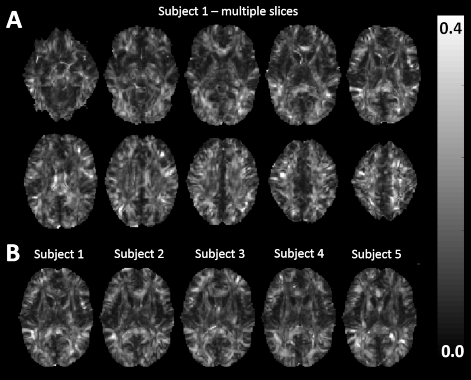
For the cohort of healthy volunteers, we computed maps of voxel-wise angular dissimilarities between the propagators of each subject and the average propagators. For the longitudinal dataset, we computed maps of voxel-wise angular dissimilarities between propagators at the three timepoints.
Results and Discussion
MAP-MRI propagators and parameters derived from the registered datasets showed values consistent with previous in vivo findings2,7. Maps of voxel-wise angular dissimilarities between propagators from healthy volunteers and template (i.e., average) propagators (Fig.2) show a high sensitivity to inter-subject anatomical variability with the largest differences in regions of white matter and tissue interfaces. Fig.2B shows maps of voxel-wise angular dissimilarities between diffusion propagators of each volunteer and template propagators in a representative slice. The largest values can be seen again at tissue interfaces, due to large intersubject neuroanatomical variability and partial volume effects.
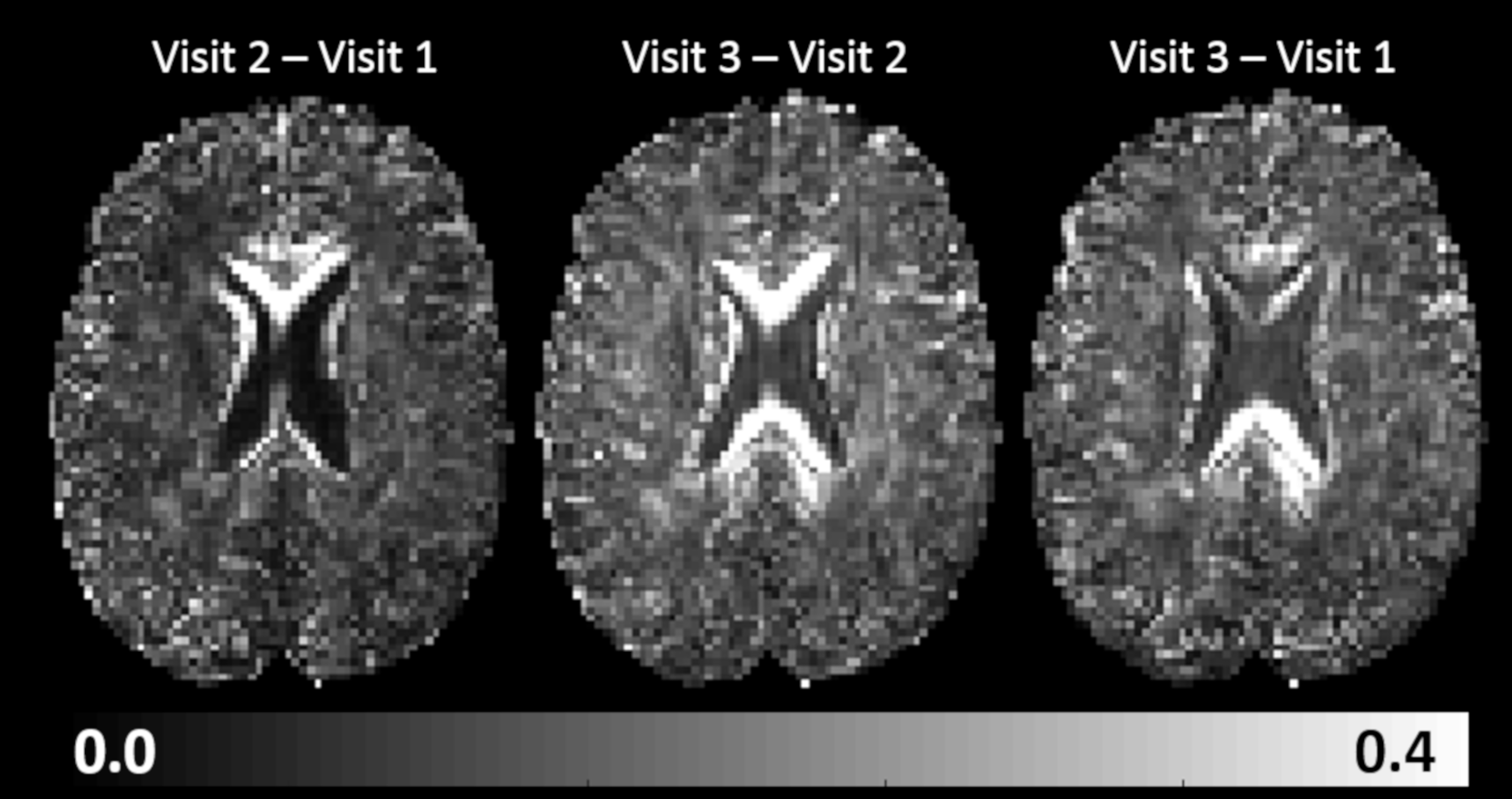
DTI and MAP parameters revealed no distinguishable differences between the timepoints of the longitudinal dataset. We found, however, notable localized changes between propagators at the three timepoints which stand out against the relatively low background values of angular dissimilarity in the rest of the brain (Fig.3). These preliminary results call for a more extensive clinical investigation to determine whether the observed changes are reproducible and related to the surgery. Future proof-of-principle and clinical validation studies will establish the sensitivity of the proposed MAP analysis framework for detecting local tissue changes longitudinally by quantifying propagator differences voxel-wise.
Conclusion
Our preliminary results indicate that voxel-wise analysis of diffusion propagators across multiple datasets within the powerful framework of MAP-MRI could provide a sensitive clinical tool for detecting microstructural differences that are too subtle to observe with conventional methods such as DTI. The proposed framework may improve the sensitivity to fine microstructural alterations in single-subject longitudinal studies aimed at detecting pathologies with focal or diffuse presentations, monitoring treatment response, or characterizing neuronal plasticity, healthy development, and aging.Acknowledgements
This work was supported by the Center for Neurodegeneration and Regenerative Medicine (CNRM), under the auspices of the Department of Defense (DoD) and the Henry Jackson Foundation (HJF) grant number #308049-8.01-60855, and by the Intramural Research Program (IRP) of the National Institute of Biomedical Imaging and Bioengineering (NIBIB) and the Eunice Kennedy Shriver National Institute of Child Health and Human Development (NICHD) within the National Institutes of Health (NIH). We also thank the University of Arizona for providing the longitudinal data set used in this study. The carotid endarterectomy work was supported by the University of Arizona Translational Imaging Program Project Stimulus (TIPPS) grant.References
1 Özarslan, E. et al. Mean apparent propagator (MAP) MRI: A novel diffusion imaging method for mapping tissue microstructure. NeuroImage 78, 16-32 (2013).
2 Avram, A. V. et al. Clinical feasibility of using mean apparent propagator (MAP) MRI to characterize brain tissue microstructure. NeuroImage 127, 422-434 (2016).
3 Basser, P. J., Mattiello, J. & LeBihan, D. Estimation of the effective self-diffusion tensor from the NMR spin echo. Journal of Magnetic Resonance, Series B 103, 247-254 (1994).
4 Avram, A. V. et al. Anatomical template of MAP MRI-derived 3D diffusion propagators and microstructural parameters. Proceedings of the ISMRM 27, 1577 (2018).
5 Irfanoglu, M. O. et al. DR-TAMAS: Diffeomorphic Registration for Tensor Accurate Alignment of Anatomical Structures. NeuroImage 132, 439-454 (2016).
6 Pierpaoli, C. et al. TORTOISE: an integrated software package for processing of diffusion MRI data. Proceedings of the ISMRM 18, 1597 (2010).
7 Avram, A. V., Barnett, A. & Basser, P. The Variation of MAP-MRI –derived Parameters Along White Matter Fiber Pathways in the Human Brain. Proceedings of the ISMRM 22, 2587 (2014).
Figures
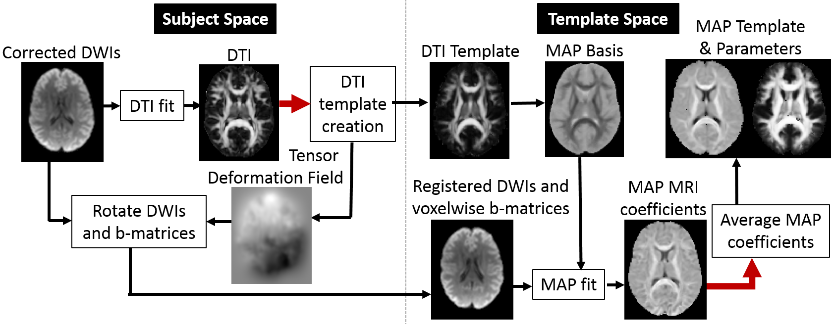
Figure 1: A flow chart for spatial standardization and analysis of multiple MAP-MRI datasets.