3458
Differentiation of Sinonasal Mucosal Malignant Melanomas from Sinonasal Carcinomas Using Whole-tumour Histogram Analysis of Diffusion Kurtosis Imaging and Intravoxel Incoherent Motion1Eye & ENT Hospital of Fudan University, Shanghai, China, 2Radiology, Eye & ENT Hospital of Fudan University, Shanghai, China, 3Shanghai Key Laboratory of Magnetic Resonance, East China Normal University, Shanghai, China, 4MR Scientific Marketing, Siemens Healthcare, Shanghai, China
Synopsis
This is the first study that used histogram metrics derived from diffusion kurtosis (DKI) and intravoxel incoherent motion (IVIM) to differentiate sinonasal mucosal malignant melanomas (SNMMMs) and squamous cell carcinomas (SCCs) which are sometimes indistinguishable with conventional MRI. Overall, histogram metrics obtained from K, D, D* and f were found to be significantly higher in SNMMMs than in SCCs. Furthermore, the combined use of the two independent indicators, the 75th percentile of K and skewness of D, can effectively differentiate between SNMMMs and SCCs.
Background and purpose
SNMMMs are commonly amelanotic (~50%),1 lacking the characteristic melanin signal on conventional MRI2-4. Thus, it is usually challenging to differentiate SNMMM from most common sinonasal malignant tumor, SCC. Recently, DKI and IVIM have been shown to be useful approaches to differentiate between benign and malignant lesions in the sinonasal region.5, 6 However, no studies applied DKI and IVIM to discriminate SNMMMs from SCCs. Moreover, previous investigations measured DKI or IVIM parameters from manually placed regions of interest (ROIs) on the largest section of tumors,5, 6 which could not reflect the characteristic of the entire tumor. Whole-tumor histogram analysis could reflect the full spectrum and heterogeneity of histology within the tumor.7, 8 Thus, the purpose of this study was to evaluate the diagnostic performance of DKI and IVIM using whole-tumor histogram analysis for differentiating SNMMMs from SCCs.Methods
DKI and IVIM were performed in 12 patients with SNMMMs and 26 patients with SCCs on a 3T MR scanner (MAGNETOM Verio, Siemens Healthcare, Erlangen, Germany). The detailed parameters were as follows: TR/TE = 5200/83 msec, δ = 27.4 msec, Δ = 39.4 msec, number of averages = 2, acquisition matrix = 120 × 120; field of view (FOV) = 220×220 mm2, slice thickness = 5 mm, intersection gap = 5 mm, parallel imaging acceleration factor = 2; 14 different b values ranging from 0 to 2500 sec/mm2 were used (b = 0, 50, 100, 150, 200, 250, 300, 350, 400, 800, 1000, 1500, 2000, and 2500 sec/mm2). The IVIM and DKI processing were performed using custom-written scripts in MATLAB (version R2016a; MathWorks, Natick, Mass) to provide ADC, D, D*, f, Dk and K parametric maps on a pixel-by-pixel basis.9 The whole-tumor histogram metrics were calculated on these parametric maps using PyRadiomics (version 1.3.0; http://github.com/Radiomics/pyradiomics) based on Python (version 3.5.4; http://www.python.org).10 The Student’s t-tests, ROC curve and multivariate stepwise logistic regression analyses were used for statistical analysis.Results
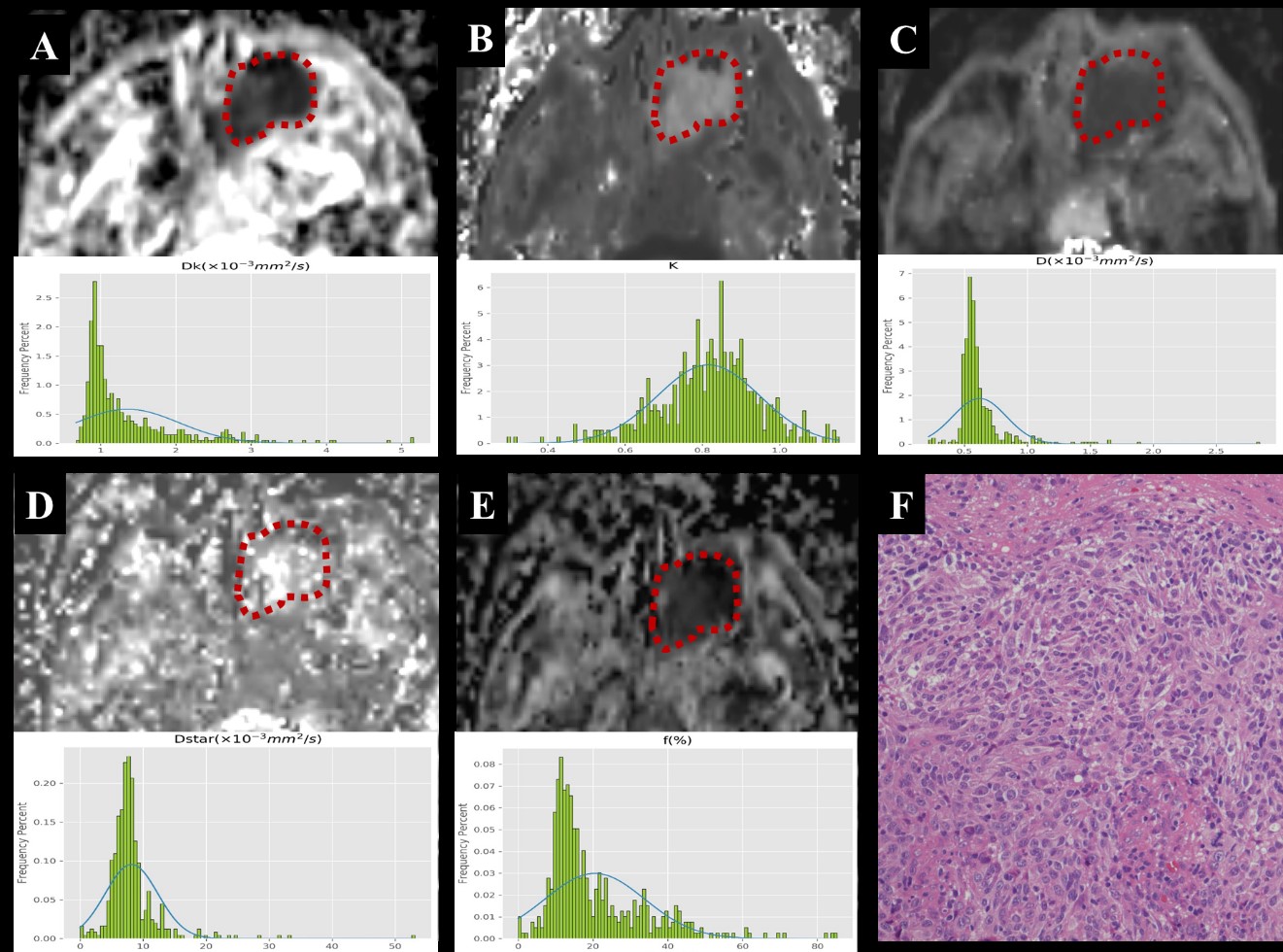
Significantly higher mean, median, 75th and 90th percentiles of K, skewness of D, mean, median, 90th percentile and kurtosis of D*, 75th and 90th percentiles of f were found in SNMMMs than in SCCs (all P < .05). Higher 75th percentile of K (β coefficient = 0.007; OR = 1.007, 95% CI = 1.001-1.012; P = 0.026) and skewness of D (β coefficient = 0.209; OR = 1.232, 95% CI = 1.001-1.518; P = 0.049) were independent indicators for the differentiation of SNMMMs for SCCs. One example of the calculated DWI parameters, histogram and haematoxylin-eosin staining of a patient is shown in Figure 1. The combination of 75th percentile of K and skewness of D can improve the diagnostic performance with a sensitivity and specificity of 75.0% and 80.8%, respectively, for differentiating these two malignant tumors. The histogram analysis tool was not given in method secitonConclusion
Whole-tumour histogram analysis of DKI and IVIM are valuable for differentiating SNMMMs from SCCs. The 75th percentile of K and skewness of D were independent predictors for distinguishing between them.Keywords
Neoplasms; Magnetic resonance imaging; Diffusion magnetic resonance imaging; Melanoma; Squamous cell carcinomaAcknowledgements
This work was supported by the Grant of Science and Technology Commission of Shanghai Municipality (Grant number: 17411962100) and Key Project of the National Natural Science Foundation of China (Grant number: 61731009).References
1.Williams MD. Update from the 4th Edition of the World Health Organization Classification of Head and Neck Tumours: Mucosal Melanomas. Head and neck pathology 2017;11:110-117
2.Kim SS, Han MH, Kim JE, et al. Malignant melanoma of the sinonasal cavity: explanation of magnetic resonance signal intensities with histopathologic characteristics. American journal of otolaryngology 2000;21:366-378
3.Xu QG, Fu LP, Wang ZC, et al. Characteristic findings of malignant melanoma in the sinonasal cavity on magnetic resonance imaging. Chin Med J (Engl) 2012;125:3687-3691
4.Yousem DM, Li C, Montone KT, et al. Primary malignant melanoma of the sinonasal cavity: MR imaging evaluation. Radiographics : a review publication of the Radiological Society of North America, Inc 1996;16:1101-1110
5.Jiang JX, Tang ZH, Zhong YF, et al. Diffusion kurtosis imaging for differentiating between the benign and malignant sinonasal lesions. Journal of magnetic resonance imaging : JMRI 2017;45:1446-1454
6.Xiao Z, Tang Z, Qiang J, et al. Intravoxel Incoherent Motion MR Imaging in the Differentiation of Benign and Malignant Sinonasal Lesions: Comparison with Conventional Diffusion-Weighted MR Imaging. American Journal Of Neuroradiology 2018;39:538-546
7.Chandarana H, Rosenkrantz AB, Mussi TC, et al. Histogram analysis of whole-lesion enhancement in differentiating clear cell from papillary subtype of renal cell cancer. Radiology 2012;265:790-798
8.Fujima N, Yoshida D, Sakashita T, et al. Prediction of the treatment outcome using intravoxel incoherent motion and diffusional kurtosis imaging in nasal or sinonasal squamous cell carcinoma patients. European radiology 2017;27:956-965
9.Xiao Z, Zhong Y, Tang Z, et al. Standard diffusion-weighted, diffusion kurtosis and intravoxel incoherent motion MR imaging of sinonasal malignancies: correlations with Ki-67 proliferation status. European radiology 2018;28:2923-2933
10.van Griethuysen JJM, Fedorov A, Parmar C, et al. Computational Radiomics System to Decode the Radiographic Phenotype. Cancer research 2017;77:e104-e107
Figures