3457
Adapting Diffusion Weighted Arterial Spin Labelling (DWASL) to use a Single Sided Bipolar Diffusion Gradient to Probe Microvascular SignalSamantha Paterson1, Antoine Vallatos2, and William Holmes3
1Neuroscience & Psychology, University of Glasgow, Glasgow, United Kingdom, 2University of Edinburgh, Edinburgh, United Kingdom, 3University of Glasgow, Glasgow, United Kingdom
Synopsis
Diffusion Weighted Arterial Spin Labelling uses a pair of bipolar diffusion gradients surrounding a spin echo pulse to image signal in the intravascular and extravascular compartments. We propose moving these gradients to a single sided gradient, reducing Δ and changing where the change to the extravascualar signal starts. Our results show that the ratio has changed from b = 50 to b = 300 s/mm2 for our new single sided sequence. This can give us more information on the intravascular signal, helping to improve the probing of blood brain barrier permeability.
Introduction
Arterial Spin Labelling (ASL) is a non-invasive perfusion technique that by labelling the arterial blood can be used to probe cerebral blood flow (CBF) changes. By applying diffusion gradients, we can probe signal from either the arteries, capillaries or tissue dependent on gradient strength or inversion delay. This information can be useful in Blood Brain Barrier (BBB) permeability studies, where a breakdown in the barrier is associated with increase water exchange into the tissue. We use the high SNR mbASL sequence, which uses adiabatic pulses to label the arterial water. This sequence has shown significantly higher SNR against the standard FAIR sequence in recent studies1. At current, bipolar gradients are applied around a 180-degree pulse and when the DWASL signal is measured at multiple b-values, a steep drop is seen from the suppressed signal before tending to tissue diffusion2. As the signal is suppressed with a small gradient value, these values tend to be noisy & unstable, losing valuable information on the exchange of water in the brain. We propose moving the bipolar gradients to reduce the time between the gradients and increases the gradient strength needed, providing a more stable sequence with the expected drop in vascular signal shifting to a higher b-value. Figure 1 demonstrates the current DWASL sequence and comparison with the modified sequence. The theoretical signal at each b value is demonstrated.Methods
Scans were performed using a Bruker PharmaScan 7T MRI scanner using a mouse surface coil. A DWASL sequence with gradients around the 180-degree pulse (Sequence A) was compared with the gradients moved to a single side (Sequence B). All other parameters were kept the same as follows: b = 0:100:500 s/mm2. NA = 10. FOV = 25x25mm. CI = 5000ms. TR = 7000ms. TI = 50ms. Np = 20. For Sequence A, Δ = 29.9ms, and Sequence B, Δ = 9.4ms. Small delta = 3.2ms. n=6 CD1 female mice were used for scanning.Data was exported in DICOM format and analysed using in-house MATLAB code. The cortex was used as the ROI region.Results & Discussion
Figure 2 is a comparison of diffusion signal from a water phantom using both DWASL sequences. This shows that both sequences produce the expected diffusion coefficient of water (2.27 and 2.3x10⁹ s/mm2 respectively). Figure 3 compares the ΔM signal from Sequence A and Sequence B. The ASL control image signal is shown which confirms the signal from Sequence A and Sequence B are from flowing blood. The rate of signal suppression has changed for sequence B with the change from intravascular to extravascular signal occurring at b = 300 in Sequence B and at b = 50 in Sequence A, confirming our theory. This is further reflected in the fitting to the bi-exponential model. Comparing the weighting factors for the two sequences, there is a large difference seen between the intravascular and extravascular signals.Conclusion
In conclusion we have developed a modified DWASL sequence that allows us to probe the intravascular signal further than previously. By changing the time delay between the bipolar gradients and changing their position in the sequence we have been able to create a more stable sequence with the change in signal between the intravascular and extravascular compartments occurring at larger b-values. This information will be vital in probing BBB permeability changes more accurately.Acknowledgements
Many thanks to Jim Mullin for his technical help in the MRI scanning and to the University of Glasgow and EPSRC for funding this project.References
1Vallatos, A et al. (2017)
2Wang, J et al. (2007)
Figures
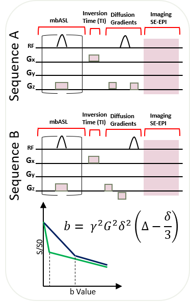
Figure 1: Schematic of the changes in the
structure of the DWASL sequence.
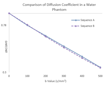
Figure 2: Comparison of control signal for Sequence A and Sequence B.
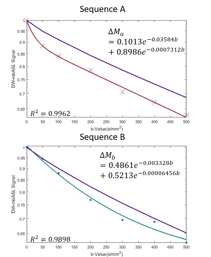
Figure 3: A comparison of the DWASL signal from Sequence A and Sequence B.