3445
Increasing reader confidence in cancer imaging using an advanced DWI processing – a prospective reader study1Radiology, German Cancer Research Center, Heidelberg, Germany, 2Medical Physics in Radiology, German Cancer Research Center, Heidelberg, Germany, 3Siemens Healthcare GmbH, Erlangen, Germany
Synopsis
Diffusion-Weighted imaging (DWI) in abdominal MRI is highly valuable for oncological follow-up-investigations. However, intrinsic limitations of the method, including susceptibility to motion artifacts and poor signal in the retroperitoneum, are of special importance regarding its usage in abdominal examinations. This prospective study compared a standard EPI-DWI with an oncologically optimized prototype DWI in 59 patients with regard to the detectability/characterization of suspicious lesions. The study demonstrated that the oncologically optimized prototype DWI sequence with complex averaging, motion correction between averages, rescaling of motion corrupted averages, and background suppression, significantly increased the reader confidence for lesion characterization/detection in oncological abdominal MRI.
Introduction
Oncological imaging commonly includes Diffusion-Weighted Imaging (DWI) sequences to identify and characterize suspicious lesions. However, image quality can be challenged by the limited SNR of DWI, especially in abdominal examinations and by motion artifacts resulting from the diaphragm, which is why DWI of the abdominal region needs to be improved [1]. This prospective study investigated an optimized DWI in oncological imaging for improved detection of malignant abdominal lesions.Methods
This IRB-approved, prospective study included 59 patients (mean age: 57 years, male/female: 22/37). Oncological follow-up MRI-examinations of the abdomen were performed with a 1.5T MRI scanner (MAGNETOM Aera, Siemens Healthcare GmbH, Germany) with both a standard EPI-DWI (“routine-DWI”, b=50,900s/mm²) and an oncologically optimized prototype DWI (“onco-DWI”) with b=0,50,900,1500s/mm² using a prototypical EPI DWI technique including complex averaging, motion correction between the averages, rescaling of motion corrupted averages, and background suppression. The following parameters were used for routine-DWI: TR=6.5 s; TE=63 ms; FOV 450x242 mm²; matrix 134x72; resolution 3.4x3.4 mm², interpolated to 1.7x1.7 mm²; slice thickness 5 mm; bandwidth 2332 Hz/Px; b-values 50 s/mm² and 900 s/mm² with 2 and 8 averages, gradient mode 3-scan trace, 90 slices in 3 steps, 11 min. For onco-DWI: TR=7.9 s; TE=57 ms; FOV 480x270 mm²; matrix 164x92; resolution 3x3 mm², interpolated to 1.5x1.5 mm²; slice thickness 5 mm; bandwidth 2540 Hz/Px; b-values 0, 50, 900, 1500 s/mm² (1, 1, 16 and 18 averages); gradient mode 3D-diagonal; 90 slices in 3 steps, 15 min. Diagnostic confidence for characterization/detection of suspicious lesions was evaluated by two independent readers using a 5-point Likert Scale [2]. Statistics included Wilcoxon signed rank tests, interreader agreement was analyzed by kappa-statistic (p<0.05).Results
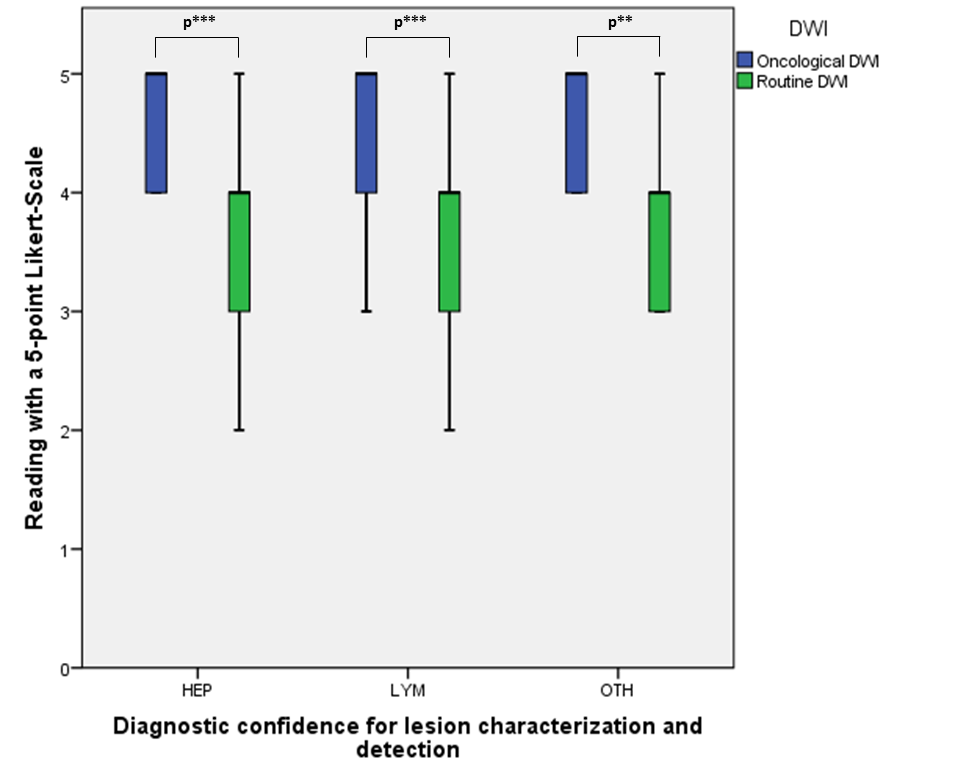
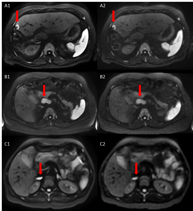
Sixty-one lesions were detected (20 hepatic, 16 lymphatic, and 25 lesions of other origins). Reader confidence for characterization/detection of malignant lesions was significantly improved using the onco-DWI (4.6±0.5) as compared to the routine-DWI (3.7±0.7) (p<0.001). The increased confidence of lesion recognition in the onco-DWI remained significant in all subgroups of hepatic, lymphatic and lesions of other origins (p<0.002) (Figure 1 and Figure 2) with an overall good interreader agreement (kappa = 0.744) (p<0.001).Discussion
This prospective study demonstrated the potential of an oncologically optimized DWI with regard to lesion detection and characterization. To date, DWI MRI is already a highly valuable tool in oncological staging examinations. However, compared to standard DWI the optimization of the DWI sequence significantly improved the reader confidence in lesion detection and characterization by providing an improved image resolution, motion correction between the averages and complex averaging, which is why DWI optimization for the specific purposes such as oncologic imaging [3] should be investigated in future studies.Conclusion
The oncologically optimized body diffusion prototype sequence with an improved image resolution, motion correction between the averages and background suppression increased the reader confidence for lesion characterization/detection in oncological abdominal MRI. This was achieved both with regard to screening and follow-up examinations while preserving a high interreader agreement with only limited extra time needed for the examination. Optimization of DWI specifications with regard to the clinical task should therefore be considered for future studies.Acknowledgements
Special acknowledgments and thanks to Siemens Healthineers for providing the optimized protoype sequence and protocol.References
1. Taron, J., et al., Clinical Robustness of Accelerated and Optimized Abdominal Diffusion-Weighted Imaging. Invest Radiol, 2017. 52(10): p. 590-595.
2. de Winter, J.C.F., Dodou, D., Five-point Likert items: T test versus Mann–Whitney–Wilcoxon. Practical Assessment, Research & Evaluation, 2010. 15(11): p. 16.
3. Zhang, T.T., et al., Differentiation of pancreatic carcinoma and mass-forming focal pancreatitis: qualitative and quantitative assessment by dynamic contrast-enhanced MRI combined with diffusion-weighted imaging. Oncotarget, 2017. 8(1): p. 1744-1759.
Figures