3437
Readout segmented EPI based Relative apparent diffusion coefficient in early monitoring the treatment effect of low-intensity transcranial ultrasound: evaluation in a rat permanent occlusion modelLanxiang Liu1, Juan Du2, Tao Zheng1, Xuemei Wang1, Huiling Yi1, Shuang Wu1, Qinglei Shi3, and Shuo Wu2
1Department of Magnetic Resonance Imaging, Qinhuangdao Municipal No. 1 Hospital, Qinhuangdao, China, 2Graduate School of Hebei Medical University, Shijiazhuang, China, 3MR Scientific Marketing, Siemens Healthcare, Beijing, China
Synopsis
Acute ischemic stroke is a common and frequently occurring disease that severely harms human health and has high morbidity and mortality. Low-intensity transcranial ultrasound (LIPUS), due to its advantages of higher spatial resolution and greater penetration depth, has emerged as a new modality for noninvasive neuromodulation. This study indicated that the rADC based on readout segmented EPI sequence is a good indicator to evaluate the curative effect of LIPUS on acute cerebral infarction treatment.
Objective
To explore the feasibility of relative apparent diffusion coefficient (rADC) values in evaluation the treatment effect of low-intensity transcranial ultrasound (LIPUS) on rat models of acute ischemic stroke at different time points. The results were confirmed by using immunohistochemistry and histopathological examinations.Methods
Sixty Sprague-Dawley rats (weight, 250 ± 10 g) were randomly divided into six groups to establish permanent models of distal middle cerebral artery occlusion (dMCAO). Five of these groups were stimulated with ultrasound at 0.5 h, 1 h, 3 h, 6 h, and 9 h after dMCAO and one left group was regarded as control group. Diffusion weighted imaging based on readout-segmented EPI sequence were acquired at 0.5 h and 1 h after dMCAO and then at 1-hour intervals until 12 hours at a 3T a scanner (MAGNETOM Verio, Siemens Healthcare, Erlangen, Germany). The rADC values were then calculated and the pathological results from the rat brains were obtained after completing the MR examination.Results
The rADC values in the infarcted areas of rat brains in the LIPUS group (0.5 h) were significantly higher than in the Control group 12 h after dMCAO [(0.56 ± 0.26) vs (0.24 ± 0.15), p > 0.05] (table.1). The rADC values of rat brains in the LIPUS group (1 h), LIPUS group (3 h), and LIPUS group (6 h) were slightly higher than that in the Control group 12 h after dMCAO. No significant difference in rADCs between the LIPUS group (9 h) and the Control group 12 h after dMCAO [(0.23 ± 0.18) vs (0.24 ± 0.15), p < 0.05].Conclusion
The rADC values demonstrated a high sensitivity in monitoring of the early therapeutic changes of LIPUS on rat models of acute ischemic stroke. In future, it could be used as a diagnostic biomarker in evaluation the treatment effect of LIPUS about acute ischemic stroke.Acknowledgements
No acknowledgement found.References
No reference found.Figures
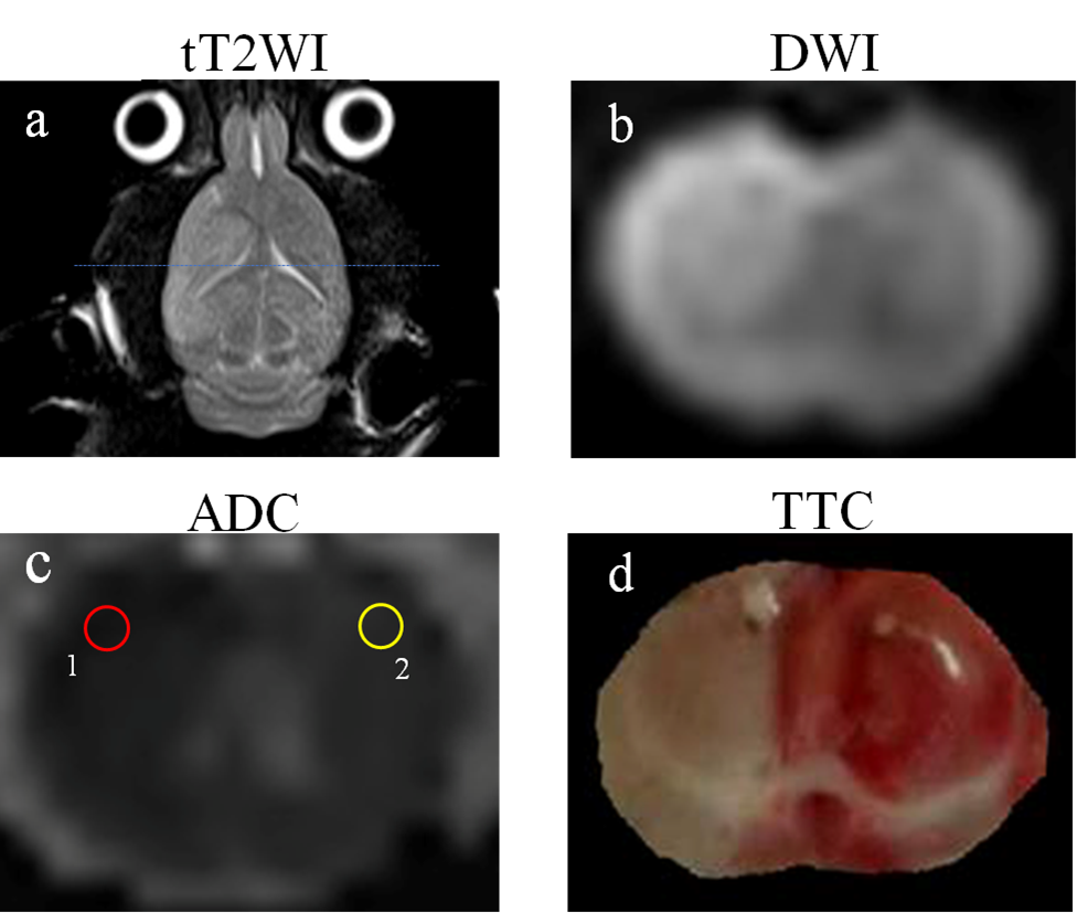
Fig.1. (a) Axial-T2WI at onset moments of 6
h, (b) Coronal-RESOLVE-DWI at onset moments of 0.5 h, (c) ADC at onset moments
of 0.5 h, (d) TTC stained images at onset moments of 13 h. rADC = ADC 1/ ADC 2×100%.
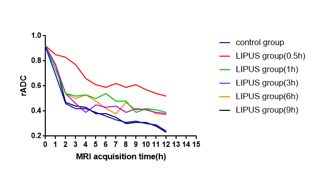
Fig. 2. The variation trends of rADC
values of every group.
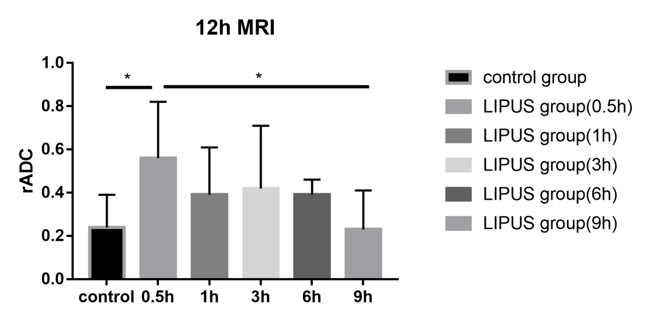
Fig.
3. The rADC values of
each group at 12h after dMCAO.
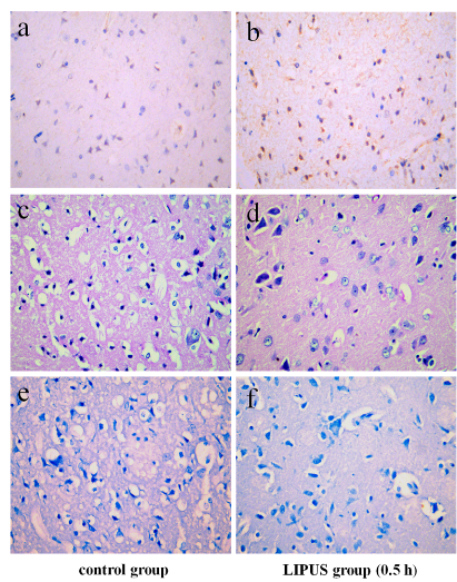
Fig. 4 . BDNF+ neurons were more
frequently and easily detectable in the LIPUS group (0.5 h) compared to Control group (a-b). The
cerebral cortex of cerebellum of LIPUS group (0.5 h) showed the minimal,
unilateral, multifocal neuronal necrosis and vacuolation of neuropils compared
to Control group (c-d). Nissl
staining revealed that neuronal degeneration and neuronal loss were greater
with larger observable damaged area in the Control group than that of the LIPUS
group (0.5 h) (e-f).
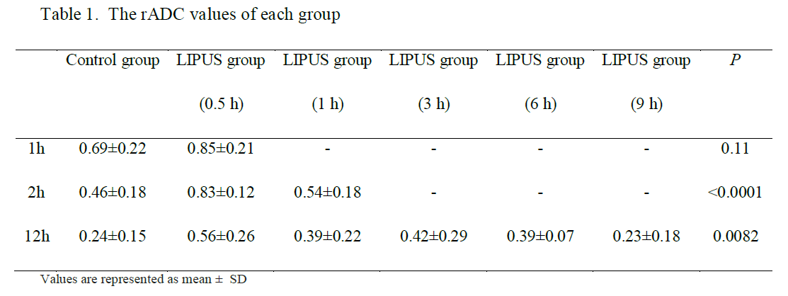
Table 1. The rADC values of each group
Table
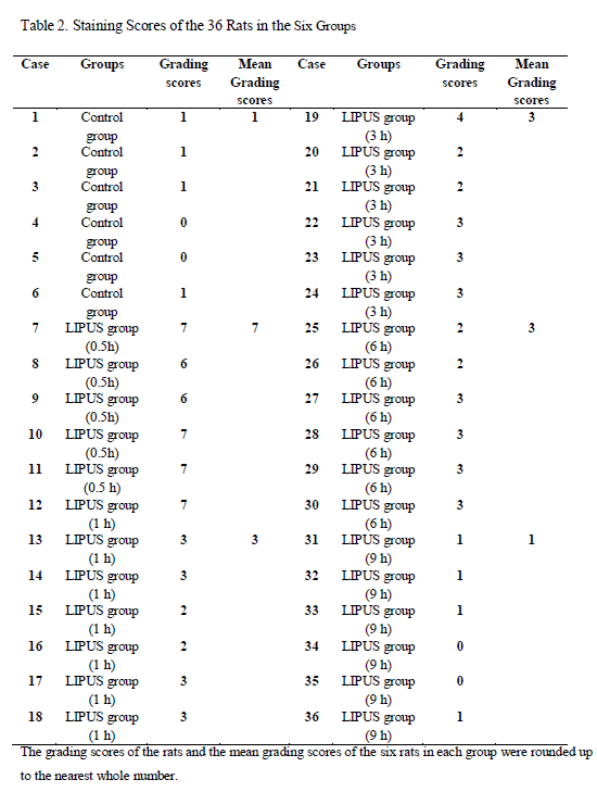
2. Staining Scores of the 36 Rats in the Six Groups