3434
Functional and microstructural alterations in the rat hippocampus after repetitive mild traumatic brain injury1Clinical Medicine, Aarhus University, Århus C, Denmark, 2Department of Physics and Astronomy, Aarhus University, Aarhus C, Denmark, 3John D. Dingell VA Medical Center, Wayne State University, Detroit, MI, United States
Synopsis
Repetitive mild traumatic brain injury (
INTRODUCTION
Repetitive mild traumatic brain injury (mTBI) is a silent epidemic and increases the risk of developing neurological disorders [1]. Conventional neuroimaging (CT and MRI) often report normal brain scans, posing significant challenges in treatment planning for mTBI patients. Conversely, histology of cadaver brains from patients and animal models of repetitive mTBI consistently reveal axonal injury, dendrite beading, astrogliosis, capillary retraction, and cell swelling [2, 3]. Recent advancement in MRI modalities (diffusion and perfusion MRI) offers to probe microstructural and functional alterations. The present study employed advanced diffusion MRI methods viz. diffusion kurtosis imaging (DKI), double diffusion encoding (DDE) to probe microstructural alterations and, flow-sensitive alternating inversion-recovery echo planar imaging (FAIR-EPI) technique to investigate cerebral blood flow (CBF) alterations in rats before and after repetitive mTBI. Longitudinal multimodal imaging provides dynamic and complementary information about functional and microstructural alterations of the tissue after repetitive mTBI, which may help in early diagnosis of repetitive mTBI.Material and Methods
Male rats (Fisher 344, n=5, age =12 weeks) were employed after an ethical approval by the Danish Animal Ethical Inspectorate (2016-15-0201-00877). The repetitive mTBI protocol was adopted from Kane et al [4] and extended to adult rats. The mTBI were performed on day1 (TBI1), day3 (TBI2) and day7 (TBI3). Rats were scanned using a 9.4 T MRI system with a cryo-surface coil for rats before and immediately after each mTBI. DKI data were acquired as described previously [5]. DDE data were acquired in the axial-plane using double-pulsed field gradient diffusion experiment (d-PFG) [6] with identical encodings using following parameters (diffusion gradient separation (Δ) =10 ms, diffusion gradient duration (δ)=3 ms, with b values 0.5, 1, and 2 ms/μm2, TR/TE = 2 sec/ 65.9 ms, slice thickness=1.2 mm and, in-plane resolution = 0.29 mm). Diffusion images were registered to the respective b0 images and were further corrected for denoising and Gibbs ringing artifacts. DKI data were used to compute axially symmetric kurtosis metrics (AK, RK, and MK), kurtosis tensor metrics (MKT, WL, and WT) and DTI metrics (AD, RD. MD and FA). DDE data was used to compute DTI metrics and eccentricity (ε), fractional eccentricity (FE) and microscopic anisotropy (MA) metrics [6, 7]. Perfusion MRI data were acquired in axial-plane using a FAIR-EPI, spin-echo sequence with 16 slice-selective (flow dependent) and 16 non-slice-selective (flow independent) images and the following parameters (TI = 100-1600 ms, with 100 ms time interval, TR ≈12 sec, matrix size = 164x96, 15 slices with 1mm thickness). Before parameter estimation, denoising and Gaussian smoothing were performed to the inversion recovery (IR) data. T1 maps were generated voxel-wise by non-linear least square fitting of the IR signal equation to the selective and non-selective IR data thus obtaining T1 maps from both preparations. Using these, CBF maps were generated in a voxel-wise manner using measured T1, and blood tissue partition coefficient (0.89 ml/gm) as described elsewhere [8]. The ROIs were placed manually on the ipsilateral and contralateral side of the mTBI viz. motor cortex (IM and CM), corpus-callosum (CCC, ICC), hippocampus (IHP, CHP), caudate-putamen (ICP, CCP) and internal-capsule (IIC, CIC) according to the rat brain atlas. Statistical analysis was performed in Matlab using linear mixed model analysis, where groups are considered as a fixed effect and animals as a random effect and level of significance was assessed using an F-test followed with FDR correction as described previously [5].
Result and Discussion
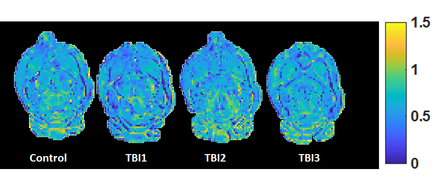
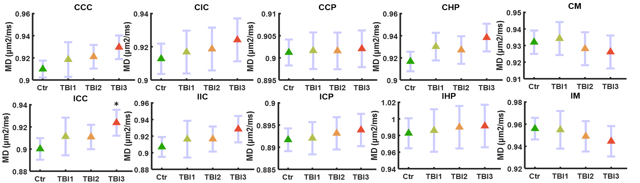
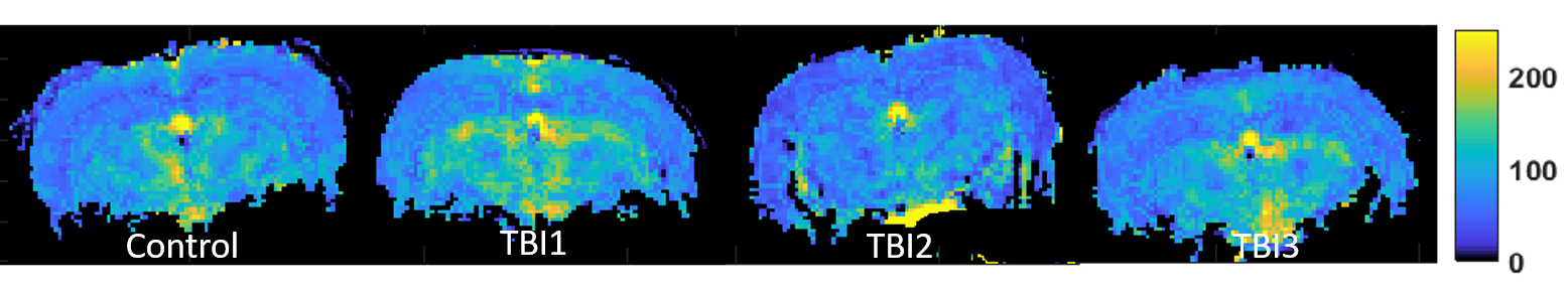
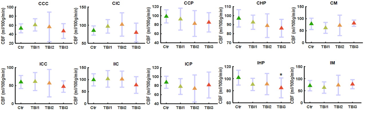
The diffusion kurtosis metric, WT (Fig. 1(a)) shows a significant decrease in the ipsilateral hippocampus (IHP) region after the third mTBI at day 7 in comparison to control (p<0.01). (Fig. 1(b)). No other DKI metrics showed any significant alterations in any other ROIs of the brain after repetitive mTBI. Among DDE metrics only MD showed a significant increase in the ipsilateral corpus callosum (ICC) after the third mTBI on day 7 (p<0.05) (Fig 2). Changes in AD and MD in CHP, MD in CCC, FA in IHP and RD in ICC and IIC did not survive FDR correction. CBF maps (Fig 3 (a)) also shows a significant reduction in the IHP after the third mTBI at day7 in comparison to control (p<0.05) (Fig 3(b)). Previous studies observed a significant alteration in kurtosis metrics [9]. Such changes may be due to axonal injury and/or gliosis in the hippocampus, and reduction in CBF may reflect mTBI induced ischemia [2, 10]. The significant decrease in MD of ICC may indicate reduced cell density in the ICC. This finding also supports previous observations regarding the sensitivity of DTI metrics to axonal injury [11]. In conclusion, our data suggest that the ICC and IHP are sensitive to repetitive mTBI and that multimodal imaging is useful to assess its functional and microstructural repercussions.Acknowledgements
Lundbeck Foundation grant and SimonFougner Hartmanns Familiefond. AC, ARK, and BH acknowledge support from NIH 1R01EB012874-01. The authors wish to thank Lippert’s Foundation, Korning’s Foundation and the Augustinus Foundation for financial support. The 9.4T lab was made possible by funding from the Danish Research Council's Infrastructure program, the Velux Foundations, and the Department of Clinical Medicine, AU. Centre for Stochastic Geometry and Advanced Bioimaging is supported by Villum FoundationReferences
1. Angoa‐Pérez, M., et al., Animal models of sports‐related head injury: bridging the gap between pre‐clinical research and clinical reality. Journal of Neurochemistry, 2014. 129(6): p. 916-931.
2. Østergaard, L., et al., Capillary transit time heterogeneity and flow-metabolism coupling after traumatic brain injury. Journal of Cerebral Blood Flow & Metabolism, 2014. 34(10): p. 1585-1598.
3. Budde, M.D., and N.P. Skinner, Diffusion MRI in acute nervous system injury. J Magn Reson, 2018. 292: p. 137-148.
4. Kane, M.J., et al., A mouse model of human repetitive mild traumatic brain injury. Journal of neuroscience methods, 2012. 203(1): p. 41-49.
5. Khan, A.R., et al., Diffusion MRI and MR spectroscopy reveal microstructural and metabolic brain alterations in chronic mild stress exposed rats: A CMS recovery study. Neuroimage, 2018. 167: p. 342-353.
6. Jespersen, S.N., et al., Orientationally invariant metrics of apparent compartment eccentricity from double pulsed field gradient diffusion experiments. NMR in Biomedicine, 2013. 26(12): p. 1647-1662.
7. Lawrenz, M., M.A. Koch, and J. Finsterbusch, A tensor model and measures of microscopic anisotropy for double-wave-vector diffusion-weighting experiments with long mixing times. Journal of Magnetic Resonance, 2010. 202(1): p. 43-56.
8. Tsekos, N.V., et al., Quantitative measurements of cerebral blood flow in rats using the FAIR technique: Correlation with previous lodoantipyrine autoradiographic studies. Magnetic resonance in medicine, 1998. 39(4): p. 564-573.
9. Zhuo, J., et al., Diffusion kurtosis as an in vivo imaging marker for reactive astrogliosis in traumatic brain injury. Neuroimage, 2012. 59(1): p. 467-477.
10. Graham, D., J.H. Adams, and D. Doyle, Ischaemic brain damage in fatal non-missile head injuries. Journal of the neurological sciences, 1978. 39(2): p. 213-234.
11. Mac Donald, C.L., et al., Diffusion tensor imaging reliably detects experimental traumatic axonal injury and indicates approximate time of injury. Journal of Neuroscience, 2007. 27(44): p. 11869-11876.
Figures