3430
Abnormal brain white matter volume underlying methamphetamine abusers: A machine learning approach1Department of Radiology, Shenzhen Mental Health Center, Shenzhen Kangning Hospital, Shenzhen, China, 2Department of Drug Dependence, Shenzhen Kangning Hospital, Medicine Division of Shenzhen University, Shenzhen, China, 3GE Healthcare, MR Research China, Beijing, China
Synopsis
The disruption of specific microstructural features of white-matter (WM) has been observed in methamphetamine (MA) abusers. However, it remains unknown whether WM volume is abnormal in MA abusers. To address this issue, a machine learning approach was applied in this study to differentiate between 21 MA abusers and 13 age- and gender- healthy controls. Our results showed that a linear support vector machine classifier achieved an accuracy of 73.53% using the white matter volume as input features. Particularly, the most discriminative WM regions included pontine crossing tract, motor system and the reading related network.
Introduction
The impairment of micro-structural changes in white-matter (WM) tracts/regions have been reported in MA abusers 1-5. Reduced fractional anisotropy (FA) may occur as a result of demyelination, as well as from axonal damage. However, previous studies focused on the deficits of microstructural features (i.e., WM diffusion parameters) and ignored macrostructural features, such as WM volume (WMV). In addition, it remains unknown whether WMV can be used to discriminate MA abusers from healthy controls at the individual level, which is an important clinical problem for identifying MA individuals. Therefore, a linear support vector machine (LSVM) method was applied to classify MA abusers from healthy controls using the WMV as input features in this study.Method
21 MA abusers and 13 age- and gender- healthy controls were recruited through advertisements on the website and at the Kangning Hospital Drug Dependence Department. T1-weighted images were acquired using 3D BRAVO sequence (Discovery MR750, GE, WI) equipped with a standard 8-channel head coil. DTI data was acquired with the following parameters: 64 non-collinear directions using b-value images 10 and 1000 s/mm2; TR= 8724 ms, TE= 81.4 ms, matrix =112 × 112, FOV = 224 mm × 224 mm, 75 contiguous slices acquired interleaved; Each slice was 2 mm thick, no gap.Inclusion criteria for MA abusers were: (1) aged 19–55 years, (2) lifetime diagnosis of DSM-IV MA dependence, as determined by the Structured Clinical Interview for DSM-IV (SCID-IV). Exclusion criteria for MA abusers and healthy controls were: (1) lifetime significant medical illness such as hypertension, hepatitis, and diabetes mellitus, (2) comorbid Axis I psychiatric disorders, as determined by SCID-IV, (3) antisocial or borderline personality disorders, as identified by the Personality Disorder Questionnaire-4, (4) lifetime exposure to any other DSM-IV dependence- or abuse-related drugs, (5) Contraindications to MR scanning. The WMV maps of all subjects were estimated using the VBM8 toolbox (http://dbm.neuro.uni-jena.de/vbm/). After that, a machine approach was applied to those WMV maps using the PRoNTo toolbox (http://www.mlnl.cs.ucl.ac.uk/pronto/) with a LSVM method. The leave-one-out cross-validation (LOOCV) was adopted to evaluate the classification performance, which provides a good estimation for the generalizability of the classifiers, particularly when the sample size is small 6. The whole procedure above was performed according to the previous study 7.Results
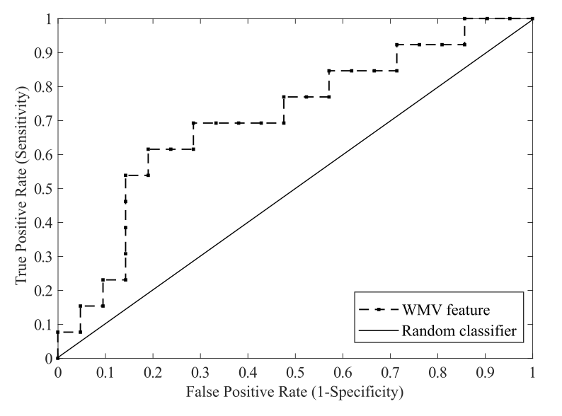
Our results showed that the LSVM classifier discriminated MA abusers from controls using the WMV achieved an accuracy with 73.53%. The classification results are shown as an ROC curve using each subject’s classification score as a threshold in Figure 1. The AUC was 0.71, which was significantly higher than chance (P < 0.001), indicating a worthy discriminative power (Figure 1). Furthermore, the top 10 discriminative WMV regions were shown in the Table 1, including two bilateral WM regions (pontine crossing tract, genu of corpus callosum and body of corpus callosum), two right WM regions (corticospinal tract and sagittal stratum) as well as five left WM regions (retrolenticular part of internal capsule, posterior thalamic radiation, superior longitudinal fasciculus, superior corona radiata and uncinate fasciculus).Discussion
Our study demonstrated that MA abusers could be distinguished from healthy controls by using WMV, indicating the effect of WMV and supporting the deficit theory for MA abusers. Moreover, this finding also provided us an alternative way for identifying MA abuse individuals. In addition, the discriminative WM regions detected in our data were mainly associated with the reading-related network (the left superior longitudinal fasciculus, left superior corona radiata, and corpus callosum), which involving the frontal, temporo-parietal, occipito-temporal, and thalamocortical regions 8. These regions were found to be highly associated with reading development in normal children 9. A meta-analysis revealed the neuropsychological effects of MA abusers, showing deficits in motor skills, language, and visuoconstructional abilities 10. Therefore, the abnormalities of discriminative WM regions may be considered as potential explanatory factors of cognitive dysfunction in the MA abusers. Otherwise, our study was achieved with a small cohort and should be contained more WM features to offer an optimal implication in clinical setting.Conclusion
Our results demonstrated that the WMV might be a useful feature for the detection of MA abuse individuals.Acknowledgements
References
1. Chung A, Lyoo IK, Kim SJ, Hwang J, Bae SC, Sung YH, Sim ME, Song IC, Kim J, Chang KH et al: Decreased frontal white-matter integrity in abstinent methamphetamine abusers. Int J Neuropsychopharmacol 2007, 10(6):765-775.
2. Chang L, Alicata D, Ernst T, Volkow N: Structural and metabolic brain changes in the striatum associated with methamphetamine abuse. Addiction 2007, 102 Suppl 1:16-32.
3. Kim IS, Kim YT, Song HJ, Lee JJ, Kwon DH, Lee HJ, Kim MN, Yoo DS, Chang Y: Reduced corpus callosum white matter microstructural integrity revealed by diffusion tensor eigenvalues in abstinent methamphetamine addicts. Neurotoxicology 2009, 30(2):209-213.
4. Salo R, Nordahl TE, Buonocore MH, Natsuaki Y, Waters C, Moore CD, Galloway GP, Leamon MH: Cognitive Control and White Matter Callosal Microstructure in Methamphetamine-Dependent Subjects: A Diffusion Tensor Imaging Study. Biological Psychiatry 2009, 65(2):122-128.
5. Salo R, Fassbender C: Structural, functional and spectroscopic MRI studies of methamphetamine addiction. Curr Top Behav Neurosci 2012, 11:321-364.
6. Pereira F, Mitchell T, Botvinick M: Machine learning classifiers and fMRI: a tutorial overview. Neuroimage 2009, 45(1 Suppl):S199-209.
7. Cui Z, Xia Z, Su M, Shu H, Gong G: Disrupted white matter connectivity underlying developmental dyslexia: A machine learning approach. Hum Brain Mapp 2016, 37(4):1443-1458.
8. Ben-Shachar M, Dougherty RF, Wandell BA: White matter pathways in reading. Curr Opin Neurobiol 2007, 17(2):258-270.
9. Myers CA, Vandermosten M, Farris EA, Hancock R, Gimenez P, Black JM, Casto B, Drahos M, Tumber M, Hendren RL et al: White matter morphometric changes uniquely predict children's reading acquisition. Psychological science 2014, 25(10):1870-1883.
10. Scott JC, Woods SP, Matt GE, Meyer RA, Heaton RK, Atkinson JH, Grant I: Neurocognitive effects of methamphetamine: a critical review and meta-analysis. Neuropsychol Rev 2007, 17(3):275-297.
Figures