3429
A fixel-based analysis of diffusion properties of white matter fiber in Williams Syndrome1Neuroscience Research Australia, Sydney, Australia, 2School of Medical Sciences, University of New South Wales, Sydney, Australia, 3Centre for Research in Atypical Neurodevelopment, Macquarie University, Sydney, Australia, 4T.Y. Nelson Department of Neurology, Children's Hospital at Westmead, Sydney, Australia
Synopsis
Williams Syndrome (WS) is a genetic neurodevelopmental disorder produced by a hemideletion of around 26 genes on chromosome 7 (7q11.23) that leads to unique changes in physical and cognitive profiles. Behavioural characteristics including hyper-sociability, excessive friendliness and empathy, sensitivity to loud noises and visual-spatial construction deficits infer unique brain structure and connections. WS is also characterised by reduced muscle tone and a decrease in motor coordination and balance1. We show significant differences in the corticospinal tract in WS participants and that white matter changes previously reported are more likely to be due to morphological changes to the fiber bundles rather than microstructural ones.
Background
Previous diffusion analyses of the structural connections of white matter in the WS brain have largely been on small cohorts (n<10) and used the tensor model to measure fractional anisotropy (FA) to infer axonal and myelination properties. However, the tensor model greatly simplifies white matter neuroanatomy and suffers from unreliable measures in regions of crossing fibers. Fixel-based analysis is a method to extract quantitative measures from white matter fiber bundles. The measures of fiber density (FD), fiber cross-section (FC) and combinations of both (FDC) are tract-specific quantities enabling direct inference on morphological and microstructural changes within white matter fiber bundles.Aim
The aim of the study was to apply a fixel-based analysis to a set of diffusion images from a cohort of WS participants to investigate these unique structural networks with these new measures of white matter integrity.Methods
Diffusion images were obtained on 21 Williams Syndrome participants aged between 17 and 53 years (median 20.0, mean 24.6) and a control group of 20 participants aged between 15 and 61 years (median 24.3, mean 26.8). High angular resolution, high b-value diffusion images (HARDI) were acquired on a Philips Achieva TX 3T system at Neuroscience Research Australia (46-directions, b-value 2400 s/mm2, TR = 8000ms, TE = 73ms, voxel size = 2.5mm isotropic, 55 slices, SENSE factor 2).
Fixel-based analysis2,3 was performed to identify and compare differences in the tract-specific quantities of FD, FC and FDC using MRTrix. Statistical analysis between WS and control groups were performed using a study-derived template of all participants. Family-wise error corrected p-values were assigned to each fixel in each voxel using non-parametric testing (5000 permutations) and results were corrected for multiple comparisons using connectivity-based fixel enhancement. Probabilistic tractography maps were cropped to display streamlines corresponding to significant fixels.
Results
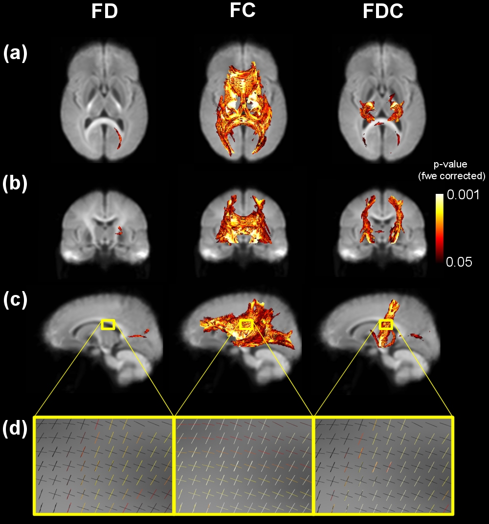
Significant differences (negative, p<0.05) in the FDC measure were found between WS and controls in the corticospinal tract as shown in the (a) axial, (b) coronal and (c) sagittal images in Figure 1. An example magnified region of interest is shown in Figure 1d to demonstrate significant fixels are associated with the corticospinal tract, not the crossing fibres coming from the corpus callosum.
FDC is a combination of fiber density and fiber cross section and further examination of these individual measures reveal the major contribution comes directly from FC that showed significant differences across the whole brain. The only region of significant difference in FD was a small section of the inferior fronto-occipital fasciculus.
Discussion and Conclusion
This study represents the first fixel-based (and tensor-free)
analysis to measure diffusion properties of white
matter in Williams Syndrome. The large areas of significant differences in FC
support previous results4,5 that showed a decrease in overall white
matter volume in WS. These same studies using the diffusion tensor model also
showed differences in the corticospinal tract corresponding to a decrease in
fractional anisotropy.
In combination with FD measures we demonstrate that the structural differences, specifically within the corticospinal tract are due to changes in the overall cross-section of fiber bundles rather than any microstructural changes such as demyelination. These findings add new information to the unique WS structural network and the role the deleted genes play in brain formation.
Acknowledgements
This work was supported by Australia’s National Imaging Facility (NIF) and Williams Syndrome Australia Limited.References
1. Gabriella Brawn, Melanie Porter, Adaptive Functioning in Williams Syndrome: A Systematic Review, Intl Journal of Disability and Education, Volume 65, 2017, p123
2. David A. Raffelt, J.-Donald Tournier, Robert E. Smith, David N. Vaughan, Graeme Jackson, Gerard R. Ridgway, Alan Connelly, Investigating white matter fibre density and morphology using fixel-based analysis, In NeuroImage, Volume 144, 2017, p58
3. David A. Raffelt, Robert E. Smith, Gerard R. Ridgway, J-Donald Tournier, David N. Vaughan, Stephen Rose, Robert Henderson, Alan Connelly, Connectivity-based fixel enhancement: Whole-brain statistical analysis of diffusion MRI measures in the presence of crossing fibres, In NeuroImage, Volume 117, 2015, p40
4. Andreia V. Faria, Barbara Landau, Kirsten M. O’Hern, Xin Li, Hangyi Jiang, Kenichi Oishi, Jiangyang Zhang, Susumu Mori, Quantitative Analysis of Gray and White Matter in Williams Syndrome, Neuroreport, Volume 23, 2012, p283
5. Stefano Marenco, Michael A. Siuta, J. Shane Kippenhan, Samuel Grodofsky, Wei-li Chang, Philip Kohn, Carolyn B. Mervis, Colleen A. Morris, Daniel R. Weinberger, Andreas Meyer-Lindenberg, Carlo Pierpaoli, Karen Faith Berman, Genetic contributions to white matter architecture revealed by diffusion tensor imaging in Williams syndrome, PNAS, Volume 104, 2007, p15117
Figures