3428
High Spatial Resolution Multi-shell DTI in Children and Young Adults Prenatally Exposed to Alcohol1Biomedical Engineering, University of Alberta, Edmonton, AB, Canada
Synopsis
Diffusion tensor imaging (DTI) has identified white matter differences in individuals with prenatal alcohol exposure (PAE). The current study uses higher spatial resolution of 1.5 mm isotropic (3.4 mm3 voxels) and larger b-values of 2000 s/mm2 (in addition to typical 1000 s/mm2) to assess the brain in children to young adults with PAE. Using the ICBM DTI template regions-of-interest for white matter, the b2000 tensor metrics (but not b1000) showed group differences in the left medial lemniscus and left stria terminalis, the latter also showing correlations with age in the control group but not the PAE group.
Introduction
Prenatal alcohol exposure (PAE) during pregnancy has been shown by MRI, including diffusion tensor imaging (DTI), to negatively impact brain structure such as white matter [1,2]. Most DTI studies in PAE have used low spatial resolution (≧2 mm isotropic voxels) or single-shell acquisitions with a typical b-value of 1000 s/mm2. One PAE study in 8-18 year old used a TBSS skeleton approach on the tensor calculated from images with b-value of 2000 s/mm2 and 4 mm slices (~14 mm3 voxels) to demonstrate widespread reductions of fractional anisotropy (FA) in white matter, particularly in those diagnosed with the most ‘severe’ diagnosis of fetal alcohol syndrome (FAS) [3]. The purpose here is to assess white matter in PAE using diffusion tensor parameters derived from either b-values of 1000 or 2000 s/mm2 acquired with a high spatial resolution of 1.5 mm isotropic.Methods
Children and young adults with PAE (n=18; 11 males; 14.4±3.7 years, 8.4.- 23.2 years) and controls (n=25; 12 males; 15.0±4.4 years, 8.2 – 23.6 years) were scanned on a Siemens PRISMA 3T with the following diffusion protocol: 5:59 min scan time, 90 1.5 mm axial slices, 1.5x1.5 mm2 in-plane zero-filled to 0.75x0.75mm2, multiband 2, TR 4700 ms, TE 64 ms, 6 b0, 30 b 1000 s/mm2 and 30 b 2000 s/mm2. Diffusion data was preprocessed for Gibbs ringing, motion/distortion, and eddy current correction (ExploreDTI v4.8.6). An automated ROI approach using the 1 mm ICBM template provided in ExploreDTI was used to extract FA, mean (MD), axial (AD) and radial diffusivity (RD) from 48 white matter regions (21 bilateral, 6 commissural). The same procedure was used for the b1000 and b2000 shells separately; linear correlations of the tensor parameters from b1000 were compared to b2000 in all 43 participants. An ANOVA was used for group comparisons (false discovery rate, FDR, corrected p-value threshold range of 0.0002-0.002). Linear correlations of diffusion metrics versus age were evaluated per group on ROIs with significant group differences in diffusion parameters (p<0.05, no FDR correction applied since ROIs defined by previous analysis).Results
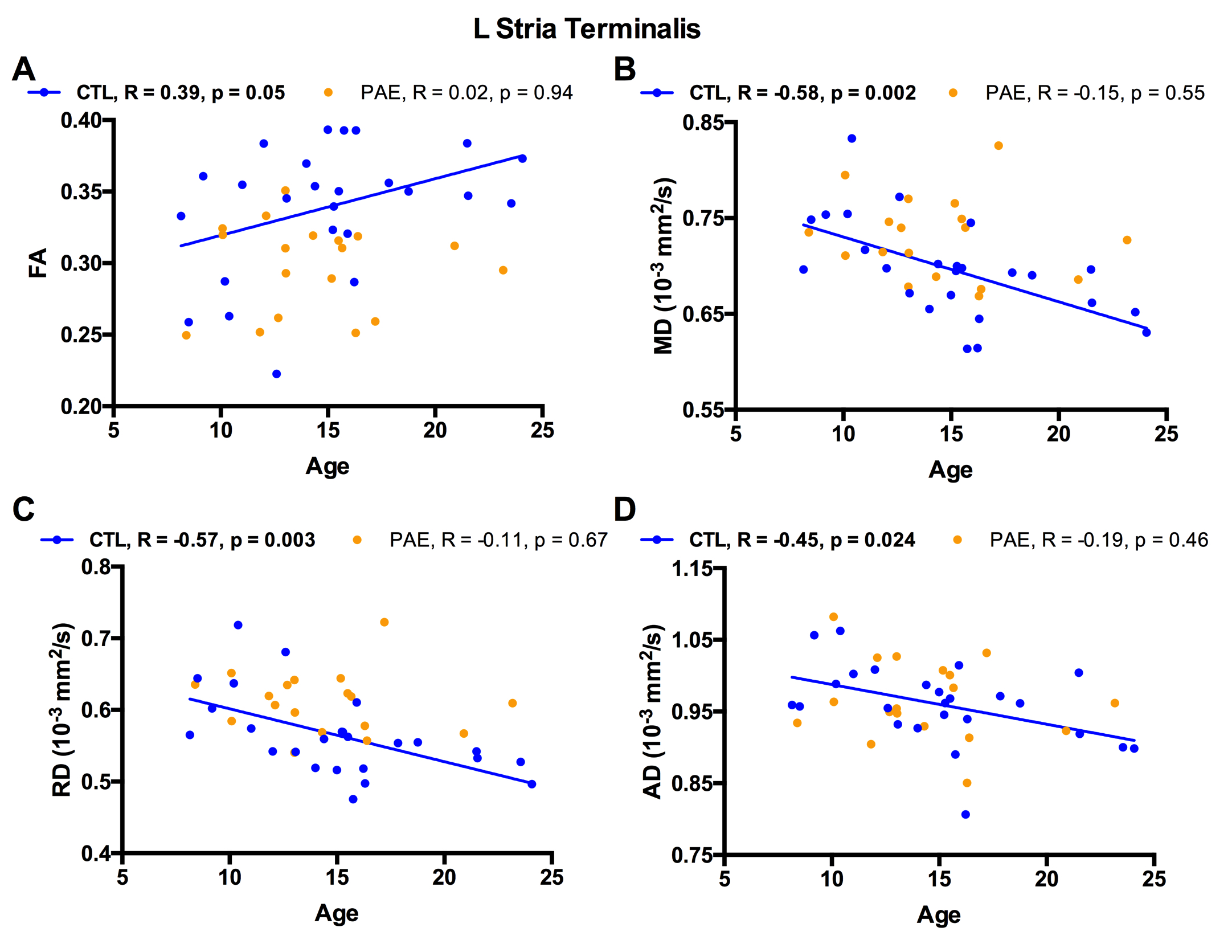
The b2000 shell yielded tensor parameters that correlated highly with the same metrics derived from the b1000 shell (FA: R=0.89±0.05, MD: R=0.87±0.07, RD: R=0.90±0.06, AD: R=0.83±0.08 over all 48 regions) in all the participants; however, all metrics were systematically underestimated on b2000 (Figure 1 for FA and MD). PAE showed group differences with higher FA and higher AD of the left Medial Lemniscus (left ML; Figure 2A) and lower FA of the left Stria Terminalis (left ST; Figure 2B) obtained from the b2000 shell; note that the b1000 shell did not show any FDR-corrected significant group differences. All four b2000-derived diffusion parameters of the left ST were significantly correlated with age in controls, while no age correlations were found in PAE (Figure 3). There were no significant correlations of diffusion parameters in the left ML with age in either group.Discussion and Conclusions
The only other study that used b2000 on a PAE population found widespread FA reductions throughout the white matter [3] unlike our findings of only two left regions surviving FDR correction. However, that study had a 5x worse spatial resolution and was therefore more sensitive to partial volume effects [4], especially problematic in a population where brain atrophy can be present [5]. Also, this earlier study showed that those diagnosed with the most severe FAS form had greater FA reductions from controls than their PAE group [3]. The 3D ROI atlas-based approach used here (with the ICBM-Mori DTI template [6]) provides an increase SNR as it averages diffusion parameters over a larger number of voxels. The proper alignment of ROIs over individually registered brains is a constant challenge in automated methods. Manual inspection was performed here and deemed sufficient despite a wide age range over a critical brain development period. Although we are the first to report abnormalities in left ML and left ST in PAE, converging results with b2000 can be observed as the Fryer study showed decreased FA in left sagittal stratum, a region in very close proximity to the left ST region where we also show a decrease in FA in the PAE group. The lack of age correlation in PAE participants also supports previous longitudinal findings of altered white matter development in this population [7]. Finally, this study further highlight the heterogeneity of metrics and regions that differs from controls, which may stem from methodological issues, but mostly diffuse effect of alcohol on brains and variability within this population.Acknowledgements
Canada Research Chairs, Canadian Institutes of Health Research (CIHR)References
[1] Lebel C, Roussotte F, Sowell ER. Imaging the impact of prenatal alcohol exposure on the structure of the developing human brain. Neuropsychol Rev 2011;21:102–18. doi:10.1007/s11065-011-9163-0.
[2] Ghazi Sherbaf F, Aarabi MH, Hosein Yazdi M, Haghshomar M. White matter microstructure in fetal alcohol spectrum disorders: A systematic review of diffusion tensor imaging studies. Hum Brain Mapp 2018;598:143. doi:10.1002/hbm.24409.
[3] Fryer SL, Schweinsburg BC, Bjorkquist OA, Frank LR, Mattson SN, Spadoni AD, et al. Characterization of white matter microstructure in fetal alcohol spectrum disorders. Alcohol Clin Exp Res 2009;33:514–21. doi:10.1111/j.1530-0277.2008.00864.x.
[4] Vos SB, Jones DK, Viergever MA, Leemans A. Partial volume effect as a hidden covariate in DTI analyses. Neuroimage 2011;55:1566–76. doi:10.1016/j.neuroimage.2011.01.048.
[5] Treit S, Chen Z, Zhou D, Baugh L, Rasmussen C, Andrew G, et al. Sexual dimorphism of volume reduction but not cognitive deficit in fetal alcohol spectrum disorders: A combined diffusion tensor imaging, cortical thickness and brain volume study. Ynicl 2017;15:284–97. doi:10.1016/j.nicl.2017.05.006.
[6] Mori S, Oishi K, Jiang H, Jiang L, Li X, Akhter K, et al. Stereotaxic white matter atlas based on diffusion tensor imaging in an ICBM template. Neuroimage 2008;40:570–82. doi:10.1016/j.neuroimage.2007.12.035.
[7] Treit S, Lebel C, Baugh L, Rasmussen C, Andrew G, Beaulieu C. Longitudinal MRI reveals altered trajectory of brain development during childhood and adolescence in fetal alcohol spectrum disorders. J Neurosci 2013;33:10098–109. doi:10.1523/JNEUROSCI.5004-12.2013.
Figures