3427
Alterations of brain white matter tracts in children with abnormal intelligence quotient1Department of Radiology, the First Affiliated Hospital, Xi’an Jiaotong University, Xi'an, China, Xi’an, China, 2the First Affiliated Hospital, Xi’an Jiaotong University, Xi'an, China, Xi'an, China, 3MR Research China, GE Healthcare, Bei Jing, People's Republic of China, Bei Jing, China
Synopsis
Studies have shown that brain white matter microstructure is of crucial importance for intelligence. Here, we detailed the alterations of brain white matter tracts in children with abnormal intelligence quotient by comparing the difference of brain white matter tract between the two groups and exploring correlations with intelligence scores based on DTI parameters. Significant differences of ILF, IFO and SLF were found in children with abnormal intelligence quotient. And all these tracts were strongly correlated with intelligence quotient, suggesting the underlying structural origins of lower cognitive function.
Introduction
Intelligence
is a very general capability that involving the ability to reason, plan, solve
problems, think abstractly, comprehend complex ideas, learn quickly and learn
from experience, which are associated with important life outcomes, including
school and occupational attainment, social mobility and better health1.
In particular, early childhood studies are of crucial importance for their
long-term prognosis. Studies on healthy children have shown that brain white
matter tract (corticospinal tract2, inferior fronto-occipital
fasciculus3, inferior longitudinal fasciculus3, superior
longitudinal fasciculus2 et al) is critical for intelligence. While
less is known regarding alterations of brain white matter microstructure in
children with abnormal intelligence quotient (AI), which was evaluated in this
study.Materials and Methods
The Institutional Review Broad of the first author’s affiliation approved this study and written informed consent were obtained from parents of the children. Subjects Forty-one children with no abnormality on MRI participated in the study, including 13 AI (abnormal intelligence quotient) children and 28 controls. AI children were defined as full scale IQ <85 scores. Intellectual ability was assessed by the Chinese Wechsler Young Children scale of Intelligence (C-WYCSI) for children aged 4 to 6 years and Chinese Wechsler Intelligence Scale for Children (C-WISC) children aged 6 to 18 years. MR Protocols All subjects were examined by using a 3.0T scanner (Signa HDxt, General Electric Medical System, Milwaukee, WI, USA) with an 8-channel head coil. Data acquisition included three-dimensional fast spoiled gradient-echo T1-weighted sequence (TR/TE, 10.2ms/4.6ms; NEX, 1; isotropic 1×1×1mm3; FOV, 24cm) and transverse fast spin-echo T2-weighted sequence (TR/TE, 4200ms/113ms; NEX of 1.5; matrix, 320×320; thickness, 4mm; FOV, 24cm), followed by a DTI (30 directions; b value, 600s/mm2; TR/TE, 11000ms/67.4ms; NEX, 1; thickness, 2.5mm; FOV, 24cm; matrix, 172×172). Data and statistical analysis DTI raw data were preprocessed by FMRIB software library (FSL; http://www.fmrib.ox.ac.uk/fsl) and four parameters of fractional anisotropy (FA), mean diffusivity (MD), axial diffusivity (AD), and radial diffusivity (RD) were calculated by using the FMRIB Diffusion Toolbox (http://fsl.fmrib.ox.ac.uk/fsl/fslwiki/FDT). Here we use the Johns Hopkins white matter tracts atlas as a reference. Between-group differences in the DTI parameters of the white matter tracts were calculated by means of independent samples t test. Considering the possible impacts of age, partial Pearson’s correlation analysis were used to assess the correlation between FSIQ scores and DTI parameters of these tracts in total children (controlling for age).
All statistical analysis was performed by using SPSS 18.0 (SPSS, Chicago, IL, USA); p<0.05 was considered as statistically significant difference.
Results
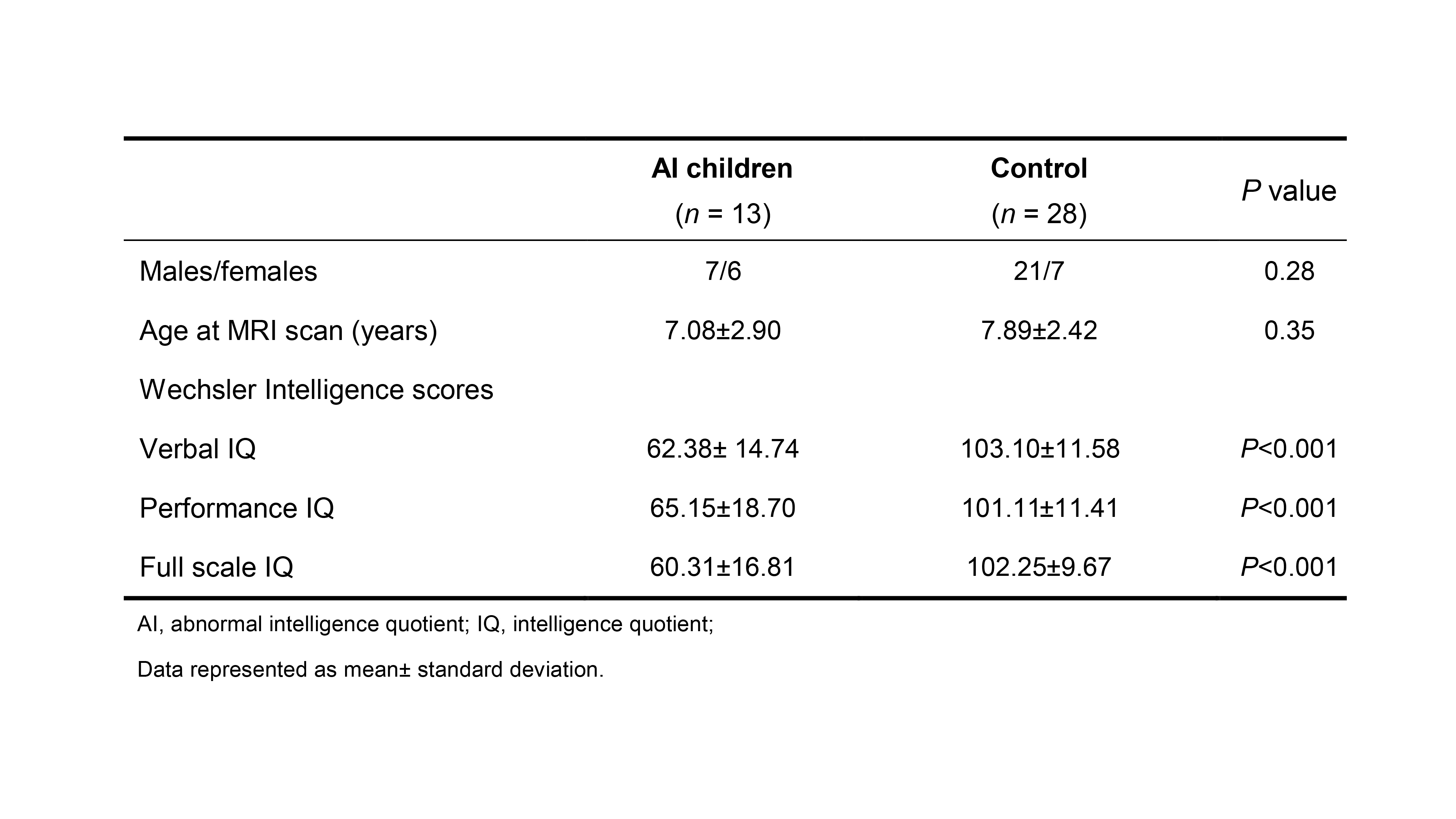
Forty-one children were included in this study. Significant differences presented in VIQ (verbal intelligence quotient), PIQ (performance intelligence quotient) and FSIQ between the two groups, while no differences were observed in age at MRI scan and sex (Table 1).
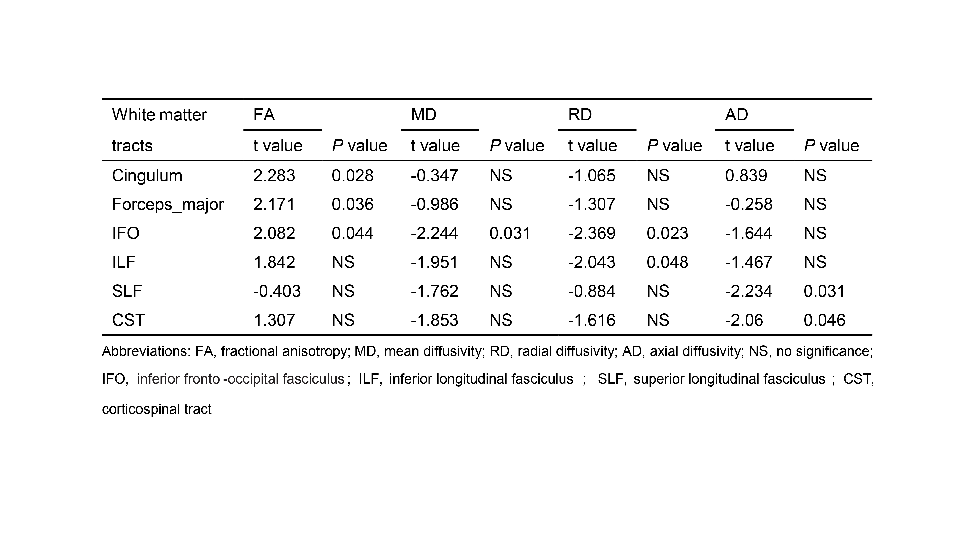
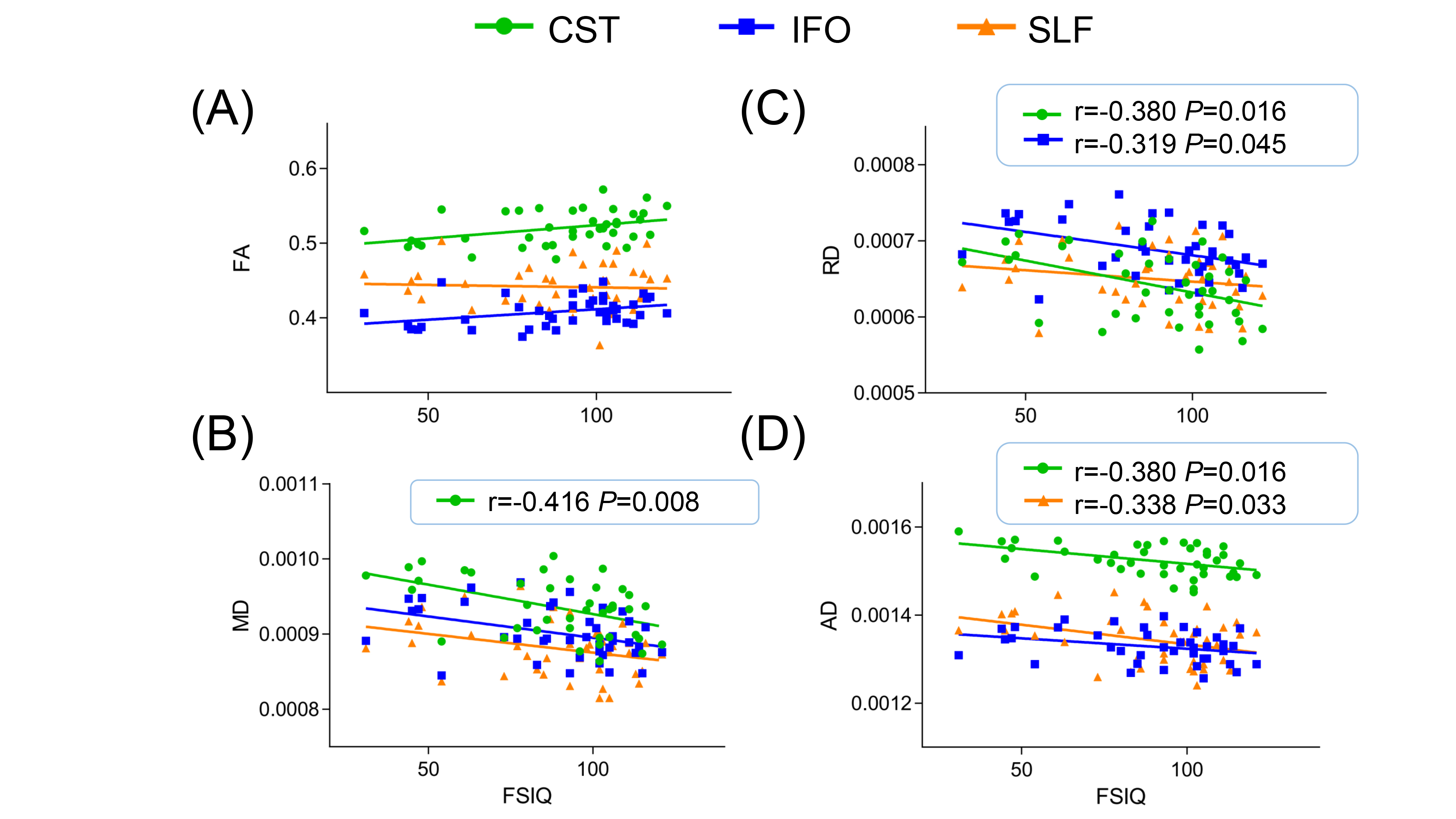
Table 2 shows the differences between children with abnormal intelligence quotient and control group in the four parameters of the white matter tracts. There was a significant group effect on the FA of the cingulum, IFO and forceps-major, and RD of IFO and ILF. Some differences presented on AD in the SLF and CST. Slightly weak correlation between RD and FSIQ score was found in IFO (r=-0.319, P=0.045) and CST (r=-0.380, P=0.016). And similar relationships between AD and FSIQ score can be found in SLF (r=-0.338, P=0.033) and CST (r=-0.380, P=0.016). Significant negative correlation between MD and FSIQ score was found in CST (PCC=-0.416, P=0.008). While no correlation between FA and FSIQ score was found. (Figure 1)
Discussion
This study detailed that significant differences on the FA of the cingulum, IFO and forceps-major, and RD of IFO and ILF, indicating that impaired axonal fiber integrity3 and myelin loss4 in children with abnormal intelligence quotient. While some differences on AD in the SLF and CST may suggest that alterations in overall fiber density and complexity5. Furthermore, all above tracts correlated with intelligence scores. The results are consistent with previous studies2,3. Although the results of DTI parameters are not completely consistent, they still reflect the differences between the two groups. In addition, no relationship was found in FA and FSIQ score. The small sample size may account of these results, so a larger one may expect more powerful evidence for supporting this research.Conclusions
Children with abnormal intelligence quotient show alterations of brain white matter microstructure especially in ILF, SLF and IFO, suggesting the underlying structural origins of lower cognitive function. Alterations of these white matter tracts in the early stage may be a neural foundation for abnormal intelligence (intellectual disability). Therefore, early detection of lesions and implementation of relevant interventions can initiate brain plasticization to raise the level of cognitive function.Acknowledgements
This work was supported by the National Key Research and Development Program of China (2016YFC0100300), National Natural Science Foundation of China (No. 81471631, 81771810 and 51706178), the 2011 New Century Excellent Talent Support Plan of the Ministry of Education, China (NCET-11-0438) the Clinical Research Award of the First Affiliated Hospital of Xi’an Jiaotong University (No.XJTU1AF-CRF-2015-004)References
- Deary I J, Penke L, Johnson W. The neuroscience of human intelligence differences. Nat Rev Neurosci, 2010, 11(3): 201.
- Tamnes C K, Østby Y, Walhovd K B, et al. Intellectual abilities and white matter microstructure in development: a diffusion tensor imaging study. Human brain mapping, 2010, 31(10): 1609-1625.
- Chiang M C, Barysheva M, Shattuck D W, et al. Genetics of brain fiber architecture and intellectual performance. J Neurosci, 2009, 29(7): 2212-2224.
- Song S K, Sun S W, Ju W K, et al. Diffusion tensor imaging detects and differentiates axon and myelin degeneration in mouse optic nerve after retinal ischemia. Neuroimage, 2003, 20(3): 1714-1722.
- Dubois J, Dehaene-Lambertz G, Kulikova S, et al. The early development of brain white matter: a review of imaging studies in fetuses, newborns and infants. Neuroscience, 2014, 276: 48-71.
Figures