3425
Reproducibility of the diffusion of the perivascular space in older adults with dementia.1University of Melbourne, Melbourne, Australia, 2Royal Melbourne Hospital, Melbourne, Australia
Synopsis
Recently, there has been interest in the glymphatic system and its role in flushing amyloid ß along with other waste products in the brain. New imaging techniques are being developed to try and measure such activity. Recently, diffusion tensor MRI was used to construct an index "DTI-ALPS" to help link dementia burden with diffusion in the perivascular space. We aimed to replicate this study in 36 patients (16 AD, 16 MCI, and 4 SMC). Significant correlations were found between DTI-ALPS and stratified Mini-Mental State Examination score. Further work is needed to evaluate the feasibility of MRI to measure glymphatic activity.
Introduction
Recently, there has been considerable interest in the glymphatic system [1-3] (the functional waste clearance pathway for the CNS) and its role in flushing solutes (such amyloid ß and tau), metabolic and other cellular waste products in the brain. It has been proposed that a failure in the clearance of soluble β-amyloid from the brain’s interstitial space contributes to accumulation of amyloid plaques and AD progression [4-6] and may be used as a biomarker of AD. Measurements of glymphatic activity however have been primarily restricted to animal studies due to the need for intrathecal administered tracers.However, a range of new techniques are being investigated to help grade glymphatic function [7-11]. One recent study [12] applied MRI diffusion tensor imaging. It identified projection and association fibres orthogonal to the perivascular space at the level of the lateral ventricle body, and developed an index “DTI-ALPS” that measured water diffusion along this space which could be used as a proxy marker to assess glymphatic function. They showed significant positive correlations between DTI-ALPS and Mini-Mental State Examination (MMSE), as well significant negative correlations between the mean diffusivities of the projection and association fibres vs MMSE. The purpose of this study was to evaluate and replicate this method for measuring diffusion along the perivascular space in 36 patients (mean age 73.2, 16 AD, 16 MCI and 4 SMC) and investigate its association with cognitive function.Methods
All 36 participants underwent MR imaging following recruitment on a Siemens 3.0T Tim Trio as part of the AIBL-Active study [13] and Velacor study [14]. Single-SE diffusion weighted EPI were acquired: TR/TE = 8700/92 ms, FOV 240 x 240 mm, matrix 96 x 96, b = 1000, voxel size 2.5 mm3, 30 directions and SWI (3D GRE, TR=40ms, TE=30ms, matrix 336x512, ⍺=15°). Diffusion MRI data was pre-processed using the TORTOISE software package v3.1.1 [15,16]. The DWIs were AC-PC corrected and deskulled using AFNI [17], corrected for Gibbs ringing, subject motion and eddy-current artefacts, and for EPI susceptibility distortions using DIFFPREP [16]. The diffusion tensor was calculated (informed RESTORE) and both it and the SWI images were then warped to the AC-PC corrected T2 data using ANTS [18]. With FSLEyes [19], the SWI and V1 (primary eigenvector) images were loaded. Two qualified neuro-radiologists with over 30 years experience marked two 3mm spherical ROI’s in the region of the projection and association fibres for both hemispheres and adjacent to the medullary veins at the level of the lateral ventricle. MMSE scores ranged from 14 to 30. Statistical analysis was performed with SPSS 25.0 [20]Results
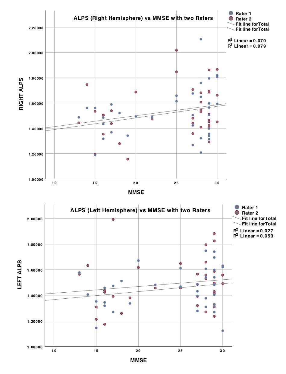
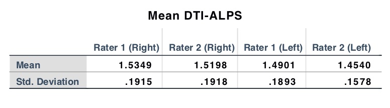
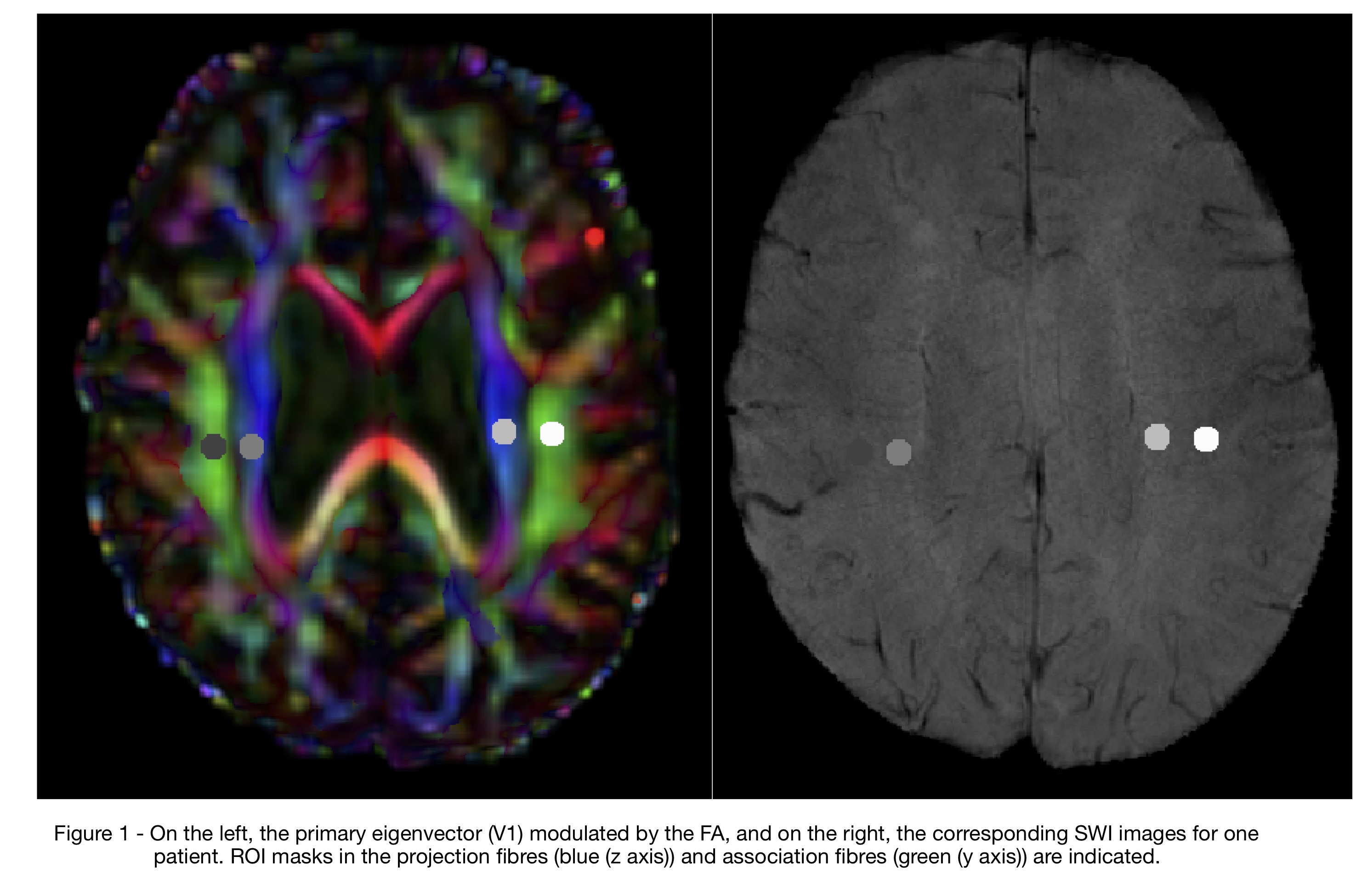
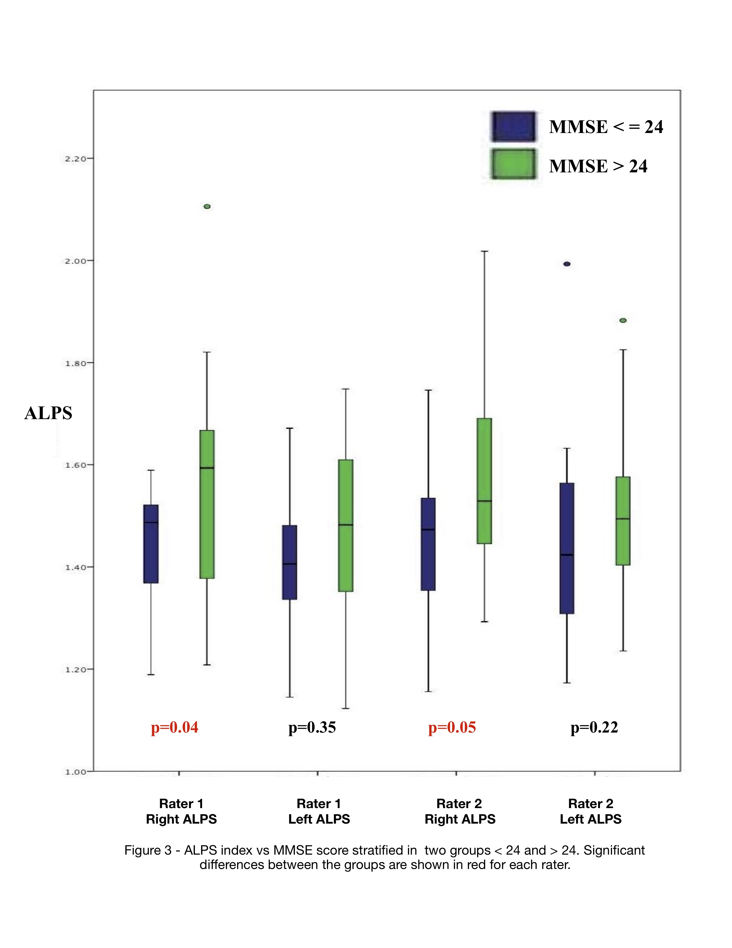
Figure 1 shows the primary eigenvector (V1) modulated by the FA, and overlaid on the SWI images for one patient. ROI masks in the projection fibres (blue (z axis)) and association fibres (green (y axis)) are indicated. Table 1 contains the Mean Values of DTI-ALPS for both raters over the entire cohort. In Figure 2, the DTI-ALPS vs MMSE for both hemispheres and both raters are displayed. No significant correlations between DTI-ALPS and MMSE [as a continuous variable] in either hemisphere were found. In addition, no definite dependencies in mean diffusivity and MMSE in either projection or association fibres were observed, irrespective of rater input. However, stratifying the groups into MMSE > 24 and < 24 [21] yielded significant correlations between the right DTI-ALPS index and MMSE for both raters (see Figure 3). No other significant effects were observed.Discussion
The diffusion along the perivascular space was calculated in 36 patients either with AD at risk of developing it by measuring the DTI-ALPS index. High quality pre-processing of the DTI data and accurate registrations are both paramount in providing reliability of the diffusion tensor input to this index. Some evidence correlating DTI-ALPS with cognition was found but only upon stratification of the MMSE score. There was some variability in placing the location of ROI masks between raters (dice coefficients, 0.2 - 0.9) but which still satisfied the same location criteria. This contributed to small differences in the DTI-ALPs results between raters. It is not clear why correlations with MMSE were only found in one hemisphere.Conclusion
There were significant correlations between MMSE vs DTI-ALPS upon stratification of MMSE. Weak positive trends were also found in DTI-ALPS vs MMSE which may reflect the varying levels of white matter degeneration in the AD/MCI/SMC groups. Further studies are needed to assess the nature of transport in the glymphatic system and whether DTI models are useful in evaluating efflux activity from the perivascular spaceAcknowledgements
No acknowledgement found.References
1/ Jessen, N.A., Munk, A.S.F., Lundgaard, I., Nedergaard, M., 2015. The glymphatic system: a beginner's guide. Neurochem. Res. 40, 2583–2599. https://doi.org/10.1007/ s11064-015-1581-6.
2/ J. J. Iliff, M. Wang, Y. Liao, B. A. Plogg, W. Peng, G. A. Gundersen, H. Benveniste, G. E. Vates, R. Deane, S. A. Goldman, E. A. Nagelhus, and M. Nedergaard, “A Paravascular Pathway Facilitates CSF Flow Through the Brain Parenchyma and the Clearance of Interstitial Solutes, Including Amyloid,” Science Translational Medicine, vol. 4, no. 147, pp. 147ra111–147ra111, 2012.
3/ Kress BT, Iliff JJ, Xia M, Wang M, Wei HS, Zeppenfeld D, Xie L, Kang H, Xu Q, Liew JA, Plog BA, Ding F, Deane R, Nedergaard M: Impairment of paravascular clearance pathways in the aging brain. Ann Neurol 2014; 76: 845–861.
4/ Iliff JJ, Wang M, Liao Y, Plogg BA, Peng W, Gundersen GA, et al. A paravascular pathway facilitates CSF flow through the brain parenchyma and the clearance of interstitial solutes, including amyloid β. Sci Transl Med (2012) 4:147ra111. doi:10.1126/scitranslmed.3003748
5/ Iliff JJ, Lee H, Yu M, Feng T, Logan J, Nedergaard M, et al. Brain-wide pathway for waste clearance captured by contrast-enhanced MRI. J Clin Invest (2013) 123:1299–309. doi:10.1172/JCI67677 6/
6/ Mawuenyega KG, Sigurdson W, Ovod V, Munsell L, Kasten T, Morris JC, Yarasheski KE, Bateman RJ. Decreased clearance of CNS beta-amyloid in Alzheimer's disease. Science. 2010; 330:1774. [PubMed: 21148344]
7/ Yang et al, Evaluating glymphatic pathway function utilizing clinically relevant intrathecal infusion of CSF tracer.J Transl Med. 2013 May 1; 11():107.
8/ Eide PKet al, MRI with intrathecal MRI gadolinium contrast medium administration: a possible method to assess glymphatic function in human brain.Ringstad GActa Radiol Open. 2015 Nov; 4(11)
9/ Huffman J et al, The emerging field of perivascular flow dynamics: biological relevance and clinical applications. Technol. Innov. 18, 63–74.
10/ Kiviniemi Vet al - Ultra-fast magnetic resonance encephalography of physiological brain activity - Glymphatic pulsation mechanisms?J Cereb Blood Flow Metab. 2016 Jun; 36(6):1033-45
11/ Rivera-Rivera LAet al, 4D flow MRI for intracranial hemodynamics assessment in Alzheimer's disease.J Cereb Blood Flow Metab. 2016 Oct; 36(10):1718-173
12/ Taoka T, Masutani Y, Kawai H, Nakane T, Matsuoka K, Yasuno F, et al. Evaluation of glymphatic system activity with the diffusion MR technique: diffusion tensor image analysis along the perivascular space (DTI-ALPS) in Alzheimer’s disease cases. Jpn J Radiol (2017) 35:172–8. doi:10.1007/ s11604-017-0617-z
13/ Aibl active (Cyarto EV et al) BMC Psychiatry, Vol 12, Iss 1, p 167 (2012)
14/ (Malpas C B et al), JOURNAL OF ALZHEIMERS DISEASE; 2016, 54 1, p223-p232, 10p.
15/ Irfanoglu, O., Nayak, A., Jenkins, J., Pierpaoli, C., 2017. TORTOISE v3: improvements and new features of the NIH diffusion MRI processing pipeline. In: ISMRM (Ed.), Proceedings of the 25th Annual Meeting of ISMRM. Presented at the International Society for Magnetic Resonance in Medicine. ISMRM, Hawaii, USA.
16/ Pierpaoli, C., Walker, L., Irfanoglu, M.O., Barnett, A., Basser, P., Chang, L.C., Koay, C., Pajevic, S., Rohde, G., Sarlls, J., Wu, M., 2010. TORTOISE: an integrated software package for processing of diffusion MRI data. In: ISMRM, I (Ed.), ISMRM. Presented at the ISMRM. ISMRM, Stockholm, p. 1597. 17/ RW Cox. AFNI: Software for analysis and visualization of functional magnetic resonance neuroimages. Computers and Biomedical Research, 29:162-173, 1996.
18/ Avants BB et al: Symmetric diffeomorphic image registration with cross-correlation: evaluating automated labeling of elderly and neurodegenerative brain. Med Image Anal. 12:26-41.
19/ https://zenodo.org/record/1470762#.W9-vIy9L3UI
20/ IBM Corp. Released 2017. IBM SPSS Statistics for Windows, Version 25.0. Armonk, NY: IBM Corp.
21/ Grut M, Fratiglioni L, Viitanen M, Winblad B. Accuracy of the Mini-Mental Status Examination as a screening test for dementia in a Swedish elderly population. Acta Neurologica Scandinavica. 1993;87(4):312–317.
Figures