3421
Evaluating Neurite Density and Orientation in the White Matter of Youth Born with Congenital Heart Disease1Advances in Brain & Child Development Research Laboratory, Research Institute of the McGill University Health Centre, Montreal, QC, Canada, 2Sherbrooke Connectivity Imaging Lab, Université de Sherbrooke, Sherbrooke, QC, Canada, 3MR Clinical Science, Philips Healthcare, Markham, ON, Canada, 4Division of Cardiology, Montreal Children's Hospital, Montreal, QC, Canada, 5Division of Radiology, Montreal Children's Hospital, Montreal, QC, Canada, 6School of Physical & Occupational Therapy, McGill University, Montreal, QC, Canada
Synopsis
In this study, neurite orientation dispersion and density imaging (NODDI) was used to quantify neurite density and orientation in white matter tracts in youth born with congenital heart disease (CHD). Neurite density index was significantly lower in youth born with CHD as compared to control youth in numerous, widespread association tracts. There were no regional differences in orientation dispersion index that survived correction for multiple comparisons. Our findings suggest a predominant role for lower neurite density, rather than lower neurite coherence and organization, in the white matter abnormalities observed in youth born with CHD.
Introduction
Despite having undergone open heart surgery early in life, approximately 30% of adolescents born with congenital heart disease (CHD) present with structural brain abnormalities on conventional MRI.1 In particular, there is evidence of microstructural white matter abnormalities in the brains of these youth from DTI studies.2,3 However, DTI metrics are relatively non-specific, affected by numerous microstructural factors including myelination and axon caliber, density, and orientation. As such, the precise microstructural nature of the white matter abnormalities in youth born with CHD remains unknown. Neurite orientation dispersion and density imaging (NODDI) provides an opportunity to selectively quantify neurite density and orientation.4 Therefore, we used NODDI to quantify alterations to these microstructural elements in the white matter of youth born with CHD.Methods
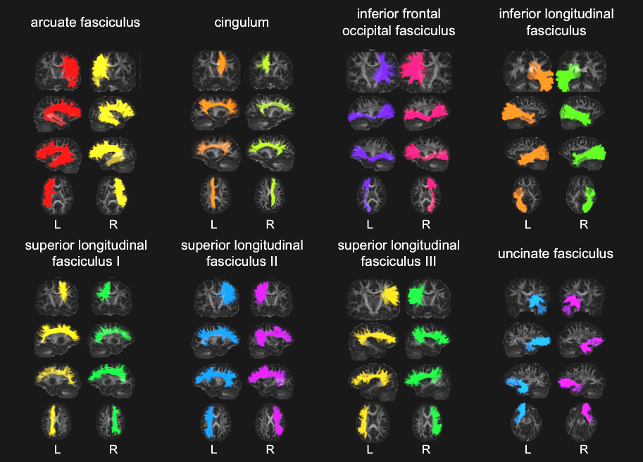
Youth aged 16 to 24 years born with complex CHD who underwent open heart surgery involving cardiopulmonary bypass during the first two years of life were recruited to participate in this cross-sectional study. Control youth of similar age and sex were also recruited. All participants completed a brain MRI including a high-resolution anatomical T1-weighted acquisition (TR = 8.1 ms, TE = 3.7 ms, flip angle = 8°, voxel size = 1.00 x 1.00 x 1.00 mm3) and a NODDI acquisition (TR = 9400 ms, TE = 78 ms, flip angle = 90°, voxel size = 2.00 x 2.04 x 2.00 mm3) on a 3T MRI System (Achieva X, Philips Healthcare, Best, The Netherlands) using a 32-channel head coil. The NODDI acquisition included a non-diffusion-weighted sequence (b = 0 s/mm2) with reversed phase encoding and two single-shell high angular resolution diffusion-weighted imaging sequences (b = 700 s/mm2 and 30 directions; b = 2000 s/mm2 and 60 directions). Data processing was accomplished with the use of Nextflow5 and Singularity.6 Diffusion-weighted images (DWIs) were denoised using Mrtrix PCA-based denoising.7 Eddy currents and susceptibility and motion artefacts were corrected using Topup and Eddy from FSL, brains were extracted using FSL Bet, and bias field correction was done using ANTs N4 correction. DWI intensities were normalized to a common range using Mrtrix. DWIs were resampled to the resolution of the T1-weighted images. Fiber ODFs were estimated with Constrained Spherical Deconvolution.8 Whole-brain tractograms were generated using probabilistic particle filter tracking,9 seeding from the white matter and white matter/grey matter interface and using 7 seeds per voxel. A modified version of RecoBundles10 was used to extract the white matter tracts shown in Figure 1. AMICO11 was used to compute maps of neurite density index (NDI) and orientation dispersion index (ODI), after which tractometry was performed to compute average NDI and ODI values for each white matter tract.12 NDI and ODI were compared between the CHD and control groups in each white matter tract using independent-samples t-tests, employing the false discovery rate correction for multiple comparisons.
Results
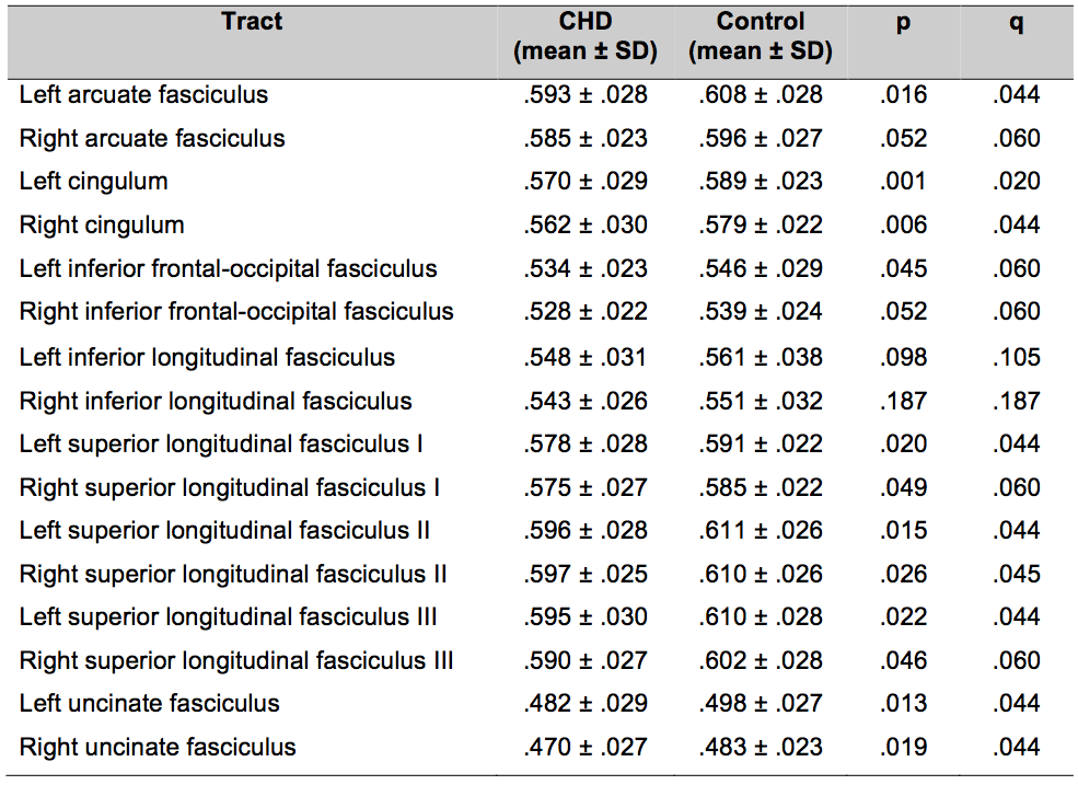
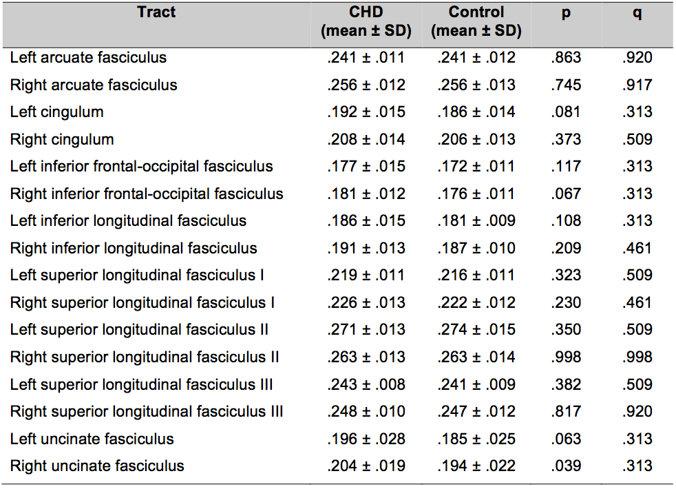
Data was collected from 43 youth born with CHD (24 females, mean age = 20.0 years) and 40 control youth (24 females, mean age = 20.6 years). The two groups did not differ significantly in terms of age (p = .253) or sex (p = .700). NDI was significantly lower in youth born with CHD as compared to controls bilaterally in the cingulum, superior longitudinal fasciculus II, and uncinate fasciculus and in the left arcuate fasciculus and left superior longitudinal fasciculus I and III (Table 1; q < .05). ODI was higher in youth born with CHD as compared to control youth in the right uncinate fasciculus (Table 2; p < .05); however, this difference did not survive correction for multiple comparisons (q > .05).Discussion
As one of the first studies to use NODDI in this population, our findings suggest that a lower density of axons within white matter tracts is a key microstructural element of the white matter abnormalities in youth born with CHD. This reduced axonal packing could be the result of axonal loss and impaired axonal regeneration following early-life critical illness. Given the role of these association tracts in various neuropsychological functions, these alterations may be related to the persistent neurodevelopmental impairments experienced by youth born with CHD. Investigations exploring the extent to which these structural abnormalities are associated with functional outcomes are currently underway.Conclusion
This study revealed a lower density of axons within numerous association tracts in youth born with CHD. These findings pave the way for future structure-function studies that may establish low neurite density as a novel neurobiomarker of neurocognitive deficits in this clinical population, and may eventually facilitate the effective diagnosis and treatment of these difficulties.Acknowledgements
This work was supported by start-up funds from the Research Institute of the McGill University Health Centre and McGill University. KE has received studentship support in the form of a Canada Graduate Scholarship – Master’s from the Canadian Institutes of Health Research (2017-2018), funding from the Canada First Research Excellence Fund awarded to McGill University for the Healthy Brains for Healthy Lives initiative (2017-2018, 2018-2019), and a Lasha Research Fellowship from the McGill University Faculty of Medicine (2018-2019). We would especially like to thank the participants and their families for taking the time to take part in the study, as well as the MRI technologists and clinicians involved in this project.References
1. Bellinger DC, Wypij D, Rivkin MJ, et al. Adolescents with d-transposition of the great arteries corrected with the arterial switch procedure: neuropsychological assessment and structural brain imaging. Circulation. 2011;124(12):1361-1369.
2. Rivkin MJ, Watson CG, Scoppettuol LA, et al. Adolescents with D-transposition of the great arteries repaired in early infancy demonstrate reduced white matter microstructure associated with clinical risk factors. J Thorac Cardiovasc Surg. 2013;146(3):543-549.
3. Watson CG, Stopp C, Wypij D, et al. Altered White Matter Microstructure Correlates with IQ and Processing Speed in Children and Adolescents Post-Fontan. J Pediatr. 2018;200:140-149.
4. Zhang H, Schneider T, Wheeler-Kingshott CA et al. NODDI: practical in vivo neurite orientation dispersion and density imaging of the human brain. NeuroImage. 2012;61(4):1000-1016.
5. Di Tommaso P, Chatzou M, Floden EW, et al. Nextflow enables reproducible computational workflows. Nat Biotechnol. 2017;35(4):316-319.
6. Kurtzer GM, Sochat V, Bauer MW. Singularity: Scientific containers for mobility of compute. PLoS One. 2017;12(5):e0177459.
7. Veraart J, Novikov DS, Christiaens D, et al. Denoising of diffusion MRI using random matrix theory. NeuroImage. 2016;142:394-406.
8. Tournier JD, Calamante F, Connelly A. Robust determination of the fibre orientation distribution in diffusion MRI: non-negativity constrained super-resolved spherical deconvolution. NeuroImage. 2007;35(4):1459-1472.
9. Girard G, Whittingstall K, Deriche R, et al. Towards quantitative connectivity analysis: reducing tractography biases. NeuroImage. 2014;98:266-278.
10. Garyfallidis E, Cote MA, Rheault F, et al. Recognition of white matter bundles using local and global streamline-based registration and clustering. NeuroImage. 2018;170:283-295.
11. Daducci A, Canales-Rodríguez EJ, Zhang H, et al. Accelerated microstructure imaging via convex optimization (AMICO) from diffusion MRI data. NeuroImage. 2015;105:32-44.
12. Cousineau M, Jodoin PM, Garyfallidis E, et al. A test-retest study on Parkinson's PPMI dataset yields statistically significant white matter fascicles. NeuroImage: Clinical. 2017;16:222-233.
Figures