3415
Structural connectivity between cerebellum and cerebral cortex in idiopathic generalized epilepsy: a diffusion tensor imaging study1University of Electronic Science and Technology of China, Chengdu, China
Synopsis
Probabilistic tracking method was applied to study the cerebellar efferent and afferent fibers in the patients with idiopathic generalized epilepsy (IGE), trying to address the structural connectivity (SC) alteration in the
Abstract
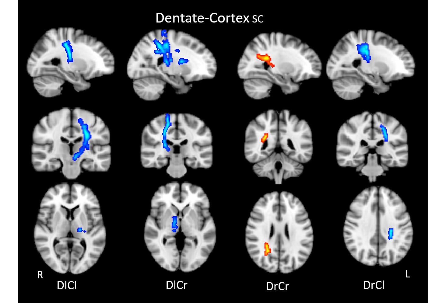
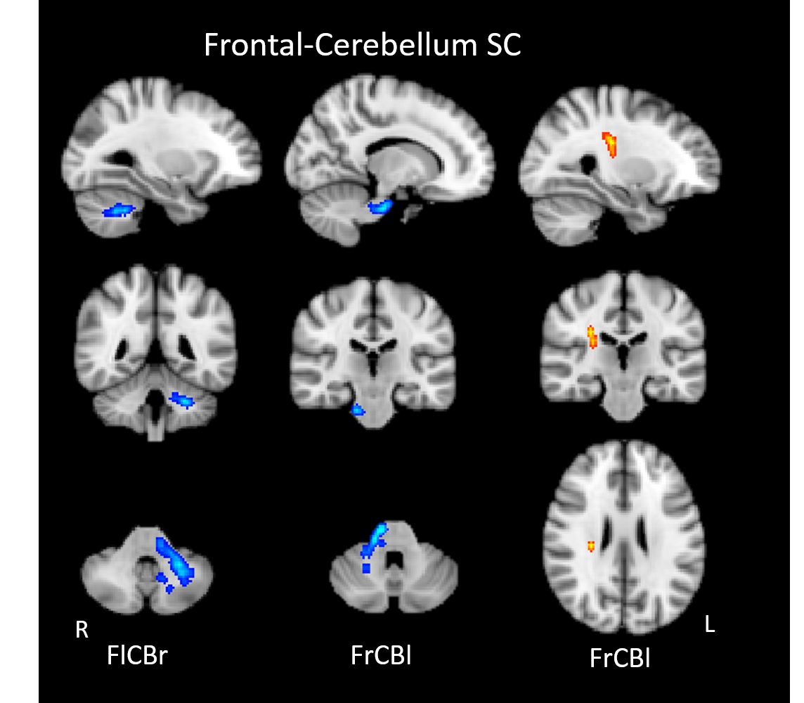
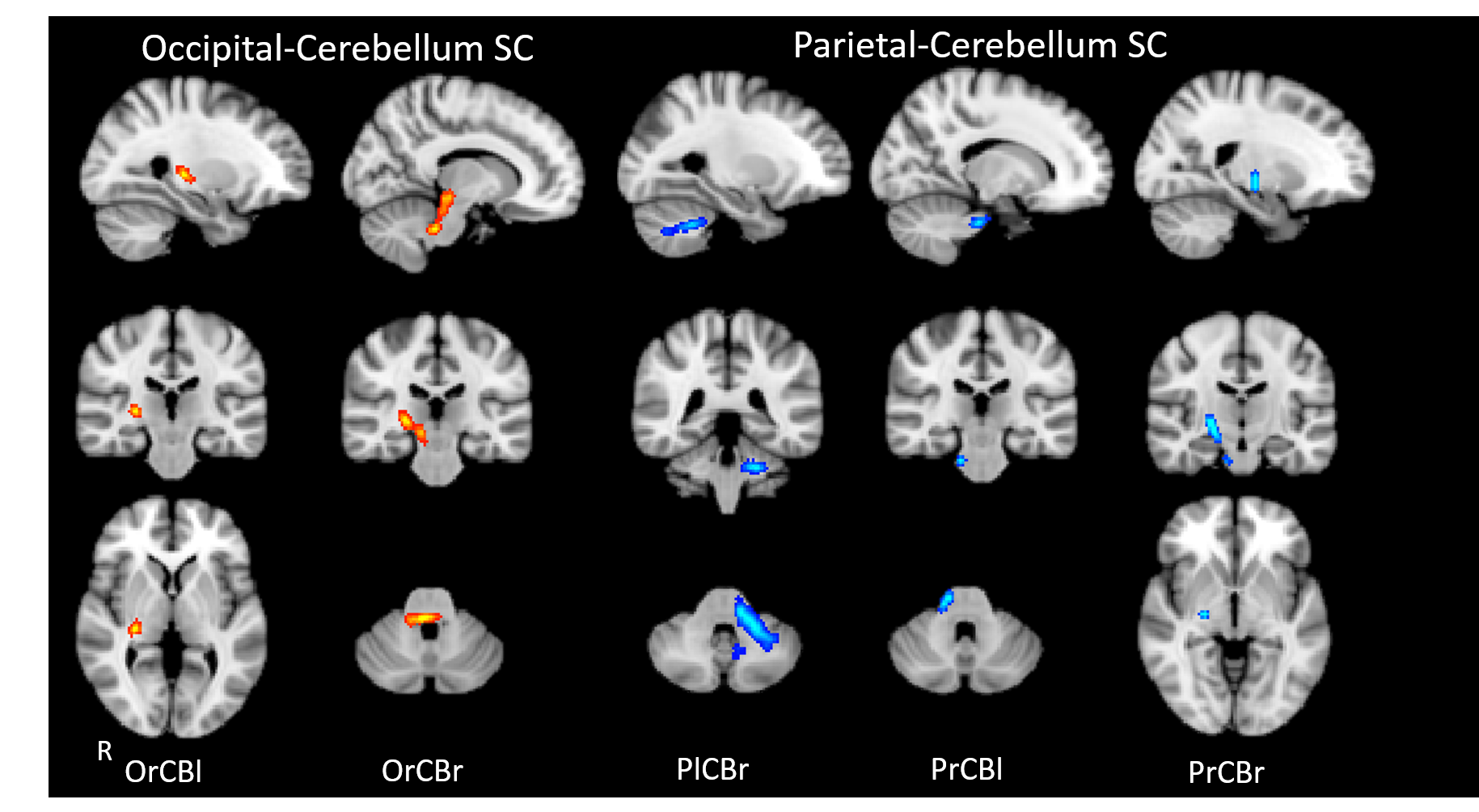
Introduction: Accumulated studies have suggested disrupted functional interaction in the cerebello-cerebral circuit, supporting a potential regulation effect of cerebellum on functional brain networks in idiopathic generalized epilepsy (IGE)1. While the underlying structural connectivity (SC) change in this circuit has not been studied systematically in IGE. Present study aims to investigate the bidirectional SC in cerebello-cerebral circuit in the patients with IGE. Methods: Sixty-six patients with IGE were collected in present study. We performed bidirectional probabilistic tracking between cerebellum and cerebral cortex for bilateral hemispheres. Dentate was suggested to be main output nucleus of cerebellum, thus it was used as seed regions in this study to describe efferent fibers of cerebellum. In detail, dentate and cerebral cortex were set as seed target regions respectively for each hemisphere, probabilistic fiber tracking was performed to describe the efferent fibers of cerebellum to cerebral cortex, with the superior cerebellar peduncle(SCP) and thalamus setting as the waypoints. For the afferent fibers of cerebellum from cerebral cortex, probabilistic tracking was performed using frontal, parietal, temporal, occipital lobe and cerebellum as seed and target regions respectively and the bilateral middle cerebellar peduncle (MCP) as the waypoint. Then, the strength of SC in 20 tracts was extracted and compared between groups using two sample t-test with TFCE correction (p<0.05). Results: The patients with IGE showed decreased SC in bilateral cerebellar efferent fibers to the sensorimotor cortex through the corticospinal tract (DlCl, DlCr and DrCl). Specifically, bilateral thalamus were involved in the abnormal SC in efferent fibers of left cerebellum to bilateral cortex (DlCl and DlCr). Increased SC was observed in the afferent fibers of right cerebellum to ipsilateral occipital cortex (DrCr)(Figure 1). For the altered SC in the cerebellar afferent fibers from cerebral cortex, consistent with the increased connectivity in DrCr, increased SC was observed in retrolenticular part of internal capsule and MCP in the afferent fibers of bilateral cerebellum from the right occipital cortex (OrCBl and OrCBr) (Figure 1). Contrary to the alterations in DlCr, increased SC was found in the afferent fibers from the sensorimotor cortex (FrCBl) (Figure 2). Besides, both frontal and parietal cortex were involved in the decrease of intercerebellar SC in MCP (FlCBr, FrCBl, PlCBr and PrCBl) (Figure 2 and Figure 3). Moreover, decreased SC in posterior limb of internal capsule in the afferent fibers from right parietal cortex (PrCBr) was also observed in IGE (Figure 3). Discussion: In line with previous findings in functional studies, the disrupted SC of cerebellum with motor cortex and basal ganglia suggested its participation in the abnormality of motor system in IGE1, 2. Besides, the involvement of thalamus in decreased SC in the cerebellar efferent fibers might be responsible for deficient functional inhibition in distributed cerebral cortex, especially the frontoparietal regions, which was associated with hyper excitability in IGE. Frontoparietal cortex related disruption of intercerebellar SC might contribute to the impaired frontoparietal cognitive function3, 4. Specifically, contrary alteration of SC in efferent and afferent fibers between frontal cortex and contralateral cerebellum was presumed to indicate abnormally unmatched ascending and descending communication in IGE, which might be a specific feature for understanding the pathological mechanisms of IGE. Finally, increased SC in the fibers between occipital and cerebellum might be related with defective visual information process in patients5. Conclusion: This study further identified the effects of cerebellum on the ganglia-thalamo-cortical circuit, which associated with the motion abnormality in IGE. Frontoparietal cortex related disruption of SC in cerebellum might contribute the cognitive impairments in patients with IGE. More important, present findings demonstrated unbalanced SC in the efferent and afferent fibers between cerebellum and frontal cortex and presumed it might be a specific feature of SC in IGE. In all, present findings provided new clues for further understanding physiopathologic mechanism in IGE from structure aspect.Acknowledgements
No acknowledgement found.References
1. L. Dong, C. Luo, Y. Zhu, et al., Complex discharge-affecting networks in juvenile myoclonic epilepsy: A simultaneous EEG-fMRI study. Hum Brain Mapp, 2016. 37(10): p. 3515-29.
2. C. Luo, Q. Li, Y. Xia, et al., Resting state basal ganglia network in idiopathic generalized epilepsy. Hum Brain Mapp, 2012. 33(6): p. 1279-94.
3. R.L. Buckner, F.M. Krienen, A. Castellanos, et al., The organization of the human cerebellum estimated by intrinsic functional connectivity. J Neurophysiol, 2011. 106(5): p. 2322-45.
4. J. Gong, X. Chang, S. Jiang, et al., Microstructural alterations of white matter in juvenile myoclonic epilepsy. Epilepsy Res, 2017. 135: p. 1-8.
5. G. Strigaro, P. Prandi, C. Varrasi, et al., Defective visual inhibition in photosensitive idiopathic generalized epilepsy. Epilepsia, 2012. 53(4): p. 695-704.
Figures